. 2024 Dec 10;121(50):e2410680121.
doi: 10.1073/pnas.2410680121. Epub 2024 Dec 4.

Alternative splicing of Clock transcript mediates the response of circadian clocks to temperature changes

Affiliations

Alternative splicing of Clock transcript mediates the response of circadian clocks to temperature changes

Yao D Cai et al. Proc Natl Acad Sci U S A. .

Abstract

Circadian clocks respond to temperature changes over the calendar year, allowing organisms to adjust their daily biological rhythms to optimize health and fitness. In Drosophila, seasonal adaptations are regulated by temperature-sensitive alternative splicing (AS) of period (per) and timeless (tim) genes that encode key transcriptional repressors of clock gene expression. Although Clock (Clk) gene encodes the critical activator of circadian gene expression, AS of its transcripts and its potential role in temperature regulation of clock function have not been explored. Here, we observed that Clk transcripts undergo temperature-sensitive AS. Specifically, cold temperature leads to the production of an alternative Clk transcript, hereinafter termed Clk-cold, which encodes a CLK isoform with an in-frame deletion of four amino acids proximal to the DNA binding domain. Notably, serine 13 (S13), which we found to be a CK1α-dependent phosphorylation site, is deleted in CLK-cold protein. We demonstrated that upon phosphorylation at CLK(S13), CLK-DNA interaction is reduced, thus decreasing transcriptional activity of CLK. This is in agreement with our findings that CLK occupancy at clock genes and transcriptional output are elevated at cold temperature likely due to higher amounts of CLK-cold isoforms that lack S13 residue. Finally, we showed that PER promotes CK1α-dependent phosphorylation of CLK(S13), supporting kinase-scaffolding role of repressor proteins as a conserved feature in the regulation of eukaryotic circadian clocks. This study provides insights into the complex collaboration between AS and phospho-regulation in shaping temperature responses of the circadian clock.

Keywords: alternative splicing; circadian rhythm; phosphorylation; post-translational modification; temperature.


Conflict of interest statement

Competing interests statement:The authors declare no competing interest.

Figures
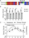
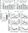
Fig. 1.
Fig. 1.
Cold induces alternative splicing of Clk. (A) Alignment of amino acid sequences encoded by two Clk transcripts isolated from heads of w1118 flies. Four amino acids (aa 13-16) are absent in CLK-cold. (B and C) The ratio of Clk-long to Clk-cold was measured in heads of w1118 flies by quantitative RT-PCR. Flies were entrained in 12 h:12 h LD at indicated constant temperature in (B) and 14 h:10 h LD at natural daily temperature cycles in (C). Flies were collected on LD3 at indicated time-points (ZT). Error bars indicate ± SEM (n = 3), **P < 0.01, *P < 0.05, two-way ANOVA and Šídák’s post hoc test.
Fig. 2.
Fig. 2.
Elevated CLK-DNA binding contributes to increased mRNA level of CLK targets at low temperature. (A and B) ChIP assays using fly head extracts comparing daily CLK occupancy at the per and vri promoter in w1118 flies collected at 25 and 10 °C. CLK-ChIP signals were normalized to % input. ChIP signals for an intergenic region were used for nonspecific background deduction. Flies were entrained in 12 h:12 h LD and collected on LD3 at indicated time-points (ZT) (n = 5). Error bars indicate ± SEM, **P < 0.01, *P < 0.05, two-way ANOVA and Šídák’s post hoc test. (C) per-E-box-luciferase (per-luc) reporter assay performed in Drosophila S2 cells. Luciferase activity was normalized to CLK-long and expressed as fold change relative to CLK-long. Error bars indicate ± SEM (n = 12), ***P < 0.001, two-tailed Student’s t test. (D and E) Steady state daily mRNA expression of CLK targets (per, vri, cwo, and dgol) and non-CLK targets (Clk and cry) in heads of w1118 flies. Flies were entrained in 12 h:12 h LD and collected on LD3 at indicated temperatures and time-points (ZT) (n = 3). Error bars indicate ± SEM, ***P < 0.001, **P < 0.01, *P < 0.05, two-way ANOVA and Šídák’s post hoc test.
Fig. 3.
Fig. 3.
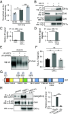
CLK(S13) is a substrate of CK1α. (A) per-E-box-luciferase (per-luc) reporter assay performed in Drosophila S2 cells. Luciferase activity was normalized to CLK(WT) and expressed as fold change relative to CLK-long. Error bars indicate ± SEM (n = 4), ***P < 0.001, one-way ANOVA and Dunnett post hoc test. (B) Western blots showing reciprocal coIPs to examine the interactions of CLK and CK1α. S2 cells were cotransfected with 0.8 μg of pAc-Clk-V5-His and 0.8 μg of pMT-ck1α-6xc-myc or transfected with a single plasmid for control experiments. Protein extracts were divided into two equal aliquots, and each aliquot was independently incubated with either α-c-myc beads or α-V5 beads. Immuno-complexes were analyzed by western blotting in the presence of the indicated antibody. (C and D) Bar graphs displaying quantification of reciprocal coIPs (B). Values for binding are normalized to amount of bait detected in the IPs and expressed as relative signal intensity (maximum value = 1). Error bars indicate ± SEM (n = 3), two-tailed Student’s t test. (E) Western blots showing mobility shift of CLK on a Phos-tag SDS-PAGE. S2 cells were transfected with 0.8 μg of pAc-Clk-V5 together with 0.6 μg of either pMT-ck1α-FH, pMT-ck1α(K49R)-FH, or pMT-FH. (F) Quantification of phosphorylated/total CLK in (E). Error bars indicate ± SEM (n = 3). ***P < 0.001, **P < 0.01, one-way ANOVA and Tukey’s post hoc test. (G) Schematic diagram depicting Drosophila melanogaster CLK (amino acid 1 to 1027) adapted from Mahesh et al. (51), and CK1α-dependent phosphorylation sites identified by mass spectrometry. Previously described domains of CLK: bHLH (aa 17-62) (35, 36); PAS-A (aa 96-144) (35, 36); PAS B (aa 264-309) (35, 36); C-terminal of PAS domain (PAC) (aa 315-379) (35); NLS (aa 480-494) (31); PER binding domain (PER BD) (aa 657-707) (52); Q-rich regions (aa 546-575, aa 957-1027), Poly-Q (aa 552-976) (35, 36) and NES (aa 840-864) (31). (H) Drosophila S2 cells were transfected with pAc-Clk(WT)-FLAG or pAc-Clk(S13A)-FLAG and cotransfected with an empty plasmid (pMT-cmyc-His) or pMT-ck1α-cmyc. Protein extracts were incubated with α-FLAG resin. Total CLK isoforms, CLK(pS13), and CK1α protein levels were analyzed by Western Blotting with the indicated antibodies. (I) Bar graph showing relative CLK pS13 levels in (H) normalized to total CLK isoforms. Error bars indicate ± SEM (n = 3), ***P < 0.001, one-way ANOVA and Dunnett post hoc test.
Fig. 4.
Fig. 4.
Flies expressing CLK(S13) variants display altered circadian behavioral and molecular output at 25 °C. (A) Double-plotted actograms of flies harboring two copies of transgenes expressing Clk(WT) or Clk with altered S13, a CK1α-dependent phosphorylation site, in Clkout background. Average activity of each genotype was plotted using FaasX. n represents the sample size; Tau (τ) represents the average period length of the indicated group of flies in constant darkness (DD). R represents percentage of flies that are rhythmic. Flies were entrained for 4 d in 12 h:12 h LD and then Left in DD for 7 d. (BD) Steady state daily mRNA expression of CLK targets (per, tim, and vri) in heads of Clk(WT); Clkout and Clk(S13D); Clkout flies. Flies were entrained in 12 h:12 h LD cycles at 25 °C and collected on LD3 at indicated time-points (ZT) (n = 3). Error bars indicate ± SEM. (E) Representative confocal images of dorsal projection of sLNvs neurons in adult fly brains stained with α-PDF (C7). Scale bar [merged image in Clk(WT) ZT3] represents 10 μm. Flies were entrained for 4 d in 12 h:12 h LD cycles and collected at the indicated times on LD4 for fixation and immunofluorescence analysis. (F) Box plot showing the quantification of PDF intensity in (E). Error bars indicate min to max. (GI) Steady state mRNA expression of CLK targets (per, tim, vri) in heads of Clk(WT); Clkout and Clk(S13A); Clkout flies. Flies were entrained in 12 h:12 h LD cycles at 25 °C and collected on LD3 at indicated time-points (ZT) (n = 3). Error bars indicate ± SEM, ***P < 0.001, **P < 0.01, *P < 0.05, two-way ANOVA and Šídák’s post hoc test.
Fig. 5.
Fig. 5.
CLK(S13) phosphorylation is required to maintain behavioral rhythmicity at colder temperature. (A and B) Double-plotted actograms of flies harboring two copies of transgenes for Clk(WT), Clk(S13A), or Clk(S13D). Average activity of each genotype was plotted using FaasX. n represents the sample size; Tau (τ) represents the average period length of the indicated group of flies in constant temperature. R represents percentage of flies that are rhythmic. For (A), flies were entrained for 4 d in 10 h:14 h 25 °C/17 °C (DD) and then released into constant 17 °C (DD) for 7 d. For (B), flies were entrained for 4 d in 12 h:12 h LD and then released into the indicated temperature for 7 d in DD. (C) Quantification of PDF intensity in dorsal projection of sLNvs neurons in adult fly brains stained with α-PDF (C7). Flies were entrained for 4 d in 12 h:12 h LD cycles and collected at the indicated times and temperature on LD4 for fixation and immunofluorescence analysis. Error bars indicate min to max, *P < 0.05, **P < 0.01, ***P < 0.001, two-way ANOVA.
Fig. 6.
Fig. 6.
CLK(S13) phosphorylation modulates CLK-DNA binding. (A) Model of Drosophila CLK (cyan)-CYC (yellow) bHLH heterodimer in complex with DNA (orange), with the side chain of S13 and the phosphate backbone of a proximal E-box nucleotide shown as spheres. The S13 hydroxyl oxygen atom, along with the oxygen atoms in the phosphate backbone, are shown in red. The Inset shows a close-up view and the estimated distance between the two oxygen atoms. The model was generated by superimposing an AlphaFold-predicted structure of Drosophila CLK-bHLH (aa 1-71) with the crystal structure of human CLK and BMAL1 in complex with E-box DNA (PDB 4H10). A cartoon representation is shown in the Upper Right. (B) Design of the Biolayer Interferometry (4) experiment. Biotin-labeled per promoter DNA was immobilized onto streptavidin-coated biosensors. CLK-bHLH-DNA interactions led to an increase in the effective thickness in the biolayer and a change in interference wavelength. (C) Quasi-steady state signal response of CLK-bHLH-DNA binding in the presence (black, filled) and absence (gray, hollow) of the phosphomimetic S13D mutation. Solid lines and shaded areas show fits and 95% prediction interval to the 4-parameter Hill equation for CLK-bHLH-WT and nonbinding baseline for CLK-bHLH-S13D. (DG) ChIP assays using fly head extracts comparing CLK occupancy at per and tim promoter of indicated fly genotypes on LD3 at indicated time-points (ZT) after entrainment in 12 h:12 h LD at 25 °C. CLK-ChIP signals were normalized to % input. ChIP signals for two intergenic regions were used for nonspecific background deduction (D and E, n = 3; F and G, n = 4). Error bars indicate ± SEM, ***P < 0.005, **P < 0.01, *P < 0.05, two-way ANOVA and Šídák’s post hoc test. (H) Model describing the alteration of the molecular clock in flies expressing CLK(S13A) variant. In WT flies, upon CLK removal from DNA promoted by PER-dependent phosphorylation and CWO-dependent mechanisms, S13 phosphorylation prevents CLK from binding back to DNA. CLK then undergoes nuclear export and further phosphorylation (31). Hyperphosphorylated CLK is either targeted for degradation or dephosphorylated by CKA-PP2A complex to replenish the pool of hypophosphorylated CLK (30, 31). Dephosphorylated CLK and newly translated, hypophosphorylated CLK then promotes transcription of clock-controlled genes. In S13A flies, after initial CLK removal from DNA by CWO, increased CLK(S13A)-DNA binding affinity leads to premature binding of transcriptionally inactive CLK and DNA. This leads to a decrease in CLK(S13A) nuclear export, hence reducing the replenishment of transcriptionally active CLK in the next cycle. PER and TIM are not depicted for simplicity. Created with BioRender.com licensed to the lab of J.C. Chiu. (I) Western blots comparing CLK protein profiles in heads of Clk(WT); Clkout and Clk(S13A); Clkout. Flies were entrained for 4 d in 12 h:12 h LD and collected at the indicated times on LD3. Brackets indicate hypo- and hyperphosphorylated CLK isoforms. α-HSP70 was used to indicate equal loading and for normalization. Bottom blot also detects CLK in the same two genotypes (W for WT and A for S13A) but the samples for each timepoint (ZT) were ran side by side to facilitate comparison of mobility shift. (J) Quantification of hyperphosphorylated/total CLK. The top half of the CLK signal shown at ZT4 in Clk(WT) flies (lane 1) is used as a reference to classify CLK isoforms as hyperphosphorylated (n = 4). Error bars indicate ± SEM, **P < 0.01, two-way ANOVA and Šídák’s post hoc test.
Fig. 7.
Fig. 7.
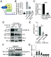
PER-DBT scaffolding promotes CK1α-dependent CLK(S13) phosphorylation. (A) Schematic illustrating the PER-DBT scaffolding model first proposed by Yu et al. (28). Created with BioRender.com licensed to the lab of J.C. Chiu. (B and C) per-E-box-luciferase (per-luc) reporter assay performed in S2 cells. (B) The fold activation of per-luc were graphed. Error bars indicate ± SEM (n = 3). One-way ANOVA and Tukey’s post hoc test. (C) Luciferase activities were normalized to CLK+DBT(K/R)+PER-NLS. Error bars indicate ± SEM (n = 3). (D) Drosophila S2 cells were transfected with indicated plasmids. Protein extracts were incubated with α-FLAG resin. Total CLK isoforms, pCLK(S13), PER, and CK1α protein levels were analyzed by Western Blotting with indicated antibodies. (E) Bar graph showing relative CLK pS13 levels in (D) normalized to total CLK level. Error bars indicate ± SEM (n = 3), ***P < 0.001, one-way ANOVA and Šídák’s post hoc test. (F and G) Fly heads of the specified genotypes were collected at the indicated times on LD3 after 2 d of entrainment. Extracted proteins (lysate) were subjected to western blotting with α-CLK(pS13) directly without IP. Total CLK proteins are shown in the Bottom panel and used for normalization and quantification shown in (G) (n = 3).
Fig. 8.
Fig. 8.
Model illustrating the regulation of the molecular clock by temperature-sensitive alternative splicing of Clk. Top panel: At warm temperature (25 °C), CLK-long isoform harboring S13 residue is expressed, due to alternative 3′ splice site selection of exon 2 of Clk. CLK-cold is also expressed but not shown to simplify the model. PER-DBT scaffolding promotes CK1α-dependent phosphorylation of CLK(S13) and reduces CLK-DNA binding. Bottom panel: At cold temperature (10 °C), expression of CLK-cold isoform lacking S13 residue increases, therefore lowering the impact of S13 inhibitory phosphorylation. As a result, this leads to elevated mRNA expression of CLK targets under low temperature. Created with BioRender.com licensed to the lab of J.C.C.

Update of

References

    1. Johnson C. H., Zhao C., Xu Y., Mori T., Timing the day: What makes bacterial clocks tick? Nat. Rev. Microbiol. 15, 232–242 (2017). - PMC - PubMed
    1. Dunlap J. C., Loros J. J., Making time: Conservation of biological clocks from fungi to animals. Microbiol. Spectr. 5, 5.3.05 (2017). - PMC - PubMed
    1. Swan J. A., Golden S. S., LiWang A., Partch C. L., Structure, function, and mechanism of the core circadian clock in cyanobacteria. J. Biol. Chem. 293, 5026–5034 (2018). - PMC - PubMed
    1. Cox K. H., Takahashi J. S., Circadian clock genes and the transcriptional architecture of the clock mechanism. J. Mol. Endocrinol. 63, R93–R102 (2019). - PMC - PubMed
    1. Patke A., Young M. W., Axelrod S., Molecular mechanisms and physiological importance of circadian rhythms. Nat. Rev. Mol. Cell Biol. 21, 67–84 (2020). - PubMed