. 2024 Dec 4;16(776):eadq5796.
doi: 10.1126/scitranslmed.adq5796. Epub 2024 Dec 4.

Diverse NKT cells regulate early inflammation and neurological outcomes after cardiac arrest and resuscitation

Collaborators, Affiliations

Diverse NKT cells regulate early inflammation and neurological outcomes after cardiac arrest and resuscitation

Tomoyoshi Tamura et al. Sci Transl Med. .

Abstract

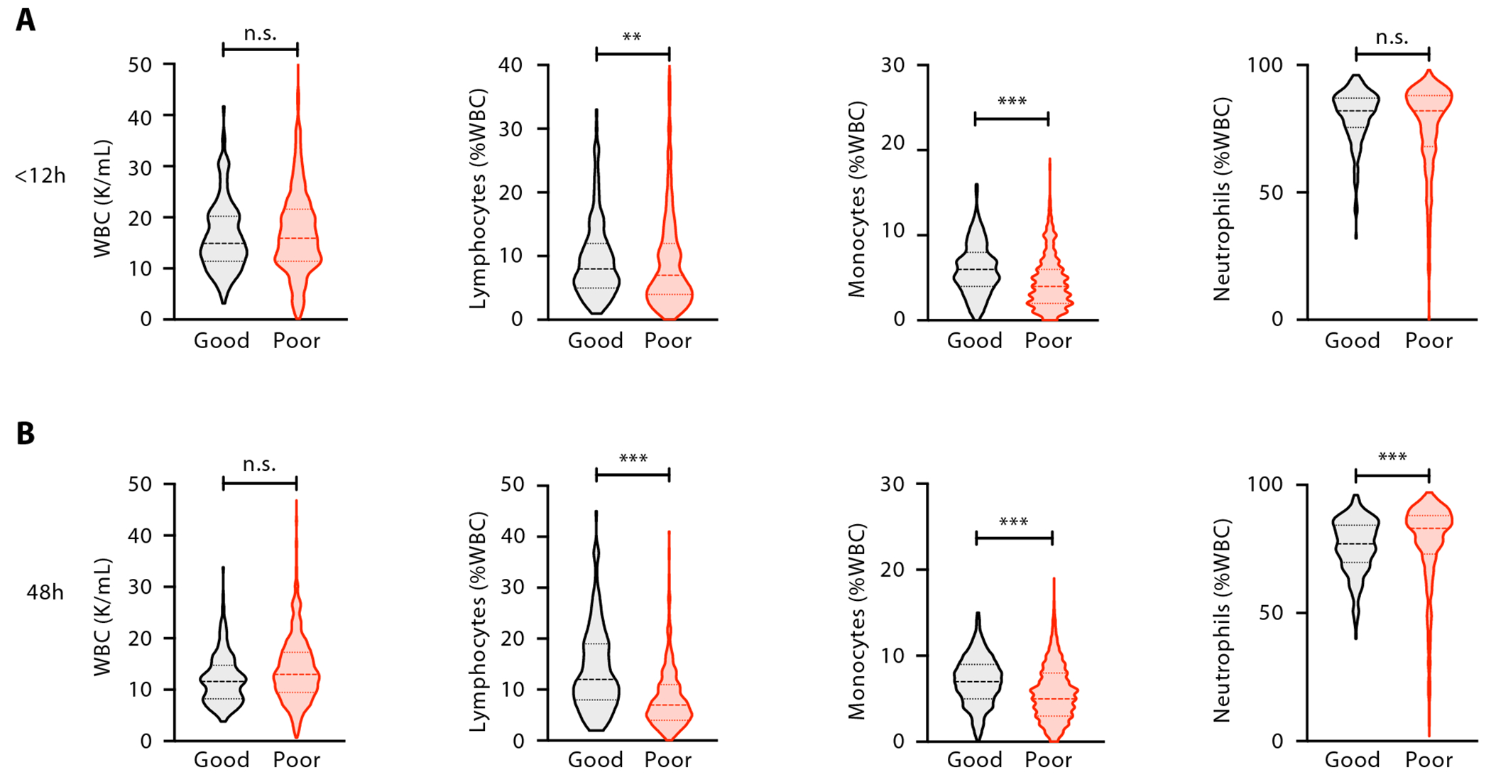
Neurological injury drives most deaths and morbidity among patients hospitalized for out-of-hospital cardiac arrest (OHCA). Despite its clinical importance, there are no effective pharmacological therapies targeting post-cardiac arrest (CA) neurological injury. Here, we analyzed circulating immune cells from a large cohort of patients with OHCA, finding that lymphopenia independently associated with poor neurological outcomes. Single-cell RNA sequencing of immune cells showed that T cells with features of both innate T cells and natural killer (NK) cells were increased in patients with favorable neurological outcomes. We more specifically identified an early increase in circulating diverse NKT (dNKT) cells in a separate cohort of patients with OHCA who had good neurological outcomes. These cells harbored a diverse T cell receptor repertoire but were consistently specific for sulfatide antigen. In mice, we found that sulfatide-specific dNKT cells trafficked to the brain after CA and resuscitation. In the brains of mice lacking NKT cells (Cd1d-/-), we observed increased inflammatory chemokine and cytokine expression and accumulation of macrophages when compared with wild-type mice. Cd1d-/- mice also had increased neuronal injury, neurological dysfunction, and worse mortality after CA. To therapeutically enhance dNKT cell activity, we treated mice with sulfatide lipid after CA, showing that it improved neurological function. Together, these data show that sulfatide-specific dNKT cells are associated with good neurological outcomes after clinical OHCA and are neuroprotective in mice after CA. Strategies to enhance the number or function of dNKT cells may thus represent a treatment approach for CA.


Conflict of interest statement

In disclosures unrelated to this work, RMB serves on Advisory Boards for Merck and Genentech. For disclosure unrelated to this work, PCH owns equity in and serve as consultant for iDoc Telehealth Solutions, Inc. and receives unrelated research funding from National Institute of Health/National Library of Medicine, Center for Disease Control and Prevention, and Day Zero Diagnostics, Inc. DAM and EAB are members of the TIMI Study Group which has received institutional research grant support through Brigham and Women’s Hospital from: Abbott Laboratories, Amgen, Anthos Therapeutics, Arca Biopharma, AstraZeneca, Bayer HealthCare Pharmaceuticals, Inc., Daiichi-Sankyo, Eisai, Intarcia, Janssen, Merck, Novartis, Pfizer, Quark Pharmaceuticals, Regeneron, Roche, Siemens, and Zora Biosciences. DAM has received consulting fees from Abbott Laboratories, InCarda, Inflammatix, Merck, Novartis, Regeneron and Roche Diagnostics. AJW receives research funding unrelated to this work from the National Institutes of Health (NHLBI, NINDS), Society for Academic Emergency Medicine Foundation, Zoll Foundation, National Aeronautics and Space Administration/Translational Research Institute for Space Health, Department of Defense, and the Centers for Disease Control and Prevention. Separately, and also unrelated to this work, AJW has received consulting fees from Inflammatix Inc. FI receives unrelated research funding from Kyowa Hakko Bio and Cyclerion. FI is a member of the Advisory Board of Nihon Kohden Innovation Center and ZOLL foundation Board of Directors. EYK received unrelated research funding from Bayer AG and 10X Genomics. EYK has an unrelated financial interest in Novartis AG. The remaining authors have no other disclosures or conflicts of interest relevant to this work.

Figures

Fig. 1.
Fig. 1.. Lymphopenia is associated with poor neurological outcomes after clinical OHCA.
(A and B) Complete blood count and cell differential (CBC diff) within 12h (A) or at 48h (B) post-OHCA. Data are presented as distribution of data along with the median and interquartile range. Data were analyzed by Mann-Whitney U test; n.s., not significant, *P < 0.05, **P < 0.01, ***P < 0.001. WBC, white blood cell. Good and Poor denotes good (n = 231) and poor (n = 1574) neurological outcomes at hospital discharge, respectively.
Fig. 2.
Fig. 2.. scRNA-seq identifies increased relative abundance of IFNG+NCAM1 (CD56)+ T cells early after OHCA in patients with good neurological outcomes.
scRNA-seq analysis of PBMC from patients at 6h (n = 7) or 48h (n = 9) post-OHCA, or healthy controls (n = 3), are shown. (A) Fractional abundance of lymphoid subsets. **P < 0.01, ***P < 0.001 compared with heathy controls. (B and C) Fine clustering of T cells shown by tSNE visualization with T cell subset or subpopulations noted (B) or separated by healthy cohort or time after OHCA (C), with neurological outcome 30 days after CA noted for post-OHCA patients. (D) Fractional abundance of T cell subsets and subpopulations. (E to G) Heatmaps of selected genes, shown by T cell clusters defined in (B). Mean gene expression of each cluster was used to calculate row Z score. Shown are marker genes associated with innate T cell subsets (E), cytotoxicity (F), or key cytokines and their associated transcription factors (G). neuro., neurological outcomes; Treg, regulatory T. Good or poor denotes good or poor neurological outcomes at 30 days after CA, respectively. Mixed effect logistic regression was used for testing the difference in cell-type frequencies among different patient groups (R package lme4).
Fig. 3.
Fig. 3.. An early increase in circulating sulfatide-specific NKT cells after OHCA is associated with good neurological outcomes 30 days after CA.
(A to D) Flow cytometry plots of peripheral blood mononuclear cells from patients after OHCA (Good outcomes, n = 14; Poor outcomes, n = 19) (or healthy controls, n = 15) are shown. Percentages of CD3+ T cell subsets are shown on the right with quantification on the left. Shown are frequencies of γδ T cells (A), MAIT cells (B), sulfatide-specific diverse NKT cells identified by CD1d tetramer loaded with sulfatide (C), and α-GalCer-specific NKT cells identified by CD1d tetramer loaded with α-GalCer (D). Median and IQR are shown; data were analyzed by Kruskal-Wallis test, *P < 0.05. α-GalCer, alpha-galactosylceramide; dTCR, delta T cell receptor.
Fig. 4.
Fig. 4.. Sulfatide-specific NKT cells improved neurological outcomes and survival after experimental CA.
Mice underwent experimental asystolic cardiac arrest followed by resuscitation. (A) Lipid antigen-specific NKT cells were assessed in the brain of WT or Cd1d−/− (CD1d KO) mice that were either naïve or at 24h post-CA. CD1d tetramer was loaded with lipid antigen (or unloaded) and conjugated to fluorophore. Flow cytometry plots and quantification at 24h post-CA are shown. (B to D) Shown are survival (B), neurological function scores (C), and fluoro-Jade staining of neuronal injury (D) for WT and CD1d KO mice after CA (n = 14, 17, respectively). Representative immunohistochemistry and quantification of neuronal injury in the hippocampus are shown for (D) (x80). (E and F) Shown are survival (E) and neurological function scores (F) for WT and Jα18 KO (Traj−/−) mice after CA (n = 11 each group). (G) Shown is qPCR assessment of chemokine Cxcl2 in whole brain homogenate of the indicated mice (n = 6 per group). Data are shown as relative fold change and were normalized to the housekeeping gene Hprt. (H) Shown are the numbers of polymorphonuclear leukocytes (PMN) in the brain of mice (n > 10, each group). Data in (A and D) were analyzed by Kruskal Walis test; data in (B and E) were analyzed by Log-rank test; data in (C,F,G,H) were analyzed by Mann-Whitney U test; for (C and F), dead mice were excluded from statistical analysis of neurological function scores. *P < 0.05, **P < 0.01, n.s., not significant. Survival and neurological function score results shown are from a representative experiment.
Figure 5.
Figure 5.. snRNA-seq reveals that NKT cells reduce the inflammatory response in hippocampal neurons and glial cells after experimental CA.
(A) Experimental design. WT and Cd1d−/− (CD1d KO) mice underwent experimental asystolic cardiac arrest for 10-minutes followed by resuscitation with CPR and epinephrine i.v. At 24 h post-ROSC, snRNA-seq was performed on the hippocampal region of the brain. (B) Shown is a t-SNE plot of cell types (by color) in the hippocampus post-CA. (C) Shown are the number of DEG between WT and CD1d KO mice, calculated by cell type (See fig. S13 and data file S4). The number of genes with log2fold-change (FC) > 0.05 and adj. p-value < 0.05 are shown. (D to F) GSEA performed by cell type with the Hallmark database. (D) Significantly different (adj. p-value < 0.05) gene sets are shown by cell type, with gene ratio per pathway (dot size) and normalized enrichment score (color). The IFN-γ response is highlighted in red. (E and F) Enrichment plots for gene sets increased in CD1d KO mice compared to WT mice are shown for inhibitory neurons (E) and microglia (F). (G) Shown is a volcano plot of gene expression in microglia comparing WT (791 cells) and CD1d KO (938 cells) mice post-CA. 115 DEG had log2FC > 0.05 and adj. p-value < 0.05. Astro, astrocytes; ChoPlex, choroid plexus; Endo, endothelium; Epend, ependymal cells; Ext, excitatory neurons; Inh, inhibitory neurons; Micro, microglia; Nb, neuroblasts; Oligo, oligodendrocytes; OPC, oligodendrocyte precursor cells; PeriFib or Perivasc Fb, perivascular fibroblasts.
Fig. 6.
Fig. 6.. NKT cells reduce Ifng expression and recruitment of myeloid cells to the brain after CA.
(A to C) qPCR assessment of cytokines and chemokines in whole brain homogenate from WT and Cd1d−/− (CD1d KO) mice that are naïve (Nv, n = 2 to 8) or post-CA (n = 4 to 8). (A to C) Shown are measurements of selected cytokines known to be dysregulated in clinical CA and regulated by NKT cells in other diseases (A), cytokines that can induce expression of Ifng (B), and monocyte/macrophage-attracting chemokines (C). Data are shown as relative fold change and were normalized to the housekeeping gene Hprt. (D) Shown are the number of monocytes/macrophages (CD45hi11b+68hi), measured by flow cytometry, in brains isolated from WT and CD1d KO mice that were either naive (n = 5) or 24h post-CA (n = 10, n = 7, respectively). Data were analyzed by Mann-Whitney U test; *P < 0.05.
Fig. 7.
Fig. 7.. Sulfatide treatment improved neurological function after experimental CA.
WT mice were treated with sulfatide at 18h pre-CA (n = 10); at 5 min post-ROSC (n = 16); or treated with vehicle at 5 min post-ROSC (n = 20). (A and B) Survival (A) and neurological function scores (B) are shown. Data in (A) were analyzed by Log-rank test. Data in (B) were analyzed by Kruskal-Wallis test. Dead mice were excluded from statistical analysis of neurological function scores. *P < 0.05, **p<0.01. Results shown are pooled from multiple independent experiments.

References

    1. Tsao CW, Aday AW, Almarzooq ZI, Alonso A, Beaton AZ, Bittencourt MS, Boehme AK, Buxton AE, Carson AP, Commodore-Mensah Y, Elkind MSV, Evenson KR, Eze-Nliam C, Ferguson JF, Generoso G, Ho JE, Kalani R, Khan SS, Kissela BM, Knutson KL, Levine DA, Lewis TT, Liu J, Loop MS, Ma J, Mussolino ME, Navaneethan SD, Perak AM, Poudel R, Rezk-Hanna M, Roth GA, Schroeder EB, Shah SH, Thacker EL, VanWagner LB, Virani SS, Voecks JH, Wang N-Y, Yaffe K, Martin SS, Heart Disease and Stroke Statistics-2022 Update: A Report From the American Heart Association., Circulation 145, e153–e639 (2022). - PubMed
    1. Yan S, Gan Y, Jiang N, Wang R, Chen Y, Luo Z, Zong Q, Chen S, Lv C, The global survival rate among adult out-of-hospital cardiac arrest patients who received cardiopulmonary resuscitation: a systematic review and meta-analysis., Crit. Care 24, 61 (2020). - PMC - PubMed
    1. Nielsen N, Wetterslev J, Cronberg T, Erlinge D, Gasche Y, Hassager C, Horn J, Hovdenes J, Kjaergaard J, Kuiper M, Pellis T, Stammet P, Wanscher M, Wise MP, Åneman A, Al-Subaie N, Boesgaard S, Bro-Jeppesen J, Brunetti I, Bugge JF, Hingston CD, Juffermans NP, Koopmans M, Køber L, Langørgen J, Lilja G, Møller JE, Rundgren M, Rylander C, Smid O, Werer C, Winkel P, Friberg H, TTM Trial Investigators, Targeted temperature management at 33°C versus 36°C after cardiac arrest., N. Engl. J. Med 369, 2197–2206 (2013). - PubMed
    1. Iadecola C, Anrather J, The immunology of stroke: from mechanisms to translation., Nat. Med 17, 796–808 (2011). - PMC - PubMed
    1. Miyatake H, Fujino K, Tanaka S, Tsujita Y, Horie M, Eguchi Y, Association between lymphocyte count and neurological outcomes in post-cardiac arrest patients treated with mild therapeutic hypothermia., Acute Med. Surg 6, 30–39 (2019). - PMC - PubMed

Publication types