. 2025 Jan:51:102199.
doi: 10.1016/j.tranon.2024.102199. Epub 2024 Dec 4.

4'-Demethylpodophyllotoxin functions as a mechanism-driven therapy by targeting the PI3K-AKT pathway in Colorectal cancer

Affiliations

4'-Demethylpodophyllotoxin functions as a mechanism-driven therapy by targeting the PI3K-AKT pathway in Colorectal cancer

Jun Liu et al. Transl Oncol. 2025 Jan.

Abstract

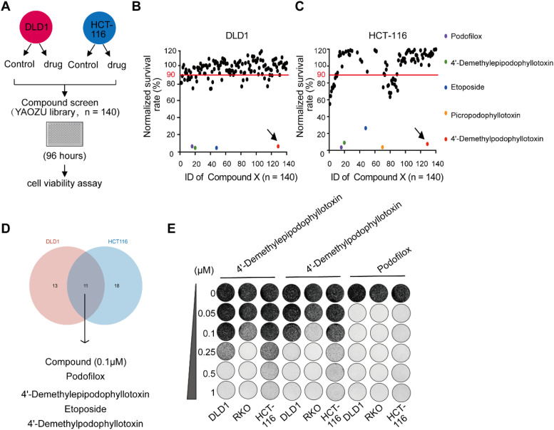
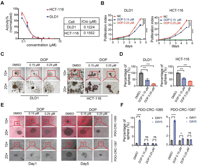
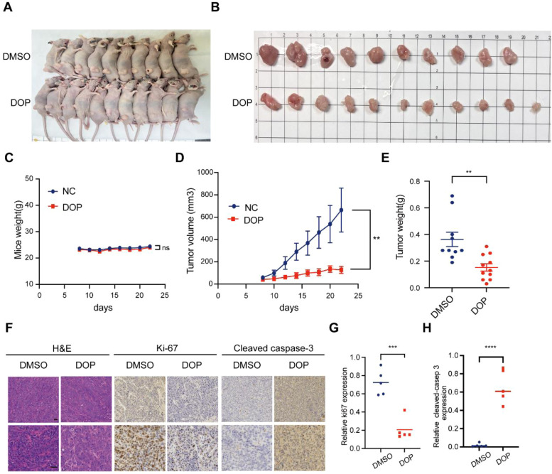
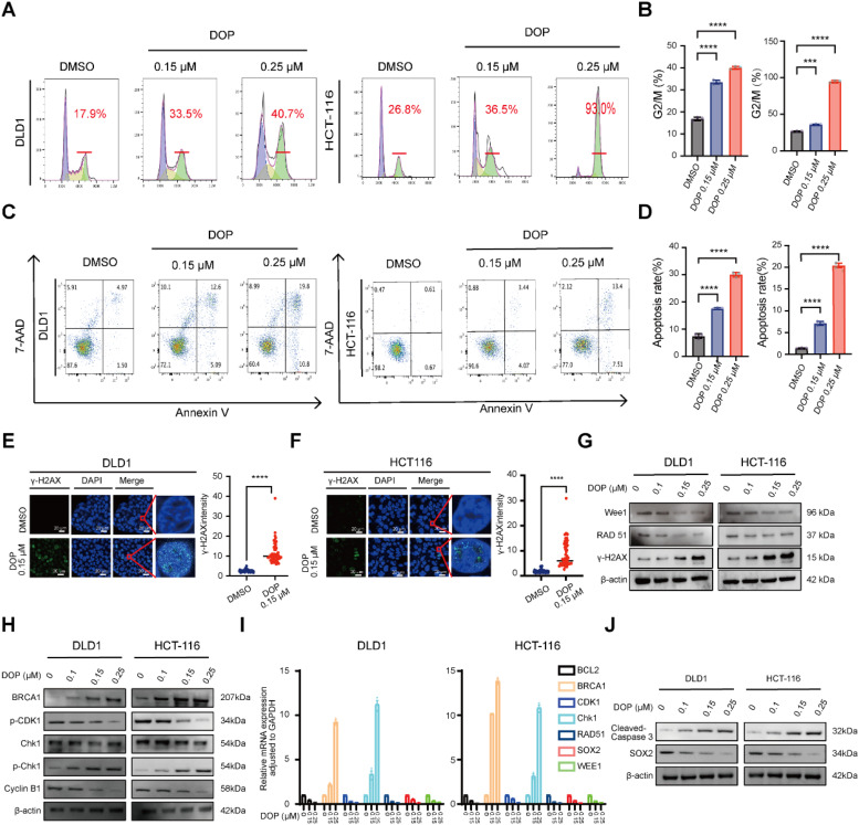
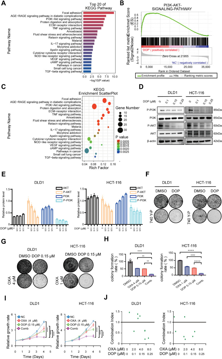
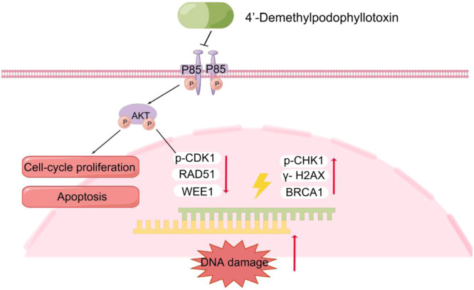
The treatment of colorectal cancer (CRC) poses significant challenges in terms of drug resistance and poor prognosis, necessitating the exploration of effective therapeutic strategies. In this study, high-throughput drug screening was utilized to identify Chinese herbal medicines with notable therapeutic effects on CRC. Among the compounds identified, 4'-demethylpodophyllotoxin (DOP), a derivative of podophyllotoxin, emerged as a potent anti-cancer compound. DOP exhibited time- and dose-dependent growth inhibition on CRC cell lines and tumor organoids derived from patients. RNA-seq revealed that DOP activated the PI3K-AKT pathway, leading to tumor cell apoptosis and cell cycle arrest at the G2/M phase. Additionally, DOP induced DNA damage in CRC cells. To further validate its therapeutic efficacy in CRC, the DLD1-derived xenograft model demonstrated that DOP effectively suppressed CRC growth in vivo. In conclusion, these findings highlight the significant therapeutic potential of DOP as an anti-tumor drug for treating CRC, thereby opening new avenues for investigating Podophyllotoxin derivatives in this specific field.

Keywords: 4′-Demethylpodophyllotoxin; Chemoresistance; Colorectal cancer; PI3K-AKT pathway.


Conflict of interest statement

Declaration of competing interest The authors declare that they have no conflict of interests.

Figures

Fig 1
Fig. 1
DOP exhibits sensitivity in the CRC Cell Line. (A) Diagram of the high-throughput compound screen (YAOZU library, n = 140). Plot of the proliferation ratio of high-throughput drug screening analysis of DLD1 (B) and HCT-116 (C), every colored point represents potential compound. (D) Wayne diagram representing potential compounds of DLD1 and HCT-116. (E) Effect of candidate compounds on the proliferation of DLD1, RKO and HCT-116 cells evaluated by colony formation assay.
Fig 2
Fig. 2
DOP suppresses the growth and self-renewal capacity of CRC in vivo. (A) IC50 values of DOP. (B) DOP treatment exhibited time- and concentration-dependent inhibition of proliferation of DLD1 and HCT-116. Representative bright field images of tumor-sphere (C) and histograms of sphere area (D) were shown (Scale bar, 200 μM). Representative bright-field microscopy images of CRC tissue-derived organoids (E) and histograms of sphere area (F). T test was used to test the difference among groups (Scale bar, 200μm).
Fig 3
Fig. 3
DOP inhibits CRC growth in vivo. (A) Representative pictures of tumors formed in nude mice bearing DLD1 cells after the treatment of DOP (25 mg/kg) and control group (DMSO, 25 mg/kg). (B) Representative images of xenograft tumors in nude mice after the treatment of DOP group (n = 11) and DMSO group (n = 10). (C) Body weight of nude mice in each group, ns means no significance. (D) Tumor growth curve in nude mice of DOP and control group after 14 days of treatment, ** means P value ≤ 0.01. (E) Tumor weight of nude mice in each group. (F) IHC staining of Ki-67 and cleaved caspase-3 in tumor tissues of nude mice (Scale bar, 200 μM). The IHC scores for Ki67 and cleaved caspase-3 expression were assessed in primary tumor tissue. These scores were determined based on the intensity score and the proportion of tumor cells that exhibited positive staining. The IHC score of Ki-67 (G) and cleaved caspase-3 (H).
Fig 4
Fig. 4
DOP caused cell cycle arrest in the G2/M phase, enhanced apoptosis and can also induce DNA damage response. (A) Flow cytometry analysis of cell cycle by PI staining after the treatment of DOP for 24 h in DLD1 and HCT-116 cells, the red horizontal line represents G2/M period, (B) corresponding quantitative alterations in the rate of G2/M period of DLD1 and HCT-116. (C) Flow cytometry analysis of apoptosis after the treatment of DOP for 24 h in DLD1 and HCT-116 cells, the red horizontal line represents G2/M period, (D) corresponding quantitative alterations in the rate of G2/M period of DLD1 and HCT-116. Representative images (left) and indirect quantitative intensity (right) of γ-H2AX immunofluorescence staining of DLD1 (E) and HCT-116 (F) treated with DMSO and DOP as indicated, the red square area represents γ-H2AX foci (Scale bar, 20 μM). (G) Cells treated with DOP as indicated were subjected to western blot analyses for DNA damage. Cells treated with DOP as indicated were subjected to western blot (H) and RT-qPCR (I) analyses for cell cycle arrest. (J) Cells treated with DOP as indicated were subjected to western blot analyses for apoptosis and stemness.
Fig 5
Fig. 5
The growth of colon rectal cells is hindered by DOP through the PI3K/AKT pathway. KEGG pathway analysis enriched by differentially expressed genes in DLD1 cell after treated with DOP (0.15 μM) for 48 h, KEGG annotation histogram (A), GSEA analysis for PI3K-AKT signaling pathway (B) and KEGG enrichment scatterplot (C). The representative (D) and quantitative image (E) for cells treated with DOP as indicated were subjected to western blot analyses. (F) The colony formation assay was conducted using PI3K-AKT agonist 740 Y-P (10 μM) in combination with DOP (0.15 μM). The representative (G) and quantitative image (H) for cells treated with oxaliplatin (OXA) at a concentration of 4 μM for DLD1 and 1 μM for HCT-116, along with DOP at a concentration of 0.15 μM, were utilized in the colony formation assay. (I) The CCK-8 assay was conducted using OXA (4μM) in combination with DOP (0.15 μM). (J) The combination index assay was conducted using OXA (2, 4, 8 μM) in combination with DOP (0.1, 0.15, 0.25 μM).
Fig 6
Fig. 6
Schematic model illustrating the regulation of DOP in PI3K/AKT axis in mediating cell-cycle proliferation, apoptosis and DNA damage in CRC.

Similar articles

Cited by

References

    1. Dekker E., Tanis P.J., Vleugels J.L.A., Kasi P.M., Wallace M.B. Colorectal cancer. Lancet. 2019;394(10207):1467–1480. - PubMed
    1. Biller L.H., Schrag D. Diagnosis and treatment of metastatic Colorectal cancer: a review. JAMa. 2021;325(7):669–685. - PubMed
    1. Ran H., Zhu Y., Deng R., et al. Stearoyl-CoA desaturase-1 promotes colorectal cancer metastasis in response to glucose by suppressing PTEN. J. Exp. Clin. Cancer Res. 2018;37(1):54. - PMC - PubMed
    1. Yang H., Yue G.G.L., Leung P.C., Wong C.K., Lau C.B.S. A review on the molecular mechanisms, the therapeutic treatment including the potential of herbs and natural products, and target prediction of obesity-associated colorectal cancer. Pharmacol. Res. 2022;175 - PubMed
    1. Shah Z., Gohar U.F., Jamshed I., et al. Podophyllotoxin: history, recent advances and future prospects. Biomolecules. 2021;11(4) - PMC - PubMed

LinkOut - more resources