. 2025 Jan;45(1):102-112.
doi: 10.1002/pd.6722. Epub 2024 Dec 4.

Role of Myofibroblasts in the Repair of Iatrogenic Preterm Membranes Subjected to Mechanical Stimulation

Affiliations

Role of Myofibroblasts in the Repair of Iatrogenic Preterm Membranes Subjected to Mechanical Stimulation

E Costa et al. Prenat Diagn. 2025 Jan.

Abstract

Objective: We examined the role of myofibroblasts in regulating Cx43 and collagen structure in iatrogenic preterm amniotic membrane (AM) defects subjected to mechanical stimulation.

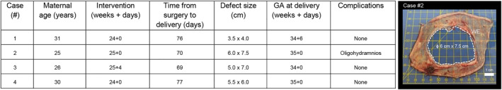
Method: Preterm AM specimens were collected from women undergoing planned preterm caesarean section after in utero intervention for correction of spina bifida by open fetal surgery (n = 4 patients; preterm delivery at 34 + 0 weeks to 35 + 0 weeks). Control specimens taken 5 cm away from the open fetal surgery defect site were compared with wound edge AM. In separate experiments, the effects of mechanical stimulation and co-treatment with Cx43 antisense on matrix and repair proteins were examined. Specimens were immunostained to detect αSMA and Cx43 in myofibroblasts and counterstained with DAPI to quantify nuclei shape. The direction of collagen fibrils in the wound edge region was examined by SHG imaging. Markers for matrix (collagen, elastin, GAG), inflammation (PGE2) and repair (TGFβ1) were examined by RT-qPCR and biochemical assays.

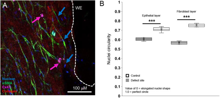
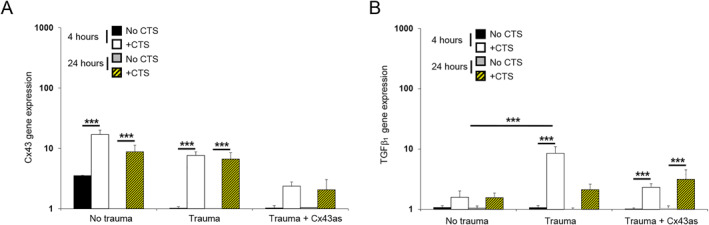
Results: In iatrogenic preterm AM specimens, the diameter of the open fetal surgery defect ranged between 3.5 and 7.5 cm. At the wound edge of the open fetal surgery defect, αSMA positive myofibroblasts had deformed nuclei and showed abundant Cx43 localized in the cell bodies or formed plaques. In the fibroblast layer, collagen had degenerated in some regions or had polarity near the wound edge. In preterm AM defects, mechanical stimulation and Cx43 antisense increased the levels of collagen and elastin but not GAG or PGE2 release. Mechanical stimulation increased Cx43 and TGFβ1 gene expression.

Conclusion: In open fetal surgery defects, myofibroblasts were elongated with collagen fibrils that either degenerated or had polarity. Whilst cells produced substantially higher Cx43 in the fibroblast than in the epithelial layer, they formed plaques, which may prevent migration and delay healing. Mechanical stimulation of preterm AM enhanced matrix repair proteins and the mechanotransduction should be explored to understand how Cx43 contributes to membrane integrity.

Keywords: Cx43; PPROM; fetal membranes; fetal surgery; iatrogenic; preterm birth.


Conflict of interest statement

The authors declare no conflicts of interest.

Figures

FIGURE 1
FIGURE 1
Clinical details for iatrogenic PPROM patients who underwent open fetal surgery for the correction of spina bifidae. Four cases of spontaneous ruptured fetal membranes were delivered preterm before 37 weeks of gestational age. The in utero intervention for correction of spina bifida neural tube defect by open fetal surgery took place between 24 + 0 and 25 + 0 weeks gestational age, creating a defect size between ∼3.0 and 7.5 cm, leading to late trimester iatrogenic preterm delivery between 34 + 0 and 35 + 0 weeks. In all cases, there was no infection after surgery or during the time up to delivery. All pregnancies were delivered by caesarean section. Image shows the size of a large diameter defect around 6 cm × 7.5 cm from a patient who delivered at 35 + 0 weeks complicated by oligohydramnios (Case #2). The approximate size of the defect is indicated for each case. Scale bar = 1 cm.
FIGURE 2
FIGURE 2
Myofibroblast morphology in human iatrogenic preterm AM. Fetal membranes were taken from a 31 year old patient who underwent open fetal surgery for correction of spina bifida neural tube defect that took place at 24 + 0 weeks gestational age leading to late trimester preterm delivery at 34 + 6 weeks (Case #1). Myofibroblast morphology was examined in αSMA expressing cells (green arrow) by IMF confocal microscopy in control AM specimens (A) and near the edge of the defect site (B, C). Cx43 was detected by immunostaining (pink arrow, C) and collagen by SHG imaging (red arrow). Control AM specimens were taken 5 cm away from the defect site. Signals for blue (DAPI), green (αSMA), pink (Cx43) and red (collagen) were detected by IMF confocal microscopy and SHG imaging. The dotted white lines show the length of the wound edge (WE) in the iatrogenic preterm AM specimen. Sale bar = 100 μM (A, B) and 300 μM (C). The white dotted box in (B) shows an enlarged image to show Cx43 localized in cell bodies.
FIGURE 3
FIGURE 3
Cx43 plaque formation in human iatrogenic preterm ruptured amniotic membrane. Representative images obtained by confocal microscopy are shown in the epithelial (A) and fibroblast layer (B) of the preterm AM defect from one late third trimester iatrogenic preterm donor (Case #1). The distribution of Cx43 was analyzed per tissue area for comparisons between the epithelial and fibroblast layers (C) or per cell nucleus (D). Error bars represent the mean and SEM values of 6 replicates from three late trimester iatrogenic preterm donors (34 + 0 weeks to 35 + 0 weeks, Case #1–3). Significant differences are indicated by ***p < 0.001. Statistical comparisons are indicated for control AM specimens and AM defect (p < 0.001***) in the epithelial layer (p < 0.001+++) and fibroblast layer (p < 0.001$$$). The dotted white and black lines show the length of the wound edge (WE) in the preterm AM specimen. Sale bar = 100 μM (A, B).
FIGURE 4
FIGURE 4
Myofibroblast nuclei deformation in human iatrogenic preterm AM. AM specimens were immunostained for αSMA to detect myofibroblast (pink arrow) nuclei (blue arrow, A). Circularity values for the nuclei shape of myofibroblasts expressing αSMA in the epithelial layer were compared to the fibroblast layer in control specimens and AM defect (B). Nuclei values close to 1 represent a perfect circle in contrast to zero, which represents a more elongated shape. The total number of nuclei counted ranged from 1202 to 1582 from 6 specimens (Case #1–3). Signals for blue (DAPI), green (αSMA) and pink (Cx43) were detected by immunofluorescence confocal microscopy. The dotted white lines show the length of the wound edge (WE) in the iatrogenic preterm AM specimen. Scale bar = 100 μM. ***p < 0.001.
FIGURE 5
FIGURE 5
Collagen organization in human iatrogenic preterm ruptured amniotic membrane. SHG imaging at the defect site showed the presence of a dense region of collagen fibers which either formed clusters (red arrow, A) or had alignment tangential to the wound edge (B) or were absent and had degenerated (red arrow, B). The organization of collagen in a basket‐like fashion in control preterm AM are shown in (C). Analysis of the direction of collagen fibers at a specific polarization angle is shown in the alignment graph (D). Error bars represent analysis of collagen fiber alignment in three regions and analysis repeated with six replicates taken from three late third trimester iatrogenic preterm donors (34 + 0 weeks to 35 + 0 weeks, Case #2–4). The dotted white lines show the length of the wound edge (WE) in the iatrogenic preterm AM specimen. Representative SHG images are shown for one field of view, where scale bar = 50 μm.
FIGURE 6
FIGURE 6
Effects of mechanical stimulation in human iatrogenic preterm AM. Preterm AM specimens were traumatized with a needed to create a 0.8‐mm defect and subjected to cyclic tensile strain (CTS) for 24 h. Mechanical stimulation was applied intermittently at 2% strain and 1 Hz frequency in the presence and absence of 50 μM Cx43 antisense (Cx43as). Absolute values for GAG (A), collagen (B) and elastin content (C) were normalized to dry tissue weight. PGE2 release was quantified in media samples (D). Explants cultured without cyclic tensile strain (−CTS) were compared to +CTS specimens. Error bars represent the mean and SEM values of 24 replicates from three late third trimester iatrogenic preterm donors (34 + 0 weeks to 35 + 0 weeks, Case #1–3). Significant comparisons are indicated for −CTS and +CTS conditions where ***p < 0.001. All other comparisons (not indicated) were not significantly different.
FIGURE 7
FIGURE 7
Effects of mechanical stimulation on gene expression in human iatrogenic preterm AM. Preterm AM specimens were traumatized with a needed to create a 0.8‐mm defect and subjected to cyclic tensile strain (+CTS) for 4 and 24 h. Mechanical stimulation was applied intermittently at 2% strain and 1 Hz frequency in the presence and absence of 50 μM Cx43 antisense (Cx43as). Gene expression of Cx43 (A) and TGFβ (B) are presented as ratio values and normalized to control values. In all cases, error bars represent the mean and SEM values of 12 replicates from three late third trimester iatrogenic preterm donors (34 + 0 weeks to 35 + 0 weeks, Case #1–3). Significant comparisons are indicated for −CTS and +CTS conditions where ***p < 0.001. All other comparisons (not indicated) were not significantly different.

Similar articles

References

    1. Devlieger D., Millar L. K., Bryant‐Greenwood G., Lewi L., and Deprest J., “Fetal Membrane Healing After Spontaneous and Iatrogenic Membrane Rupture: A Review of Current Evidence,” American Journal of Obstetrics and Gynecology 195, no. 6 (2006): 1512–1520, 10.1016/j.ajog.2006.01.074. - DOI - PMC - PubMed
    1. Deprest J. A., Lerut T. E., and Vandenberghe K., “Operative Fetoscopy: New Perspective in Fetal Therapy?,” Prenatal Diagnosis 17, no. 13 (1997): 1247–1260, 10.1002/(sici)1097-0223(199712)17:13<1247::aid-pd329>3.3.co;2-n. - DOI - PubMed
    1. Beck V., Lewi P., Gucciardo L., and Devlieger R., “Preterm Prelabor Rupture of Membranes and Fetal Survival After Minimally Invasive Fetal Surgery: A Systematic Review of the Literature,” Fetal Diagnosis and Therapy 31, no. 1 (2012): 1–9, 10.1159/000331165. - DOI - PubMed
    1. Gratacos E., Sanin‐Blair J., Lewi L., et al., “A Histological Study of Fetoscopic Membrane Defects to Document Membrane Healing,” Placenta 27, no. 4‐5 (2006): 452–456, 10.1016/j.placenta.2005.03.008. - DOI - PubMed
    1. Joyce E. M., Moore J. J., and Sacks M. S., “Biomechanics of the Fetal Membrane Prior to Mechanical Failure: Review and Implications,” supplement, European Journal of Obstetrics & Gynecology and Reproductive Biology 144 Suppl 1, no. S1 (2009): S121–S127, 10.1016/j.ejogrb.2009.02.014. - DOI - PMC - PubMed

Publication types

LinkOut - more resources