Circ_0089761 accelerates colorectal cancer metastasis and immune escape via miR-27b-3p/PD-L1 axis
- PMID: 39632246
- PMCID: PMC11617067
- DOI: 10.14814/phy2.70137
Circ_0089761 accelerates colorectal cancer metastasis and immune escape via miR-27b-3p/PD-L1 axis
Abstract
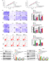
Circular RNAs have been implicated as critical regulators in the initiation and progression of colorectal cancer (CRC). This study was intended to elucidate the functional significance of the circ_0089761/miR-27b-3p/programmed cell death ligand 1 (PD-L1) axis in CRC. Our findings indicated that circ_0089761 expression was significantly elevated in CRC tissues and cell lines. Furthermore, the high expression of circ_0089761 was correlated with TNM stage and tumor size. Silencing circ_0089761 inhibited CRC cell proliferation, migration, and invasion, and increased apoptosis. Mechanistically, circ_0089761 facilitated its biological function by binding to miR-27b-3p to upregulate PD-L1 expression in CRC. Coculture experiments confirmed that low expression of circ_0089761 impeded CD8 + T cell apoptosis and depletion, activated CD8 + T cell function, and increased secretion of the immune effector cytokines IFN-γ, TNF-α, perforin, and granzyme-B. MiR-27b-3p inhibition or PD-L1 overexpression partially impeded CD8 + T cell function. The circ_0089761/miR-27b-3p/PD-L1 axis is postulated to exert pivotal functions in the mechanistic progression of CRC. Furthermore, it holds promising prospects as a feasible biomarker and therapeutic target for CRC.
Keywords: PD‐L1; circ_0089761; colorectal cancer; miR‐27b‐3p.
© 2024 The Author(s). Physiological Reports published by Wiley Periodicals LLC on behalf of The Physiological Society and the American Physiological Society.
Conflict of interest statement
The authors declare that they have no conflicts of interest to report regarding the present study.
Figures


Similar articles
-
Circ_0007422 Knockdown Inhibits Tumor Property and Immune Escape of Colorectal Cancer by Decreasing PDL1 Expression in a miR-1256-Dependent Manner.Mol Biotechnol. 2024 Sep;66(9):2606-2619. doi: 10.1007/s12033-023-01040-2. Epub 2024 Jan 22. Mol Biotechnol. 2024. PMID: 38253900
-
circ_0001006 Promotes Immune Escape in Non-small Cell Lung Cancer by Regulating the miR-320a/PD-L1 Axis.Iran J Immunol. 2025 Mar 30;22(1):58-69. doi: 10.22034/iji.2025.102661.2792. Iran J Immunol. 2025. PMID: 40055930
-
circ-Keratin 6c Promotes Malignant Progression and Immune Evasion of Colorectal Cancer through microRNA-485-3p/Programmed Cell Death Receptor Ligand 1 Axis.J Pharmacol Exp Ther. 2021 Jun;377(3):358-367. doi: 10.1124/jpet.121.000518. Epub 2021 Mar 26. J Pharmacol Exp Ther. 2021. PMID: 33771844
-
MicroRNA-124-3p suppresses PD-L1 expression and inhibits tumorigenesis of colorectal cancer cells via modulating STAT3 signaling.J Cell Physiol. 2021 Oct;236(10):7071-7087. doi: 10.1002/jcp.30378. Epub 2021 Apr 5. J Cell Physiol. 2021. PMID: 33821473
-
A scoping review on the potentiality of PD-L1-inhibiting microRNAs in treating colorectal cancer: Toward single-cell sequencing-guided biocompatible-based delivery.Biomed Pharmacother. 2021 Nov;143:112213. doi: 10.1016/j.biopha.2021.112213. Epub 2021 Sep 22. Biomed Pharmacother. 2021. PMID: 34560556
Cited by
-
LncRNA LINC01128 promotes prostate cancer cell proliferation, metastasis, and epithelial-mesenchymal transition by modulating miR-27b-3p.J Cancer Res Clin Oncol. 2025 Mar 4;151(3):98. doi: 10.1007/s00432-025-06153-6. J Cancer Res Clin Oncol. 2025. PMID: 40035871 Free PMC article.
-
CircRNAs in colorectal cancer: potential roles, clinical applications, and natural product-based regulation.Front Oncol. 2025 Jan 29;15:1525779. doi: 10.3389/fonc.2025.1525779. eCollection 2025. Front Oncol. 2025. PMID: 39944836 Free PMC article. Review.
-
miRNAs and T cell-mediated Immune Response in Disease.Yale J Biol Med. 2025 Jun 30;98(2):187-202. doi: 10.59249/PAYJ6872. eCollection 2025 Jun. Yale J Biol Med. 2025. PMID: 40589938 Free PMC article. Review.
References
-
- Bardou, M. , Barkun, A. N. , & Martel, M. (2013). Obesity and colorectal cancer. Gut, 62, 933–947. - PubMed
-
- Biller, L. H. , & Schrag, D. (2021). Diagnosis and treatment of metastatic colorectal cancer: A review. JAMA, 325, 669–685. - PubMed
-
- Chen, S. W. , Zhu, S. Q. , Pei, X. , Qiu, B. Q. , Xiong, D. , Long, X. , Lin, K. , Lu, F. , Xu, J. J. , & Wu, Y. B. (2021). Cancer cell‐derived exosomal circUSP7 induces CD8(+) T cell dysfunction and anti‐PD1 resistance by regulating the miR‐934/SHP2 axis in NSCLC. Molecular Cancer, 20, 144. - PMC - PubMed
-
- Chen, X. , Mao, R. , Su, W. , Yang, X. , Geng, Q. , Guo, C. , Wang, Z. , Wang, J. , Kresty, L. A. , Beer, D. G. , Chang, A. C. , & Chen, G. (2020). Circular RNA circHIPK3 modulates autophagy via MIR124‐3p‐STAT3‐PRKAA/AMPKα signaling in STK11 mutant lung cancer. Autophagy, 16, 659–671. - PMC - PubMed
MeSH terms
Substances
Grants and funding
LinkOut - more resources
Full Text Sources
Medical
Research Materials

