. 2024 Dec 4;15(1):10527.
doi: 10.1038/s41467-024-54110-7.

A landscape of X-inactivation during human T cell development

Affiliations

A landscape of X-inactivation during human T cell development

Björn Gylemo et al. Nat Commun. .

Abstract

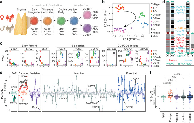
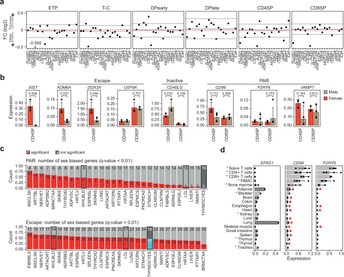
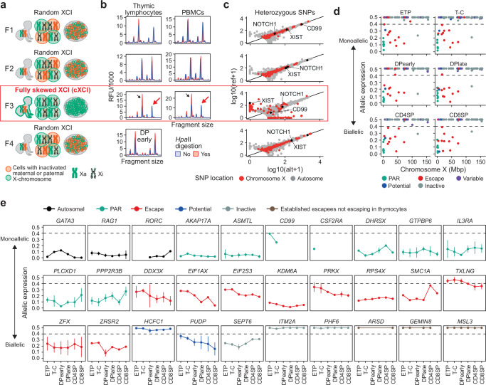
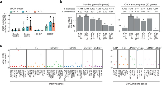
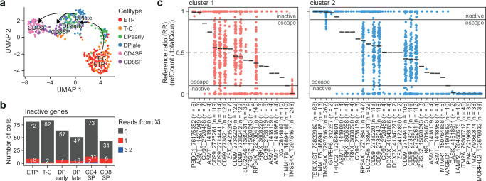
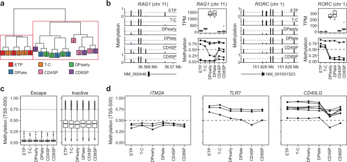
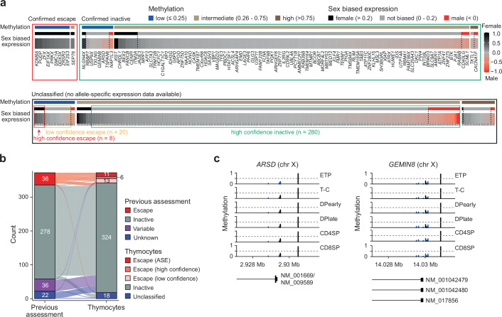
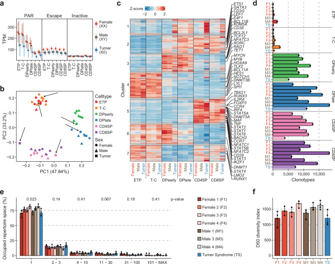
Females exhibit a more robust immune response to both self-antigens and non-self-antigens than males, resulting in a higher prevalence of autoimmune diseases but more effective responses against infection. Increased expression of X-linked immune genes in female T cells is thought to underlie this enhanced response. Here we isolate thymocytes from pediatric thymi of healthy males (46, XY), females (46, XX), a female with completely skewed X-chromosome inactivation (46, XX, cXCI) and a female with Turner syndrome (45, X0). Using whole exome sequencing, RNA sequencing and DNA methylation data, we present a sex-aware expression profile of T cell development and generate a high-resolution map of escape from X-chromosome inactivation (XCI). Unexpectedly, XCI is transcriptionally and epigenetically stable throughout T cell development, and is independent of expression of XIST, the lncRNA responsible for XCI initiation during early embryonic development. In thymocytes, several genes known to escape XCI are expressed from only one X-chromosome. Additionally, we further reveal that a second X-chromosome is dispensable for T cell development. Our study thus provides a high-resolution map of XCI during human development and suggests a re-evaluation of XCI in sex differences in T cell function.


Conflict of interest statement

Competing interests: The authors declare no competing interests.

Figures

Fig. 1
Fig. 1. Sex-biased escape gene expression in human thymocytes.
a Thymocyte development and markers used for cell sorting. b Principal component analysis (PCA) of normalized RNA-seq counts from male (triangle) and female (circle) developing thymocytes. c Expression dynamics as transcript per million (TPM) of genes associated with key transitions during thymocyte development. Boxplot representing median (central line), first and third quartiles (Q1 and Q3, respectively) (box edges) and 1.5*inter quartile range (IQR) from Q1 and Q3 (whiskers) from seven biological replicates (3 males and 4 females) are shown. d Schematic representation of selected genes escaping and inactivated by X-chromosome inactivation on the active (Xa) and inactive (Xi) X-chromosome. e Sex-biased expression of genes across the X-chromosome as log2 fold change (FC) of expression in female over male thymocytes. Horizontal red lines indicate median (dotted line) ± IQR of inactive genes (filled lines). FC (log2) values have been capped to 1 or −1 if above or below ±1, respectively. FC (log2) (dots) and standard error (vertical lines) of seven thymocyte populations from females (n = 4) and males (n = 3) shown. f Sex-biased gene expression across the X-chromosome as log2 FC of expression in female over male thymocytes by known XCI status. P-values from Benjamini-Hochberg (BH) corrected two-tailed T-test. Boxplot representing median (central line), Q1 and Q3 (box edges) and 1.5*IQR from Q1 and Q3 (whiskers) across all genes. Dots representing mean of six thymocyte populations from seven biological replicates (3 males and 4 females). e, f XCI status defined based on previous assessment with potential XCI escape genes (blue) previously not investigated or classified as unknown. a, b, c ETP, early T cell progenitors; T-C, T cell committed thymocytes; DPearly, early double positive thymocytes; DPlate, late double positive thymocytes; CD4SP, CD4 single positive thymocytes; CD8SP, CD8 single positive thymocytes. d, e, f PAR, pseudoautosomal region. Source data are provided as a Source Data file.
Fig. 2
Fig. 2. Unique expression of PAR genes in human thymocytes.
a Sex-biased expression of genes in the pseudoautosomal region (PAR) as log2 fold change (FC) in the six thymocyte subpopulations. Dashed lines indicate median ± IQR for each cell type. Red dots represent FC (log2) values outside the −0.5:0.5 range that were capped to −0.5 or 0.5 FC (log2) with exact values indicated for each capped data point. ETP, early T cell progenitors; T-C, T cell committed thymocytes; DPearly, early double positive thymocytes; DPlate, late double positive thymocytes; CD4SP, CD4 single positive thymocytes; CD8SP, CD8 single positive thymocytes. b Expression in male (brown) and female (red) CD4 and CD8 single positive (SP) thymocytes of a selection of escape, inactive and PAR genes analyzed by qPCR normalized to GAPDH. Bars indicate mean expression of biological replicates and hinges standard deviation. P-values comparing expression in males and females indicated above; unpaired, two-tailed student t-test. Standard deviation and p-value only shown where 3 biological replicates were available in each group. c Number of significant sex-biased PAR (upper) and escape genes (lower) in 30 different tissues, comparing thymocytes to GTEx data processed in Tukiainen et al.. Statistical significance for thymocytes: Wald test (BH corrected) P-value < 0.01, for GTEx data eBayes FDR < 0.01. Hematopoietic samples (whole blood, EBV-transformed lymphocytes and thymocytes) are highlighted. Tissue abbreviations can be found in Supplementary Table 3. d Gene expression of EPAS1, CD99 and P2RY8 across an independent set of 20 tissues analyzed by qPCR and normalized to GAPDH expression. Bars indicate mean expression and hinges standard deviation. Stars highlight tissues that have previously been shown to be sex-biased. Asterix and different nuances of gray depict tissues that were included (dark gray) or were not included (light gray, asterisk) in the analysis of sex-biased expression in GTEx shown in panel c. PBMC, peripheral blood mononuclear cells. Source data are provided as a Source Data file.
Fig. 3
Fig. 3. X-inactivation dynamics during human T cell development.
a Overview of X-chromosome inactivation (XCI) skewing in females included in the thymocyte data set with schematic representation of active (Xa) and inactive (Xi) X-chromosomes. b HUMARA assay of Female 1 (F1), F2 and F3 in thymic lymphocytes and peripheral blood mononuclear cells (PBMCs) and F4 in early double positive (DPearly) cells. Black arrows show peak detection in digested and undigested DNA and red arrows show complete loss of one allele after HpaII digestion (red) indicating complete XCI skewing (cXCI). c Allele-specific expression (ASE) of genes with heterozygous SNPs (hSNP) as expression from the reference (ref) and alternative (alt) allele from chromosome X and autosomal genes in F1, F2, F3 and F4. d ASE, chromosomal location and escape status of expressed X-chromosome genes with at least one hSNP in F3. e ASE of a selection of genes across thymocyte development in F3 (n = 1 for each thymocyte subpopulation). Dots and lines indicate mean and standard deviation, respectively. d, e XCI status defined based on previous assesment with potential XCI escape genes (blue) previously not investigated or classified as unknown. Source data are provided as a Source Data file.
Fig. 4
Fig. 4. Stable expression from the inactive X-chromosome.
a XIST expression in double negative (ETP, T-C), double positive (DPearly, DPlate), CD4SP and CD8SP cells based on qPCR for 3 separate qPCR probes (Supplementary Table 5). XIST expression normalized to expression of GAPDH. Bars indicate mean expression and hinges standard deviation from six biological replicates for each of the six thymocyte subpopulations. b Total minor allele read count for heterozygous SNPs in all inactive (left) and immune genes (right) in F3 (1 biological replicate for each thymocyte subpopulation). n, number of inactive or immune genes expressed from Xi in each cell type. c Minor allele read count of individual inactive or immune genes on chromosome X that are expressed from the inactive X-chromosome (Xi) in F3 (n = 1 for each thymocyte subpopulation). See “methods” for immune gene definitions. a, b, c ETP, early T cell progenitors; T-C, T cell committed thymocytes; DPearly, early double positive thymocytes; DPlate, late double positive thymocytes; CD4SP, CD4 single positive thymocytes; CD8SP, CD8 single positive thymocytes. Source data are provided as a Source Data file.
Fig. 5
Fig. 5. Single cell sequencing of human thymocyte populations.
a UMAP visualization of thymocyte subpopulations from Smart-seq2 data. b Number of cells with 0, 1 or ≥ 2 reads from inactive genes on the inactive X-chromosome (Xi). c Reference ratio (RR, reference read count / total read count) of heterozygous SNP expression from single cells pooled based on clusters identified in Supplementary Fig. 6. Black lines indicate mean RR per gene and cluster. Only SNPs with RR not equal to 0 or 1 shown. Escape from X-inactivation: reference ratio: 0.1–0.9. n, number of cells for which expression of the SNP was detected. Numbers after gene symbols are the genomic position of the heterozygous SNP. Source data are provided as a Source Data file.
Fig. 6
Fig. 6. DNA methylation on chromosome X is stable during T cell development.
a Hierarchical clustering of the 1000 most variable methylated sites in EPIC array methylation data from thymocyte populations in females F1, F3, and F4. Individuals indicated as numbers. b Methylation around transcription start sites (TSS) (left), gene expression as transcript per million (TPM) (top) and methylation of probes −500 and +1500 from TSS (bottom) in thymocyte subpopulation for genes involved in thymocyte development (RAG1 and RORC). Boxplot representing median (central line), first and third quartiles (Q1 and Q3, respectively) (box edges) and 1.5*inter quartile range (IQR) from Q1 and Q3 (whiskers) from three biological replicates (all female) are shown. c Boxplot representing median (central line), first and third quartiles (Q1 and Q3, respectively) (box edges) and 1.5*inter quartile range (IQR) from Q1 and Q3 (whiskers) of DNA methylation of all probes in −500 TSS range of escape (left) or inactive genes (right) from three biological replicates (all female), including only genes that are found to have the same XCI status in thymocytes and previous assessments. Dashed lines highlight low (methylation ≥0.25), intermediate (0.26-0.75) and high ( > 0.76) DNA methylation. d DNA methylation at TSS −500 across thymocyte development for ITM2A, TLR7 and CD40LG. a, b, c, d, ETP, early T cell progenitors; T-C, T cell committed thymocytes; DPearly, early double positive thymocytes; DPlate, late double positive thymocytes; CD4SP, CD4 single positive thymocytes; CD8SP, CD8 single positive thymocytes. Source data are provided as a Source Data file.
Fig. 7
Fig. 7. X-inactivation during T cell development.
a X-inactivation escape status as inactive (green box) or escape (red and orange box), DNA methylation (low, intermediate or high) and sex-biased expression as fold change (FC) (log2) in females over males of all expressed chromosome X genes in thymocytes. Confirmed escape status is inferred from F3 allele-specific expression analysis (inactive genes = mean allele-specific expression across thymocyte development (ASE) >0.4, escape genes = ASE < 0.4). DNA methylation for each gene as mean methylation of −500 TSS probes categorized into low (methylation ≤0.25), intermediate (0.26–0.75) and high ( >0.75) methylation. High confidence escape genes: low promoter methylation and sex-biased expression > 0.2; low confidence escape genes: low promoter methylation and sex biased expression 0-0.2; high confidence inactive genes: intermediate or high methylation and sex-biased expression < 0.2. Genes that fall outside of these thresholds are considered unclassified. See “methods” for details. b Escape status comparison between previous escape assessment and our new classification in thymocytes. Calls as escape genes were either based on allele-specific expression (ASE) or as high/low confidence according to panel a. c, DNA methylation of probes around transcription start sites of X-chromosome genes, ARSD and GEMIN8. ETP, early T cell progenitors; T-C, T cell committed thymocytes; DPearly, early double positive thymocytes; DPlate, late double positive thymocytes; CD4SP, CD4 single positive thymocytes; CD8SP, CD8 single positive thymocytes. Expression data from seven biological replicates (4 female, 3 male) and DNA methylation data from three female biological replicates. Source data are provided as a Source Data file.
Fig. 8
Fig. 8. The inactive X-chromosome does not contribute to T cell development in humans.
a Expression as transcript per million (TPM) of genes in PAR (pseudoautosomal region), escape and inactive genes for each thymocytes subpopulation in XX females (orange, 4 biological replicates), XY males (brown, 3 biological replicates) and X0 Turner syndrome patient (blue, 1 biological replicate). Points indicate mean and lines standard error. b Principal component analysis (PCA) of normalized RNA-seq counts from developing thymocytes from XX and XY karyotype females (circle) and males (triangle) as well as a Turner syndrome patient (TS) (square). TS samples are highlighted with black arrows. c Average Z-score per karyotype and cell type for each cluster of dynamic gene expression throughout thymocyte development identified in Supplementary Fig. 2c. d Clonotype count for thymocyte subpopulations in females (F1-F4), males (M1, M3, M4) and a Turner syndrome patient (TS). e Occupied repertoire space of clonotypes with 1, 2-3, 4-10, 11-30, 31-100 and 101-MAX number of clones in female, male and TS samples. P-values (Kruskal-Wallis test with adjustment for multiple testing by Holm) are indicated at the top. f Diversity score in DPearly, DPlate, CD4SP and CD8SP cells of F1, F2, F3, F4, M1, M3, M4 and TS. e, f Bars indicate mean and error bars 95% CI of four thymocyte subpopulations (DPearly, DPlate, CD4SP, CD8SP) for each individual. a, b, c, d, ETP, early T cell progenitors; T-C, T cell committed thymocytes; DPearly, early double positive thymocytes; DPlate, late double positive thymocytes; CD4SP, CD4 single positive thymocytes; CD8SP, CD8 single positive thymocytes. Source data are provided as a Source Data file.

References

    1. Fink, A. L., Engle, K., Ursin, R. L., Tang, W. Y. & Klein, S. L. Biological sex affects vaccine efficacy and protection against influenza in mice. Proc. Natl Acad. Sci. USA115, 12477–12482 (2018). - PMC - PubMed
    1. Klein, S. L. & Flanagan, K. L. Sex differences in immune responses. Nat. Rev. Immunol.16, 626–638 (2016). - PubMed
    1. Takahashi, T. et al. Sex differences in immune responses that underlie COVID-19 disease outcomes. Nature588, 315–320 (2020). - PMC - PubMed
    1. Hewagama, A., Patel, D., Yarlagadda, S., Strickland, F. M. & Richardson, B. C. Stronger inflammatory/cytotoxic T-cell response in women identified by microarray analysis. Genes Immun.10, 509–516 (2009). - PMC - PubMed
    1. Nalbandian, G. & Kovats, S. Understanding sex biases in immunity: effects of estrogen on the differentiation and function of antigen-presenting cells. Immunol. Res31, 91–106 (2005). - PubMed

Publication types