. 2024 Dec 4;15(1):10556.
doi: 10.1038/s41467-024-53848-4.

Potent AMA1-specific human monoclonal antibody against Plasmodium vivax Pre-erythrocytic and Blood Stages

Affiliations

Potent AMA1-specific human monoclonal antibody against Plasmodium vivax Pre-erythrocytic and Blood Stages

Anna C Winnicki et al. Nat Commun. .

Abstract

New therapeutics are necessary for preventing Plasmodium vivax malaria due to easy transmissibility and dormancy in the liver that increases the clinical burden due to recurrent relapse. In this manuscript we characterize 12 Pv Apical Membrane Antigen 1 (PvAMA1) specific human monoclonal antibodies from Peripheral Blood Mononuclear Cells of a Pv-exposed individual. PvAMA1 is essential for sporozoite and merozoite invasion, making it a unique therapeutic target. We show that humAb 826827 blocks the invasion of human reticulocytes using Pv clinical isolates and inhibits sporozoite invasion of human hepatocytes in vitro (IC50 of 0.3 - 3.7 µg/mL). Inoculation of human liver transgenic (FRG-humHep) female mice with humAb 826827 significantly reduces liver infection in vivo. The crystal structure of rPvAMA1 bound to 826827 shows that 826827 partially occupies the highly conserved hydrophobic groove in PvAMA1 that binds its known receptor, RON2. We have isolated a potent humAb that is isolate-transcendent, blocks both pre-erythrocytic and blood stage infection, and could be a potential therapy for Pv.


Conflict of interest statement

Competing interests: The authors declare no competing interests.

Figures

Fig. 1
Fig. 1. Sequence characterization of 12 humAbs and their selectivity, avidity, and affinity towards AMA1.
A Sequences of CDR3 IGH (orange) and corresponding IGL or IGK (yellow) from individual B cells from which humAbs were generated. The number of somatic hypermutations (SHM) of nucleotides that differ from germline sequences is shown for each clone. B 67 clonal groups were identified, from which clonal groups PvAMA1-specific humAbs were isolated is indicated. C HumAb reactivity to PvAMA1_Palo Alto, PvAMA1_PNG16, PkAMA1, PfAMA1_3D7, and TgAMA1 at varying concentrations (1.0, 0.5, 0.250, 0.125, 0.062, 0.313, 0.016, 0.008 µg/mL). D HumAb avidity as measured by reduction in binding (MFI) to AMA1 with varying concentrations of NH4SCN (0.0, 0.9, 1.2, 1.5, 1.8, 2.1, 2.4, 2.7, 3.0, 3.3, 3.6, 4.0 M). HumAbs were used at a concentration of 0.2 μg/mL. E Affinities (KDs) of PvAMA1-specific humAbs determined using SPR single-cycle kinetics. HumAbs 800801, 804805, and 808809 affinities could not be determined (N/A) in this assay. Standard error of the mean was calculated using Prism.
Fig. 2
Fig. 2. HumAbs inhibition of blood-stage infection.
A Dose response of PvAMA1-specific humAbs against Pf-PvAMA1 transgenic parasites of humAb with lowest IC50s. Each symbol represents the average of three replicates for every concentration. B The mean percentage (± SEM) of reticulocytes infected using Pv clinical isolates in short-term invasion inhibition with different humAbs at 100 μg/mL. Each dot represents a biological replicate from a different clinical isolate (n = 2–7). The flow cytometry background of target cells (reticulocytes) without parasites (mean is 9%, range 5–15% invasion) was subtracted from each experiment. Mouse mAb, 2C3 (100 μg/mL) binds to Duffy antigen on reticulocytes, thus blocking Pv invasion of reticulocytes (positive control) (p-value = 0.9891). Only humAb 826827 significantly inhibited reticulocyte invasion compared to the negative control (p-value = <0.0001). HumAb 043038 was used as a negative control for experiments represented in panel A and B. A multi-variant one-way ANOVA and Tukey’s secondary test was used to calculate the P-values compared to the positive and negative controls using Prism. C Dose response of humAb 826827 against four different Pv clinical isolates in short-term invasion inhibition cultures (Isolate 1, 2, 3, and 4 have IC50s of 39.96, 66.78, 25.22, and 61.04 μg/mL respectively. Note: These are different isolates than those used for Fig. 2B).
Fig. 3
Fig. 3. PvAMA1-specific humAbs inhibition of sporozoite invasion of human hepatocyte HCO4 cell line and primary human hepatocytes.
A IC50 of different humAbs for Pv sporozoite invasion of human HC04 hepatocytes was performed at five concentrations (0.1–1000 μg/mL). Values represent the mean (SEM) of three biological replicates, with each biological replicate performed in duplicate. 043038 was used as a negative control (p-value = 0.0045). Murine anti-CSP 2F2 was used as a positive control (p-value = >0.999). A multi-variant one-way ANOVA and Tukey’s secondary test was used to calculate the P-values compared to the negative control using Prism. B Percent inhibition of isolated Pv sporozoites (n = 5) into human primary hepatocytes using 826827 (white bars) at various concentrations. 043038 (shaded bar) was used as a negative control and was only tested at one concentration (p-value = <0.0001 at the same concentration). A multi-variant one-way ANOVA and Tukey’s secondary test was used to calculate the P-values compared to the negative control using Prism.
Fig. 4
Fig. 4. Reduction in P. vivax liver infection in FRG-humHep mice after PvAMA1 monoclonal blockaid.
Mice were injected intravenously with 30 μg and 300 μg of anti-PvAMA1 (humAb 826827, N = 4 for each concentration) and 300 μg anti-tetanus toxoid (humAb 048038, N = 4) approximately 3 hours before infection with 400,000 freshly dissected P. vivax sporozoites. A Serum concentrations of humAbs were measured 2 days after the sporozoite challenge and on day 8. B Liver sections were harvested on day 9 post-infection, weighed, homogenized, and P. vivax DNA levels were determined by RT-PCR. Each dot represents one mouse. Shown in mean ± SD. Statistics: one-tailed Mann-Whitney test.
Fig. 5
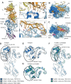
Fig. 5. Crystal structure of the PvAMA1–Fab 826827 complex.
A Ribbon representation of PvAMA1–Fab 826827 complex (PDB ID: 9DX6). The heavy chain of 826827 is orange, the light chain is yellow. PvAMA1 with N-terminal extension (residues 46-62) is white, Domain 1 (residues 63-248) light blue, Domain 2 (residues 249-385) dark blue, and Domain 3 purple (residues 386-474). Close-up views show interactions between humAb 826827 and (panel i) the mobile Domain 2 loop of PvAMA1 and (panel ii) the hydrophobic groove. For clarity, only polar interactions between side chains are indicated with dotted lines. B Five CDR loops (L1, L2, H1, H2, and H3) are involved in PvAMA1 binding. PvAMA1, in surface representation, is colored as described in panel A. The size of the buried interaction surface is indicated. C Comparison of PvAMA1 when bound by CDR-H3 of Fab 826827 or PvRON2 peptide (PDB ID: 5NQG). PvAMA1 is shown with transparency around the Domain 2 loop (dark blue). CDR-H3 and PvRON2 peptide (magenta) form disulfide-linked β-hairpin loops that bind to the hydrophobic groove on Domain 1. HumAb 826827 stabilizes the Domain 2 Ioop in a closed position on Domain 1. This loop is dislocated when PvRON2 peptide is bound to PvAMA1. D Ribbon representation of overlaid PvAMA1 structures, comprising our structure of PvAMA1 in complex with Fab 826827 (blue, PDB ID: 9DX6), PvAMA1 in complex with PvRON2 peptide (light blue, PDB ID: 5NQG), and unbound PvAMA1 (gray, PDB ID: 1W8K). This indicates that 826827 stabilizes the Domain 2 loop and allows for the refinement shown. Residues between T296 and F335 are absent in the electron density map of PDB 5NQG and 1W8K but residues 304–327 are well defined in our structure. Overlays were generated using Pymol, and refined root mean square deviation values are indicated. E Top view of the PvAMA1 overlays. F Overlay of PvAMA1 (blue, PDB ID: 9DX6) with PkAMA1 (green, PDB ID: 4UV6). The mobile Domain 2 loops adopt a similar conformation in both structures. G Top view of PvAMA1 and PkAMA1 overlay. (see Supplemental Table 4 for data collection and refinement statistics).
Fig. 6
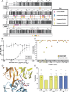
Fig. 6. Interaction residues of PvAMA1 with RON2 and 826827.
A Sequence alignment of published AMA1 amino acid sequences with the corresponding binding residues for RON2 (magenta) and humAb heavy chain (826; orange), light chain (827; yellow), or both chains (blue) arrows. The first line of the alignment represents the deposited structure 9DX6 corresponding to the PvAMA1 Palo Alto strain (ACB42438). Pv is the Sal1 variant (PVX_092275). The red box outlines the Domain 2 loop (304–327), highlighting 826827’s interactions with this region of PvAMA1. B Competition assay using 50 µg/mL of PvRON2 (Asp 2050 – Thr 2088) that competed with varying concentrations of humAb 826827 (40–0.039 µg/mL) to bind recombinant PvAMA1. 043038 was used as a negative control. Error bars indicate +/− SEM. C PvAMA1 residues contacting humAb heavy chain 826 (orange circles), light chain 827 (yellow squares), or both humAb chains (dark blue hexagons) and their mutations are displayed on the X-axis while the Y-axis shows the conservation of that position within 390 clinical isolate sequences. Highlighted amino acids represent those depicted in Fig. 6D. D Structural analysis of the interaction between 826 (orange) 827 (yellow) and PvAMA1 (blue) shows three observed polymorphisms in the binding epitope (G117R, N132D, and N130K; pink). E The pvama1 gene was sequenced in the seven Pv clinical isolates (Supplemental Table 3). Amino acids 117, 130, and 132 represent polymorphic contact residues of humAb 826827 to PvAMA1 Sal1 reference strain (PVX_092275). Bars represent the mean ( + SEM) of humAb invasion inhibition of WT compared to an isolate expressing a SNP at each of the three polymorphic residues (n = 7).

Update of

References

    1. Poespoprodjo, J. R. et al. Malaria. Lancet, 2023. - PubMed
    1. Menkin-Smith, L. & Winders, W. T. Plasmodium vivax Malaria, in StatPearls. 2023: Treasure Island (FL). - PubMed
    1. Battle, K. E. & Baird, J. K. The global burden of Plasmodium vivax malaria is obscure and insidious. PLoS Med18, e1003799 (2021). - PMC - PubMed
    1. Kho, S. et al. Evaluation of splenic accumulation and colocalization of immature reticulocytes and Plasmodium vivax in asymptomatic malaria: A prospective human splenectomy study. PLoS Med18, e1003632 (2021). - PMC - PubMed
    1. Kho, S. et al. Hidden biomass of intact malaria parasites in the human spleen. N. Engl. J. Med384, 2067–2069 (2021). - PubMed

Publication types

MeSH terms

Associated data

LinkOut - more resources