. 2025 Jan;44(2):505-533.
doi: 10.1038/s44318-024-00317-9. Epub 2024 Dec 4.

How J-chain ensures the assembly of immunoglobulin IgM pentamers

Affiliations

How J-chain ensures the assembly of immunoglobulin IgM pentamers

Chiara Giannone et al. EMBO J. 2025 Jan.

Abstract

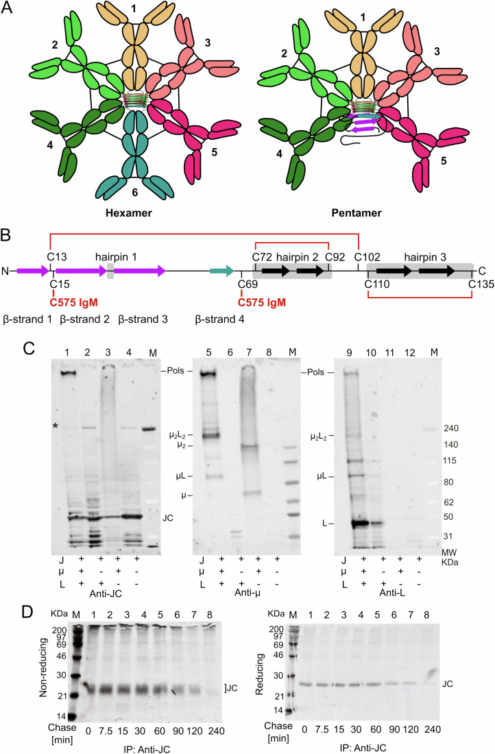
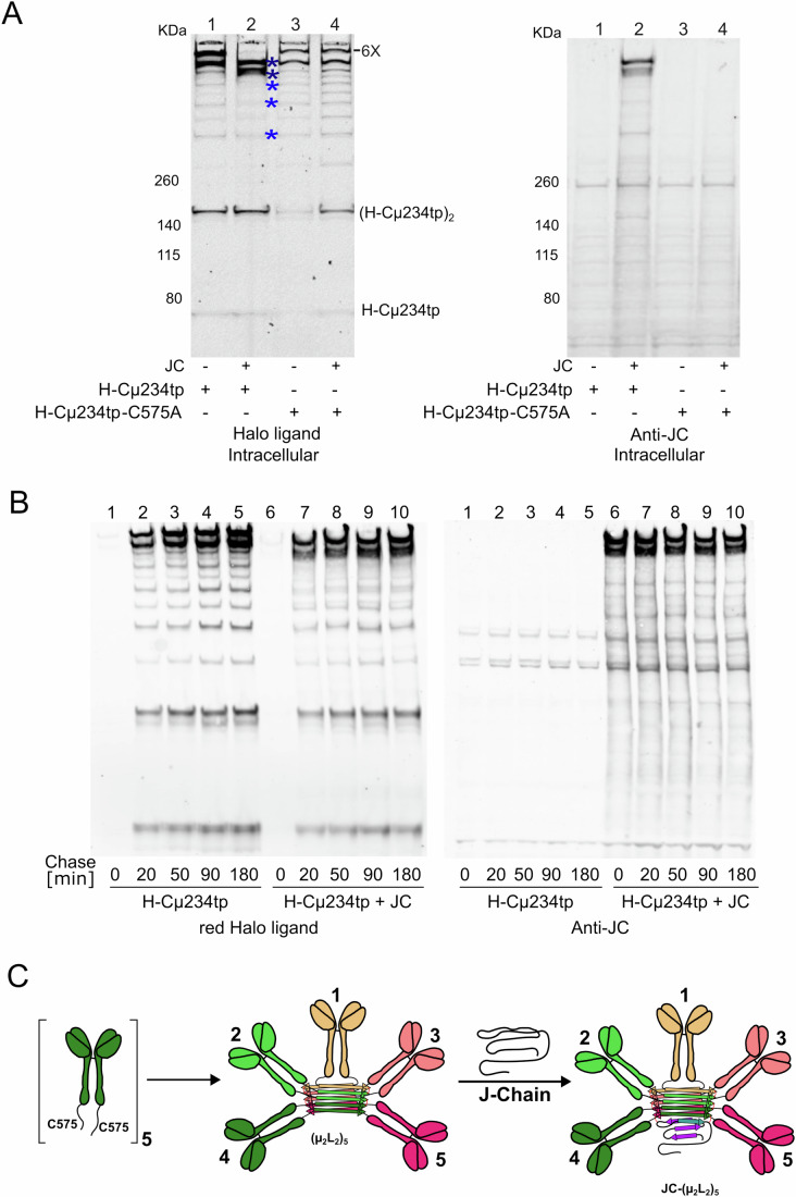
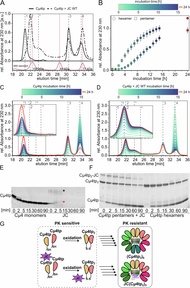
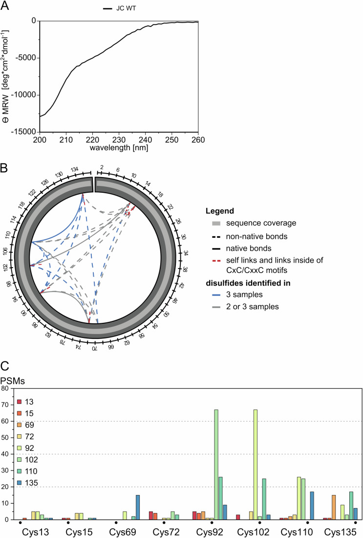
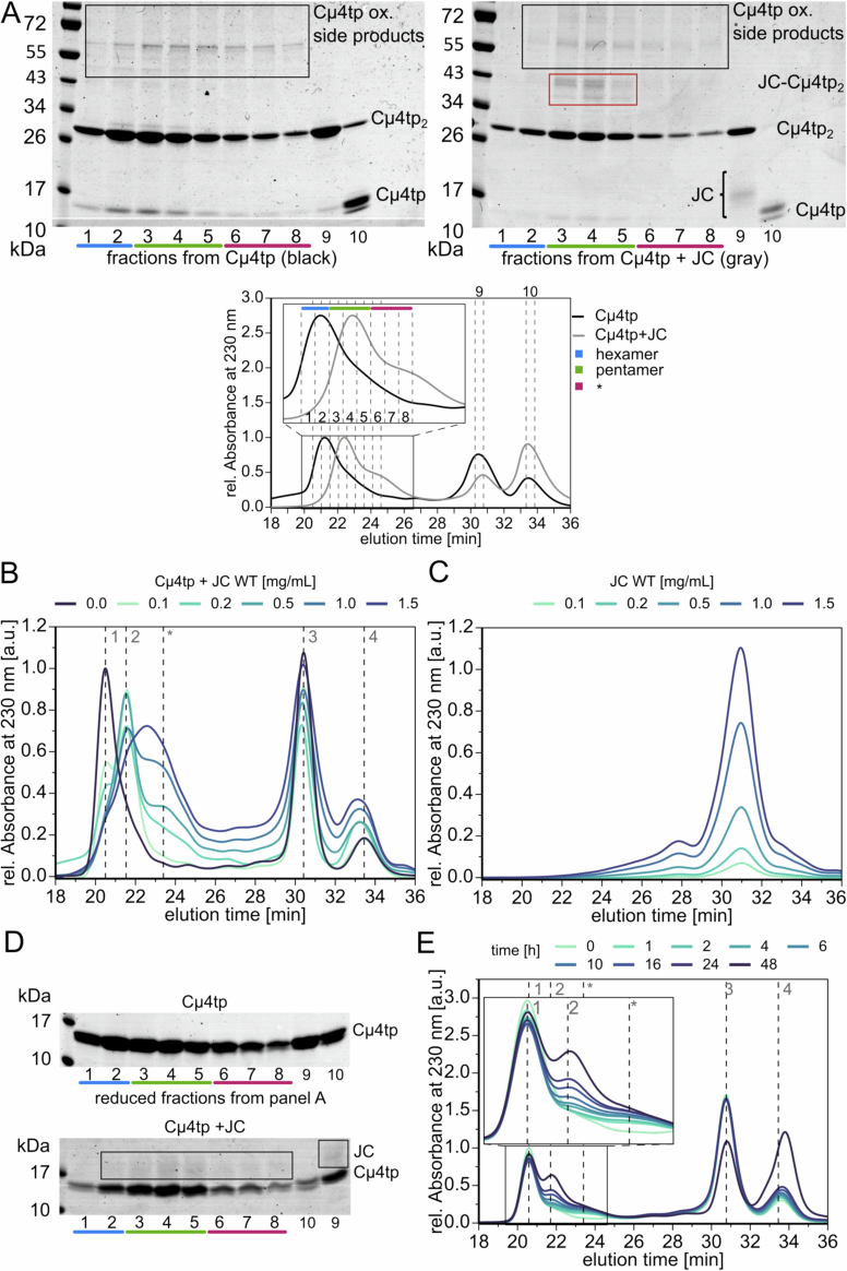
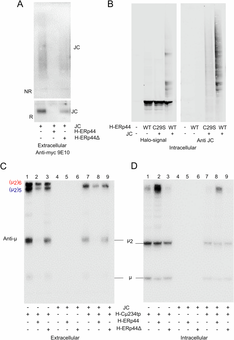
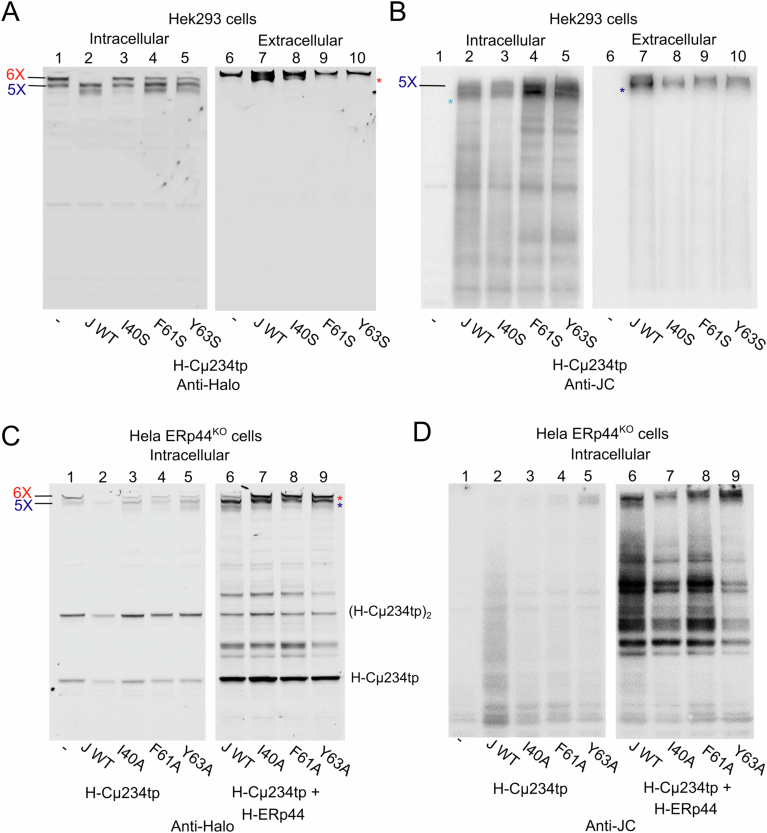
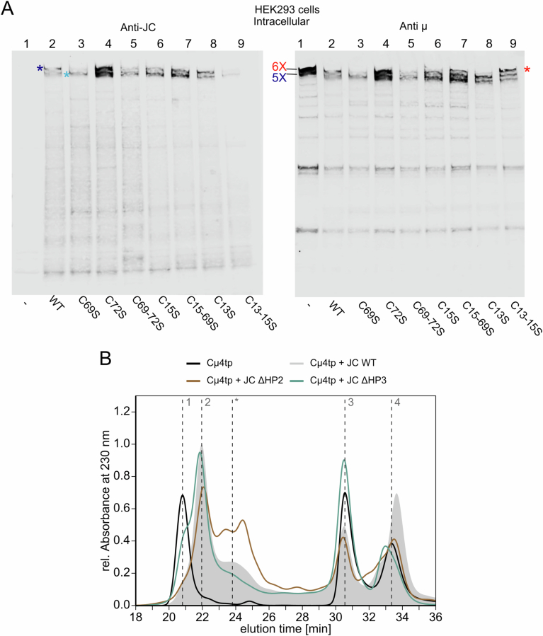
Polymeric IgM immunoglobulins have high avidity for antigen and complement, and dominate primary antibody responses. They are produced either as assemblies of six µ2L2 subunits (i.e., hexamers), or as pentamers of two µ2L2 subunits and an additional protein termed J-chain (JC), which allows transcytosis across epithelia. The molecular mechanism of IgM assembly with the desired stoichiometry remained unknown. Here, we show in vitro and in cellula that JC outcompetes the sixth IgM subunit during assembly. Before insertion into IgM, JC exists as an ensemble of largely unstructured, protease-sensitive species with heterogeneous, non-native disulfide bonds. The J-chain interacts with the hydrophobic β-sheets selectively exposed by nascent pentamers. Completion of an amyloid-like core triggers JC folding and drives disulfide rearrangements that covalently stabilize JC-containing pentamers. In cells, the quality control factor ERp44 surveys IgM assembly and prevents the secretion of aberrant conformers. This mechanism allows the efficient production of high-avidity IgM for systemic or mucosal immunity.

Keywords: Antibody Biogenesis; ERp44; Mucosal Immunity; Non-Native Disulfides; Protein Quality Control.


Conflict of interest statement

Disclosure and competing interests statement. The authors declare no competing interests.

Figures

Figure 1
Figure 1. Selective association of JC with nascent polymers.
(A) Two types of IgM polymers. IgM are secreted in two main forms, hexamers and pentamers. Both share five µ2L2 subunits (1–5) that form a β-sheet like core formed by the conserved C-terminal µtps and are stabilized by disulfide bonds linking C575 in adjacent subunits. The sixth element completing a planar hexamer can be a JC or another µ2L2 subunit. (B) JC are rich in cysteines. Schematic representation of JC and its eight highly conserved cysteines, cysteine bridges (red lines), β-sheets (arrows) and hairpins (gray boxes) that are described in this paper. β-sheets are colored complementary to Fig. 1A. (C) JC associate only to IgM polymers. Aliquots of the lysates from J558L or NS0 myeloma transfectants expressing JC, L, and/or secretory µ chains as indicated were resolved under nonreducing conditions, and blots decorated sequentially with anti-JC, anti-µ and anti-L antibodies. Arrows point at the different assembly intermediates (µ, µL, etc.). Cells producing all three subunits secrete NP-specific IgM polymers, whilst those lacking L retain and degrade both µ and JC (Sitia et al, ; Fagioli and Sitia, ; Fagioli et al, 2001). The nature of the high molecular weight band decorated by anti-JC and indicated by the asterisk, is unknown. The experiment was repeated twice. (D) Unassembled JC are rapidly degraded. N[µ1] myeloma transfectants expressing JC and µ were pulsed for 5 min with radioactive aminoacids and chased for the indicated times before immunoprecipitation with anti-JC antibodies and protein A Sepharose beads (Fagioli and Sitia, 2001). The broad and heterogeneous signals of radioactive JC observed under nonreducing conditions (left panel) collapse in a sharp band upon reduction and alkylation (right panel). In lane 1 of the top panel, DTT present in the vicinal lane containing molecular weight markers reduced partly intra-chain disulfides of JC, slowing their gel mobility. The experiment was performed once. Source data are available online for this figure.
Figure 2
Figure 2. Assembly of pentameric IgM.
(A) JC outcompete the sixth µ2L2 subunit in associating with nascent pentamers. HEK293 cells expressing H-Cµ234tp chains with a cysteine (WT) or alanine in the penultimate position (C575A) were co-transfected with JC or empty plasmid, as indicated. Aliquots of their lysates were resolved under nonreducing conditions, and blots were decorated with Halo ligands (left panel) or anti-JC (right panel). In the absence of JC, two main assemblies accumulate intracellularly (lane 1). The slower band consists of secretion-competent (H-Cµ234tp2)6 hexamers (6X) and is the only species that negotiates secretion (see Figs. 6B and  EV4A). Clearly, the presence of JC prevents the formation of hexamers and leads to the formation of low molecular weight polymers (dark blue asterisks), likely secretion-competent pentamers, and an uncharacterized form that is retained intracellularly (light blue asterisk). Note that also in HEK293 transfectants, JC interacts very weakly with H-Cµ234tp2 and other low molecular weight intermediates (light blue asterisks in lane 2). The experiment was repeated at least three times with similar results. (B) Kinetics of IgM assembly. HEK293T cells were transiently transfected to express H-Cµ234tp or H-Cµ234tp + JC. Forty-eight hours after transfections, cells were treated as specified in Fig. EV1B to conduct a Halo time-course. Aliquots of intracellular material from each time point were resolved electrophoretically under nonreducing conditions and western blots were directly analyzed by fluorography for TMR ligand, to label selectively newly made proteins. See legend to Fig. EV1B, C for further details. The experiments were repeated three times with similar results. (C) Proposed model of pentameric IgM polymerization. Five µ2L2 subunits bind covalently, forming the amyloid core. Then, an unstructured JC is incorporated which extends the amyloid core. Source data are available online for this figure.
Figure 3
Figure 3. JC drives the formation of pentameric IgM in vitro.
(A) Preferential binding of JC to (Cµ4tp2)5 pentamers in vitro. SEC chromatograms of recombinant Cµ4tp (1 mg/mL) incubated in vitro with (dashed line) or without recombinant JC (0.5 mg/mL) (solid line) at room temperature for 24 h. In these conditions, Cµ4tp monomers (peak 4, calculated mass: 14.5 kDa) oxidize to dimers (peak 3, calculated mass: 29 kDa), which further oligomerize to form hexamers (peak 1, calculated mass: 174.1 kDa, solid line). The addition of JC induces the formation of pentamers (peak 2, calculated mass: 160 kDa, dashed line), competing with the formation of hexamers. The chromatogram of the molecular mass standards (bovine γ-globulin, 158 kDa; chicken ovalbumin, 44 kDa; horse myoglobin,17 kDa) is shown in red. The complete chromatogram of the molecular mass standard is provided in Appendix Fig. S2A. All chromatograms were recorded thrice. (B) JC enhances the kinetics of oligomer formation in vitro. In the presence of JC (squares), oligomers are formed faster than in its absence (circles). After 15 h of incubation, pentamers are almost completely assembled, while hexamer formation by Cμ4tp is still ongoing. Overall, therefore, the assembly of oligomers is faster in the presence of JC. The curves and standard deviations (black error bars) are derived from triplicates of the data shown in panels (C, D) below. (C, D) Kinetic measurement of hexamer formation. Cµ4tp (1 mg/mL) incubated in PBS pH 7.4 at 25 °C forms hexamers over time (Pasalic et al, 2017). Monomers (peak 4) are gradually oxidized to dimers (peak 3), which proceed to assemble into hexamers (peak 1) (panel C). In the presence of JC (0.5 mg/mL), pentamers (peak 2) and side products (* peak) are formed (panel D), and no other intermediates of assembly are visible overtime. Measurements by HPLC-SE were performed every hour for 15 h and after 24 h h a final chromatogram was recorded. The experiments were performed in technical triplicates. (E, F) Oligomerization protects µtps and JC from proteolysis. Recombinant JC, Cµ4tp monomers, (Cµ4tp2)6 hexamers or (Cµ4tp2)5-JC (fractions 2 or 4 Fig. EV2A, respectively) were incubated with Proteinase K and samples were taken at the indicated time points (minutes). The experiment was performed once. When not part of a complex, Cµ4tp and JC are rapidly degraded (panel E). Already after 2 min, most Cµ4tp is shortened to yield a resistant fragment, while JC (black asterisk) is completely degraded into small unstable fragments (red asterisk). In contrast, Cµ4tp2 and Cµ4tp2-JC (panel F) complexes that were part of hexamers or pentamers were largely protected from degradation. (G) Schematic representation of the main assemblies formed in our in vitro experiments. Before polymerization, JC and Cµ4tp are sensitive to PK. The formation of hexamers or JC-containing pentamers confers partial protection. Owing to the absence of covalent bonds between two Cµ4 domains (apart from the inter-subunit ones between C575 in the tailpieces), hexamers and pentamers yield mainly Cµ4tp2 dimers or Cµ4tp2-JC complexes in denaturing SDS–PAGE. Source data are available online for this figure.
Figure 4
Figure 4. Recombinant JC is largely unstructured and forms native and non-native disulfides.
(A) CD spectrum of refolded and oxidized recombinant JC. The pattern obtained by CD spectroscopy shows that JC is largely unstructured. The CD was performed once. (B, C) Cross-linking mass spectrometry analysis of disulfides formed in recombinant JC. Cross-linking MS analysis of JC disulfides revealed the presence of both native and non-native disulfides as well as intermolecular links in the recombinant protein. The protein coverage was 100%. The bar graphs shown in (C) summarize the PSM counts for recombinant JC. Note that the Cys92-Cys102 has higher PSM levels, likely reflecting the susceptibility of this site to protease cleavage. Source data are available online for this figure.
Figure 5
Figure 5. Pivotal role of ERp44 in preventing the secretion of unassembled JC.
HeLa-ERp44KO cells were transfected as indicated, and aliquots from their supernatants (A, C Extracellular) or lysates (B, D Intracellular) resolved under nonreducing conditions and decorated with anti-JC or anti-ERp44, as indicated. The black brackets point at the continuous smear of JC that are secreted by (A) cells lacking ERp44. Clearly, the presence of ERp44 prevents the secretion of free JC. Blue asterisks point at JC-ERp44 covalent complexes (B). ERp44∆RDEL (lanes 3, 6, 9) is secreted (C) and fails to form covalent complexes with JC (D). The experiment was repeated at least three times with similar results. Source data are available online for this figure.
Figure 6
Figure 6. Hydrophobic residues in JC guide the interaction with nascent pentameric IgM.
(A) Contacts between JC and the µtps of subunits 4 and 5. The vicinity of JC I40, F61, and Y63 with V564, L566, and M568 in the µtps of subunits 4 and 5 points at a potential role of these residues in polymerization. The images were generated using PyMOL (The PyMOL Molecular Graphics System) and are extrapolated from the Cryo-EM structure of human IgM-Fc in complex with the J chain and the ectodomain of pIgR. (B, C). Different roles of conserved hydrophobic aminoacids in JC binding to nascent pentamers. The lysates and supernatants of HEK293 transfectants co-expressing Halo-Cµ234tp and WT, mutated (I40A, F61A, and Y63A) or no JC, were decorated with Halo ligands (B) or anti-JC (C), as indicated. Note that only WT JC efficiently prevents the formation of hexamers (6x), favoring lower-order assemblies. Unexpectedly, F61A and Y63A mutants associated more with intracellular H-Cµ234tp polymers than WT (lanes 4-5, Panel C). However, they were barely secreted (lanes 9-10, Panel B, C). The experiments were performed at least three times with similar results. (D, E) ERp44 prevents the secretion of aberrantly assembled pentamers. Aliquots of the supernatants from the indicated HeLa-ERp44KO cells transfectants were decorated with Halo ligands (D) or anti-JC (E). Note that upon ERp44 expression, only hexamers are secreted by cells expressing mutant JC, consistent with their failure to form native pentamers. The experiments were repeated at least three times with similar results. (F) Aberrant in vitro interactions of JC lacking hydrophobic residues with Cµ4tp. The HPLC-SE profiles of JC-Cµ4tp were obtained using a Superdex 200 10/300 GL column in PBS at 25 °C. The JC mutants I40A (pink profile), F61A (dark blue trace), and Y63A (light blue profile) were incubated with Cµ4tp for 24 h at room temperature at a concentration of 0.5 and 1 mg/mL, respectively. A sample volume of 10 µL was injected. The profile of Cµ4tp-JC WT, represented by a gray background, was used as a reference. The experiments were performed in technical triplicates. Source data are available online for this figure.
Figure 7
Figure 7. Disulfide rearrangements during polymerization-driven JC folding.
(A, B) Role of the JC cysteines in the assembly of secretion-competent IgM pentamers. Aliquots of the supernatants of HEK293 transfectants co-expressing H-Cµ234tp and the indicated JC mutants (C69, C72, C69-72S, C15S, C15-69S, C13S, and C13-15S) were resolved under nonreducing conditions and blots sequentially decorated with anti-JC and anti-µ, as indicated. Only the top part of the gels is shown here. Note that the C13-15S mutant is very poorly inserted in pentameric IgM and prevents only partly the secretion of hexamers. The experiments were repeated twice. (C, D) Aberrant interactions of JC lacking key cysteines with Cµ4tp in vitro. JC mutants C13S, C15S, C13S-C15S (panel C) and C69S, C72S, C15S-C69S, C69S-C72S (panel D) were incubated with Cµ4tp for 24 h at room temperature and analyzed by SE-HPLC. Their profiles are compared to WT JC, whose SEC pattern is shown as a gray background. Species: Peak 1: (Cµ4tp2)6. Peak 2: (Cµ4tp2)5-JC. The shoulder, indicated by an asterisk (*), contains uncharacterized assemblies of Cµ4-tp and JC. Peak 3:Cµ4tp2 or JC. Peak 4: Cµ4tp. The experiments were performed in technical triplicates. Source data are available online for this figure.
Figure EV1
Figure EV1. JC outcompetes the sixth µ2L2 subunit in associating with nascent pentamers.
(A) HEK293 cells expressing H-Cµ234tp chains with a cysteine (WT) or alanine in the penultimate position (C575A) were co-transfected with myc-tagged JC or empty plasmid, as indicated. Aliquots of their lysates were resolved under nonreducing conditions and blots were decorated with Halo ligands (left panel) or anti-JC (right panel). (B). Halo time-course and kinetics of IgM assembly. As schematically summarized, HEK cells were co-transfected with H-Cμ234tp with or without JC-myc. Forty-eight hours after transfection, cells were incubated with green HALOtag fluorescent ligand (R110Direct, Promega) for 75 min, to label all preexisting molecules. Then cells were washed and incubated with red HALOtag fluorescent ligand (TMRDirect, Promega) for the indicated times (minutes). Cell lysates were analyzed electrophoretically (panel (C)) under nonreducing conditions, and the gel was directly analyzed for fluorescent R110 ligand. Note that hexamers are no longer detectable upon JC addition. JC do not favor the accumulation of dimers of H-Cμ234tp dimers, consistent with their preferential binding to nascent pentamers. As expected, H-Cμ234tp assembly species labeled before the addition of the red label tend to disappear with time.
Figure EV2
Figure EV2. JC outcompete Cµ4tp2 subunits to complete stable hexamers.
(A) Composition of the fractions obtained from preparative HPLC-SE. Aliquots of the high molecular peaks obtained by incubating Cμ4tp (1 mg/mL) with (black line) or without JC (0.5 mg/mL) (gray line) were applied to SE-HPLC and 250 μL and the indicated fractions (lower panel) analyzed by SDS-PAGE under nonreducing conditions (top two panels) or protease sensitivity assays (panel D). Fractions 1–2 (blue), correspond to hexamers, fractions 3–5 (green) to pentamer, fractions 6-8 (red) to the shoulder peak *, whilst fractions 9 and 10 contain JC+Cµ4tp dimers, or mainly Cµ4 monomers, respectively. As expected, species overlap to some degree. In the presence of JC, additional bands corresponding to a Cμ4tp2-JC species are visible on SDS-PAGE (left panel, red box) in the pentamer and to a lesser extent in the * fractions. These bands are absent when Cμ4tp incubated alone. Cμ4tp on its own forms hexamers, which disassemble to dimers on SDS-PAGE (right panel). Some Cμ4tp oxidation side product bands (both panels, black boxes) are evident in the upper parts of both gels. These side products might contribute to the formation of the * shoulder. (B, C) JC outcompetes the sixth Cµ4tp2 subunit in vitro. Increasing the amount of JC (from 0 to 1.5 mg/ml) added to 1 mg/ml Cµ4, progressively inhibits the formation of (Cµ4tp2)6 “hexamers” in favor of (Cµ4tp2)5-JC “pentamers” (panel B). At their highest concentration, a broad shoulder appears that presumably corresponds to aberrant assemblies in various stoichiometries. In the absence of Cµ4tp (panel C), most JC accumulates as a broad peak. A higher molecular weight shoulder was also observed, indicative of multimeric JC species, consistent with the smeary appearance of JC in cells (see Figs. 5 and EV3, panels A, B). (D) Reducing SDS-PAGE of the fractions shown in panel A confirms that JC is covalently linked to Cμ4tp in the oligomeric assemblies. All chromatograms were recorded thrice. (E) JC cannot be inserted in preformed (Cµ4tp2)6 hexamers. Preformed (Cµ4tp2)6 hexamers were incubated with 0.5 mg/mL amounts of JC for 48 h at room temperature and finally analyzed by SEC. The size of peak 1 remains constant, indicating that once formed, hexamers are rather stable. The increase in Peak 2 reflects the formation of pentamers during the incubation.
Figure EV3
Figure EV3. ERp44KO cells secrete JC rich in heterogeneous, non-native disulfides.
(A) When aliquots from the supernatants of ERp44KO transfectants expressing myc-tagged JC are resolved electrophoretically under nonreducing conditions, anti-myc antibodies decorate a continuous smear. In contrast, DTT reduces the smear, and JC-myc chains accumulate as a 20–25 kDa band. Without alkylation, some intra-chain disulfide bonds may form during electrophoresis. Expression of WT ERp44 (center lane), but not of its ∆RDEL mutant (right lane), inhibits JC-myc secretion. All chromatograms were recorded thrice. (B) ERp44 binds JC via its C29. HeLa-ERp44KO cells were transfected as indicated, and their lysates were run under nonreducing conditions and blotted with Halo ligand (left) or anti-JC. H-ERp44 forms many abundant complexes with JC that are not formed by a mutant lacking C29. Not all bands detected by anti-JC contain H-ERp44 (right panel). These data confirm that ERp44 efficiently retains JC through C29-dependent reversible disulfide bonds. (C, D) ERp44 prevents the secretion of unpolymerized IgM. HeLa-ERp44KO cells were transfected as indicated, and aliquots from their supernatants (C) or lysates (D) resolved under nonreducing conditions and decorated with anti-µ antibodies. ERp44KO HeLa cells secrete abundant H-Cµ234tp2 subunits (D, lane 1). Co-expression of wild-type ERp44 restores retention of these incomplete subunits and promotes polymerization (panel D, compare lanes 1 and 2). H-ERp44 expression boosts also the formation of JC-containing pentamers (panel D, lanes 7-8), dampening the secretion of polymers, irrespective of the presence of JC (panel C lanes 7-8). H-ERp44∆ lacks the RDEL motif.
Figure EV4
Figure EV4. Conserved hydrophobic residues drive assembly of secretion-competent IgM pentamers.
(A, B) Essential roles of hydrophobic interactions. Replacing I40, F61, and Y63 with serine yields phenotypes similar or slightly more evident than alanine substitutions. Note that the three mutants are poorly inserted into secretion-competent pentamers (dark blue asterisk). As a result, hexamers (red asterisk) remain the most abundant intracellular species recognized by Halo ligands, and the sole one to be abundantly secreted by cells expressing ERp44. An uncharacterized form is retained intracellularly (light blue asterisk). Anti-JC antibodies intensely decorate F61S and Y63S in polymers accumulating intracellularly (lanes 4-5 panel B), but these are poorly secreted (lanes 9-10). Consequently, virtually only hexamers are detected by Halo ligands in the supernatants (lanes 9-10 panel A). (C, D). ERp44 binds JC as well as unpolymerized IgM intermediates. The lysates of ERp44KO cells expressing H-Cµ234tp, rescued with or without WT H-ERp44, were decorated with Halo ligands or anti-JC. As expected, overexpression of ERp44 prevents JC and H-Cµ234tp secretion, increasing their intracellular accumulation. The abundance of anti-JC reactive covalent complexes in lanes 6–9 (panel D) indicates that ERp44 has a high affinity for JC.
Figure EV5
Figure EV5. Reactivity of the JC cysteine mutants in cellula and in vitro.
(A) Pivotal role of C13 and C15. The lysates of HEK293 transfectants co-expressing H-Cµ234tp and the indicated JC mutants were resolved under nonreducing conditions and blots sequentially decorated with anti-JC and anti-µ, as indicated. Notably, the C13-15S mutant fails to bind IgM (lane 9) and forms mainly hexamers (red asterisk) detected with anti- µ, whilst the other mutants do bind IgM (dark blue asterisk) although with different efficiencies. An uncharacterized form is retained intracellularly (light blue asterisk). (B) The C-terminal part of JC is not essential for pentamer binding. SEC profiles on a Superdex 200 increase 10/300 GL in PBS. Cμ4tp (1 mg/mL) was incubated in vitro with WT or mutant JC (0.5 mg/mL) for 24 h at RT. Whilst the deletion of hairpin 3 and its flanking cysteines C110 and C135 (ΔHP3, turquoise) does not alter the SEC elution pattern (compare the blue trace with the gray pattern corresponding to WT JC), removal of hairpin 2 and the cysteines C72 and C92 (ΔHP2, brown trace) severely inhibits the formation of pentamers in vitro. These findings show that C110 and C135 are not vital for the formation of IgM pentamers. All chromatograms were recorded thrice.

Similar articles

Cited by

References

    1. Alberini CM, Bet P, Milstein C, Sitia R (1990) Secretion of immunoglobulin M assembly intermediates in the presence of reducing agents. Nature 347:485–487 - PubMed
    1. Anelli T, Alessio M, Bachi A, Bergamelli L, Bertoli G, Camerini S, Mezghrani A, Ruffato E, Simmen T, Sitia R (2003) Thiol-mediated protein retention in the endoplasmic reticulum: the role of ERp44. EMBO J 22:5015–5022 - PMC - PubMed
    1. Anelli T, Ceppi S, Bergamelli L, Cortini M, Masciarelli S, Valetti C, Sitia R (2007) Sequential steps and checkpoints in the early exocytic compartment during secretory IgM biogenesis. EMBO J 26:4177–4188 - PMC - PubMed
    1. Anelli T, Sitia R (2008) Protein quality control in the early secretory pathway. EMBO J 27:315–327 - PMC - PubMed
    1. Anelli T, van Anken E (2013) Missing links in antibody assembly control. Int J Cell Biol 2013:606703 - PMC - PubMed

LinkOut - more resources