

Sex dependent intergenerational effects of lead in mouse model
- PMID: 39633019
- PMCID: PMC11618497
- DOI: 10.1038/s41598-024-81839-4
Sex dependent intergenerational effects of lead in mouse model
Abstract
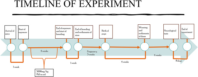
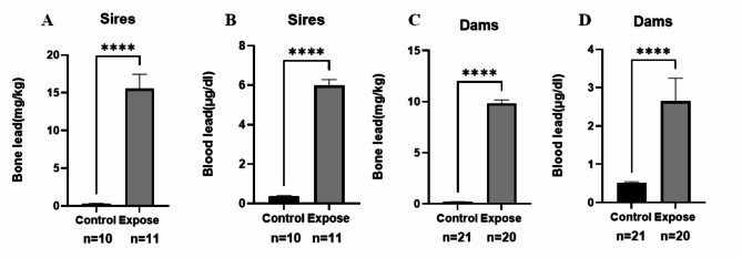
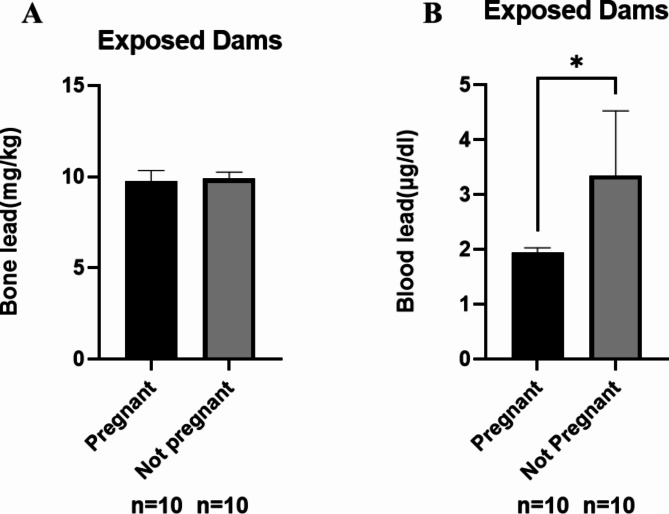
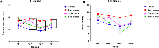
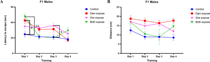
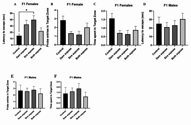
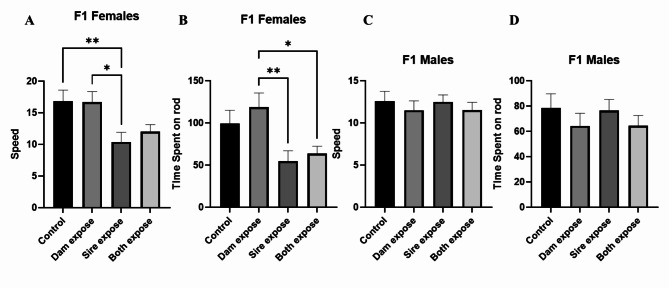
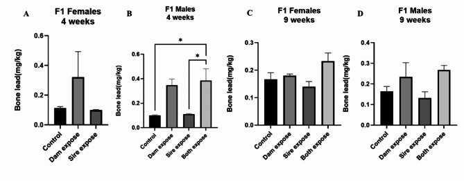
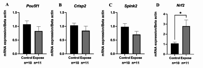
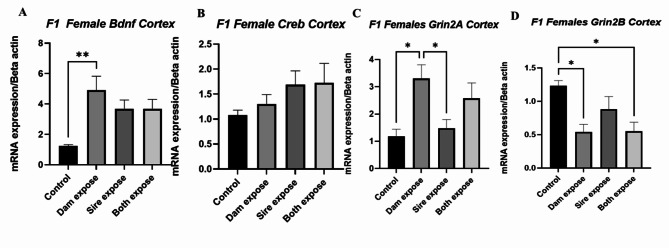
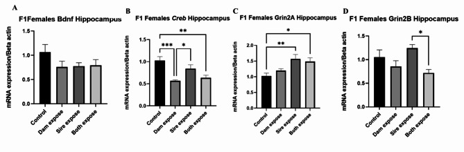
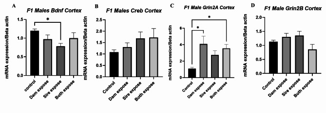
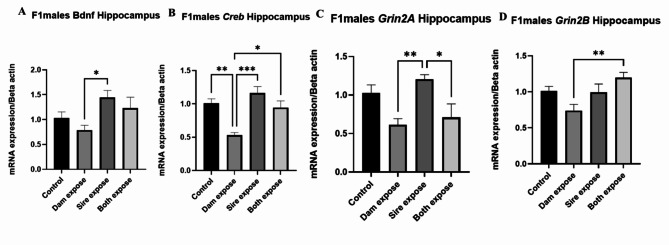
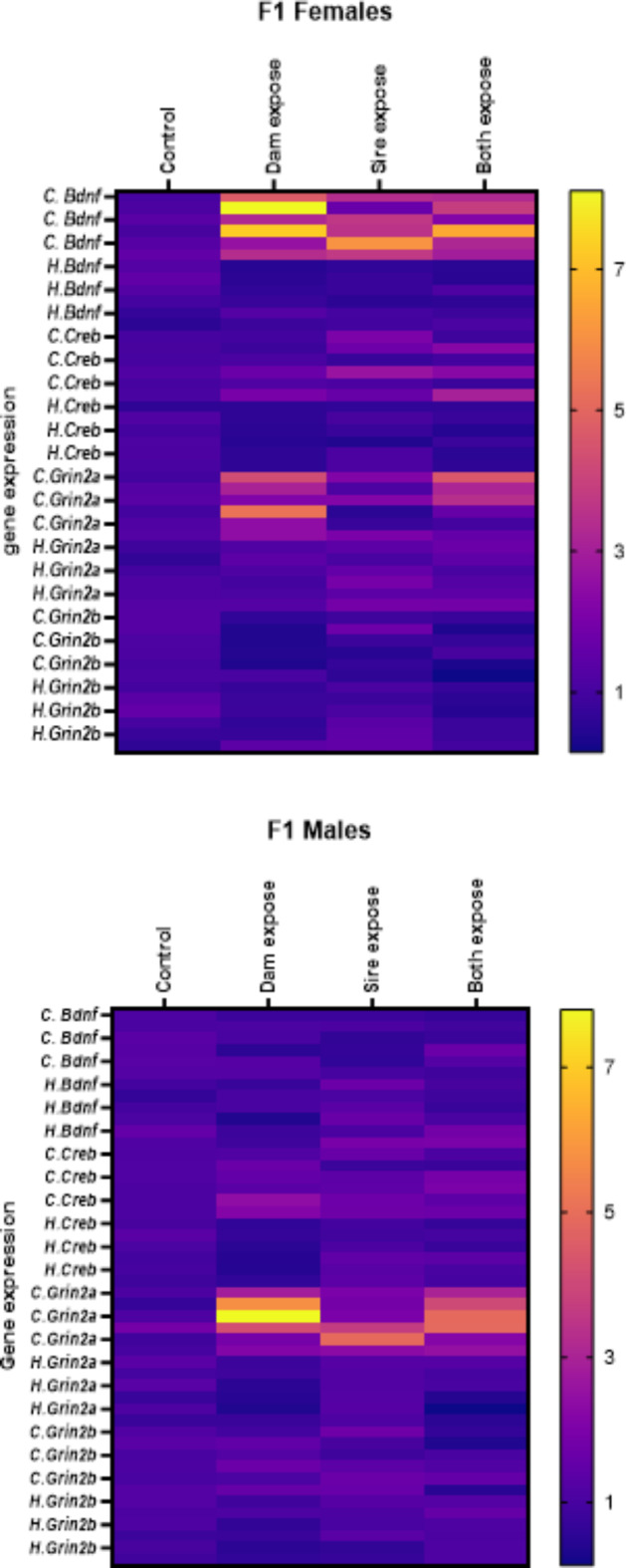
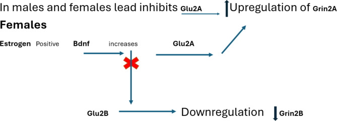
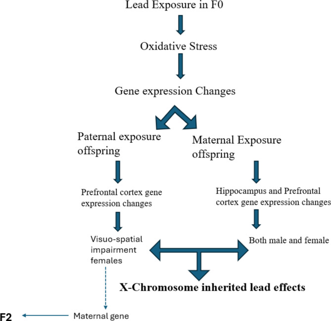
Lead (Pb) exposure negatively impacts fertility in both males and females, pregnancy outcomes, and child brain development. We investigated the reproductive and neurological effects of Pb exposure on male and female mice via Pb-contaminated soil for 4 weeks. Breeding was conducted after completion of exposure, in four groups; group 1 consisted of exposed dams and unexposed sires, group 2 consisted of exposed sires and unexposed dams, group 3 consisted of exposed sires and exposed dams and group 4 was the control. Generally, Pb exposure reduced observed conception rates, with a cumulative decrement observed when both males and females are exposed. Gene expression of the testes revealed oxidative stress as the cause of reduced conception rates. Neurological tests: Morris water maze and rotarod were conducted on F1 generation offspring. Maternally and paternally exposed F1 mice performed poorly in the Morris water maze when compared to the control. The severity of the neurological effects was also parent-dependent and sex-dependent. Paternal Pb exposure effects were more pronounced in female offspring. A comparison of gene expression changes of the hippocampus and prefrontal cortex showed paternal Pb-exposure resulted in more prefrontal cortex changes than in the hippocampus, a trend also recorded in the exposed sires. The pronounced effects in female offspring of paternal Pb exposure may suggest that Pb neurological effects may be X-chromosome-linked.
Keywords: Infertility; Maternal; Neurological; Paternal; Pb; Soil.
© 2024. The Author(s).
Conflict of interest statement
Declarations. Competing interests: The authors declare no competing interests.
Figures
Similar articles
-
Impact of parental ethanol exposure on offspring memory: Sex differences in spatial and passive avoidance tasks.Alcohol. 2025 May;124:13-21. doi: 10.1016/j.alcohol.2025.01.009. Epub 2025 Feb 2. Alcohol. 2025. PMID: 39904476
-
NTP technical report on the toxicity studies of Dibutyl Phthalate (CAS No. 84-74-2) Administered in Feed to F344/N Rats and B6C3F1 Mice.Toxic Rep Ser. 1995 Apr;30:1-G5. Toxic Rep Ser. 1995. PMID: 12209194
-
Transgenerational Effects on Lifespan and Pathology of Paternal Pre-conceptional Exposure to Continuous Low-dose-rate Gamma Rays in C57BL/6J Mice.Radiat Res. 2024 Dec 1;202(6):870-887. doi: 10.1667/RADE-24-00093.1. Radiat Res. 2024. PMID: 39471831
-
Maternal exposure to low dose BDE209 and Pb mixture induced neurobehavioral anomalies in C57BL/6 male offspring.Toxicology. 2019 Apr 15;418:70-80. doi: 10.1016/j.tox.2019.02.016. Epub 2019 Mar 2. Toxicology. 2019. PMID: 30836164
-
Sex-dependent effects of lead and prenatal stress on post-translational histone modifications in frontal cortex and hippocampus in the early postnatal brain.Neurotoxicology. 2016 May;54:65-71. doi: 10.1016/j.neuro.2016.03.016. Epub 2016 Mar 24. Neurotoxicology. 2016. PMID: 27018513 Free PMC article.
References
-
- Obeng-Gyasi, E. Sources of lead exposure in various countries. 34, 25–34 (2019). - PubMed
-
- Schachter, A. E., Gailey, A., Egendorf, S. P. & Mielke, H. W. Mechanisms of children’s soil exposure. Curr. Probl. Pediatr. Adolesc. Health Care50, 100742 (2020). - PubMed
-
- Singh Sidhu, G. P. Heavy metal toxicity in soils: sources, remediation technologies and challenges. Adv. Plants Agric. Res.5, (2016).
-
- Rawat, P. S., Singh, S., Zahid, M. & Mehrotra, S. An integrated assessment of lead exposure in children: Correlation with biochemical and haematological indices. J. Trace Elements Med. Biol.68, (2021). - PubMed
MeSH terms
Substances
LinkOut - more resources
Full Text Sources
