Review
. 2024 Dec 4;14(1):63.
doi: 10.1186/s12348-024-00442-w.

Choroidal vasculitis as a biomarker of inflammation of the choroid. Indocyanine Green Angiography (ICGA) spearheading for diagnosis and follow-up, an imaging tutorial

Affiliations
Review

Choroidal vasculitis as a biomarker of inflammation of the choroid. Indocyanine Green Angiography (ICGA) spearheading for diagnosis and follow-up, an imaging tutorial

Ioannis Papasavvas et al. J Ophthalmic Inflamm Infect. .

Abstract

Background: Indocyanine green angiography (ICGA) is the gold standard to diagnose, evaluate and follow up choroidal inflammation. It allows clinicians to precisely determine the type and extension of choroidal vasculitis in the two main choroidal structures, the choriocapillaris and the choroidal stroma. The presence of choroidal vasculitis is often overlooked by the physician who often does not include ICGA in the investigation of posterior uveitis.

Purpose: To describe choroidal vasculitis by analysing its ICGA signs in order to investigate and follow choroiditis and determine the pathophysiological mechanisms of inflammation of choroidal vessels.

Methods: The tutorial is presenting the normal findings in a non-inflamed choroid and the semiology of diverse choroidal vasculitis conditions, followed by practical illustrations using typical cases.

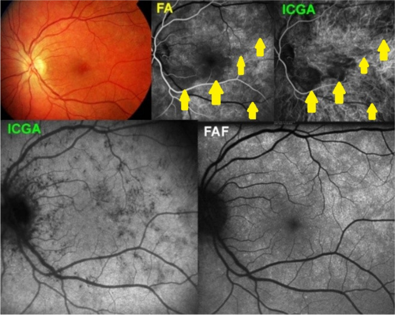
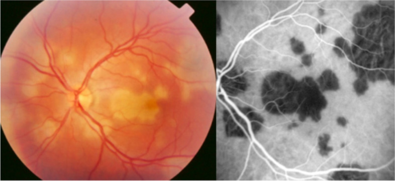
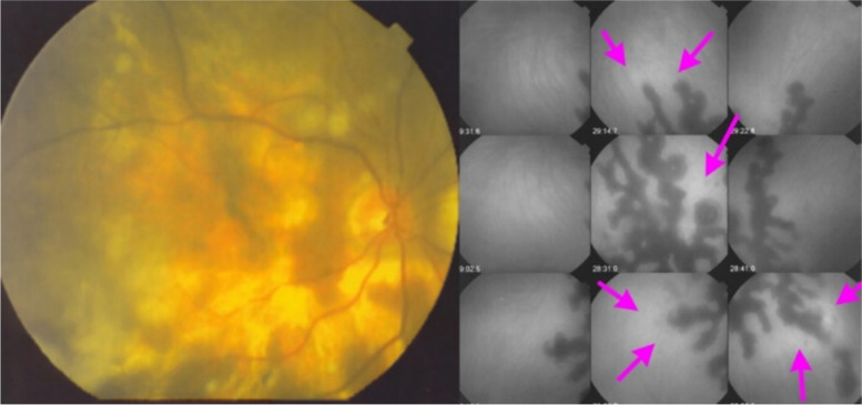
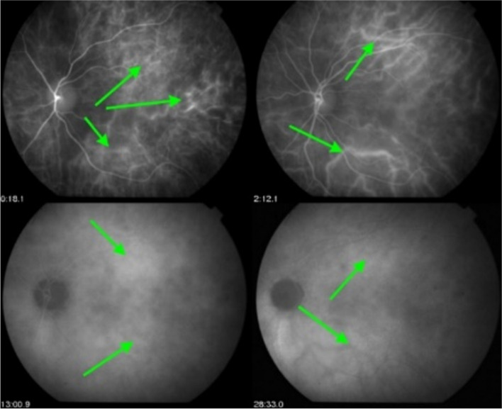
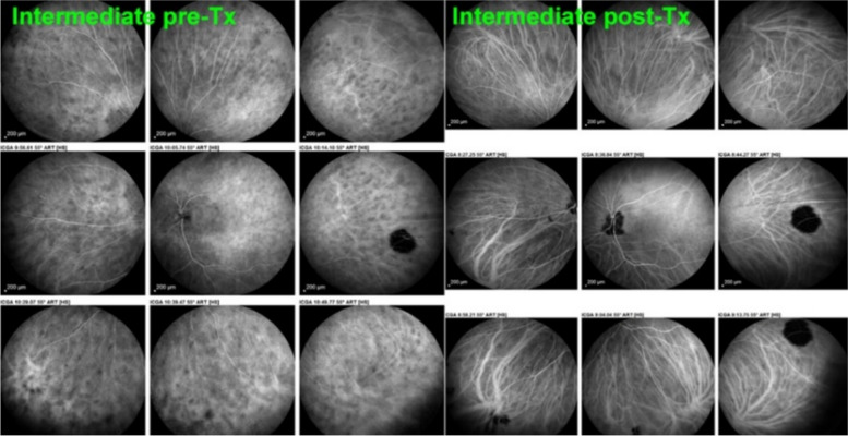
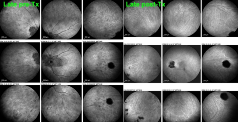
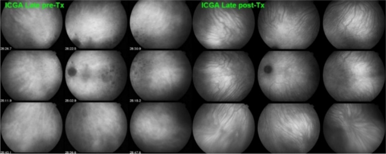
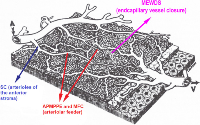
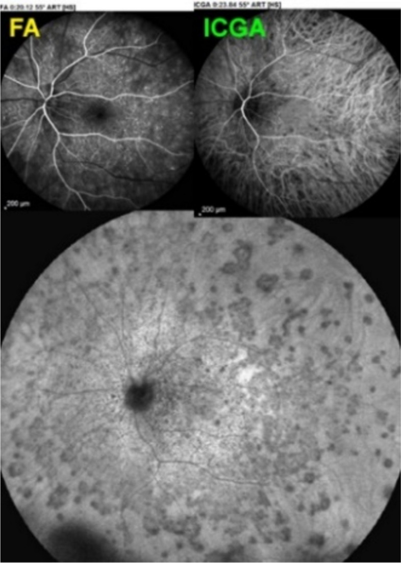
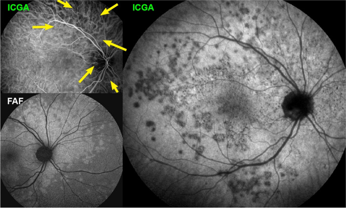
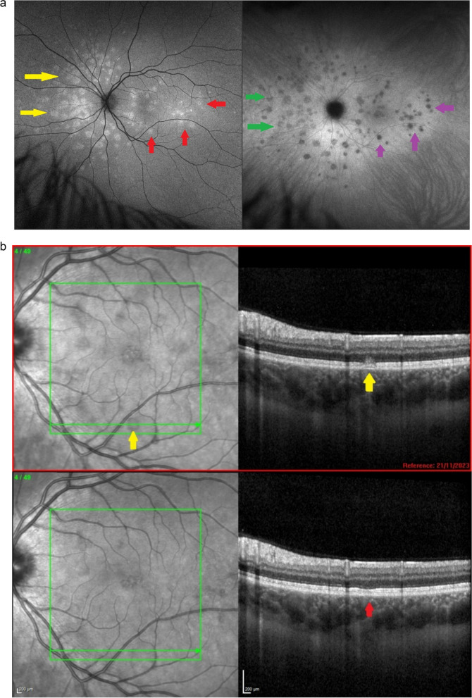
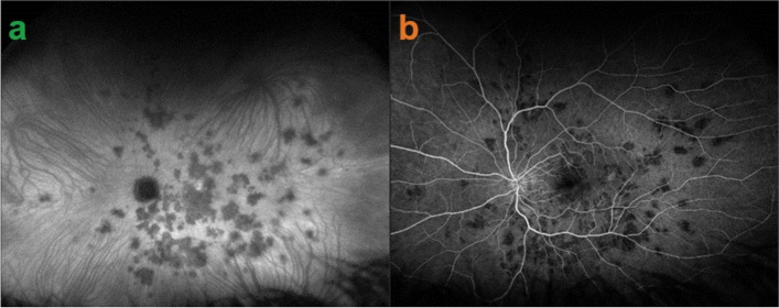
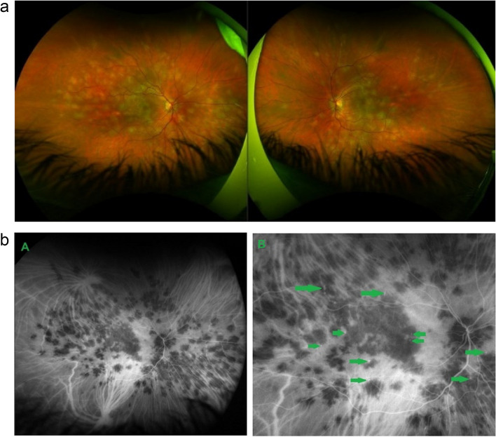
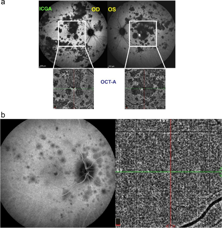
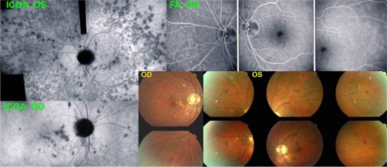
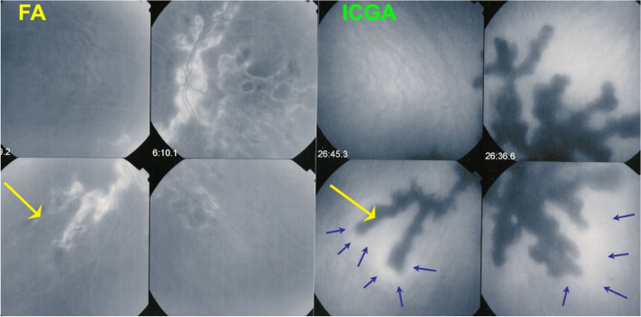
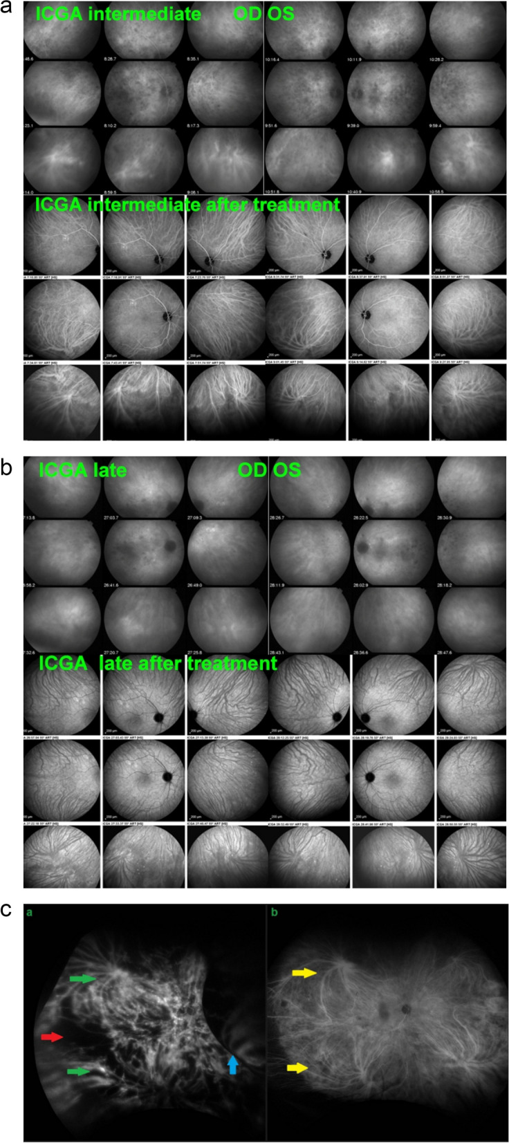
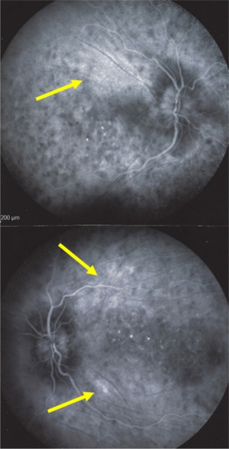
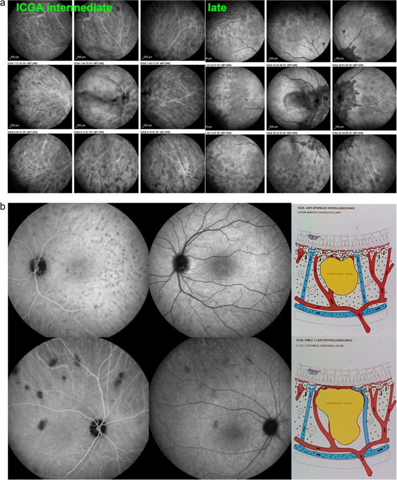
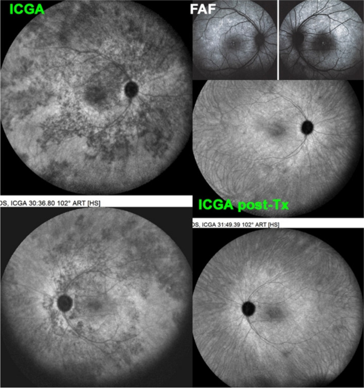
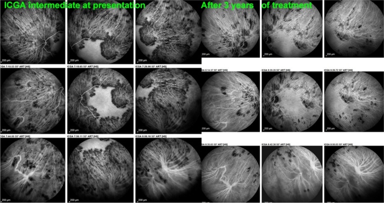
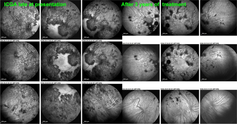
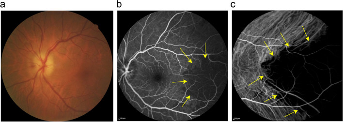
Results: The two identified patterns of choroidal vasculitis corresponded on one side to choriocapillaritis appearing as areas of hypofluorescence depicting the involvement and extension of choriocapillaris inflammatory non-perfusion. The vasculitis of the choriocapillaris goes from limited and reversible when distal endcapillary vessels are involved such as in Multiple Evanescent White Dot Syndrome (MEWDS) to more severe involvement in Acute Posterior Multifocal Placoid Pigment Epitheliopathy (APMPPE), Multifocal Choroiditis (MFC) or Serpiginous Choroiditis (SC) with more pronounced non-perfusion causing scars if not treated diligently. On the other side, stromal choroidal vasculitis is characterised by leaking hyperfluorescent vessels that appear fuzzy and at the origin of late diffuse choroidal hyperfluorescence.

Conclusion: Choroidal vasculitis is present in almost all patients with inflammatory choroidal involvement, occlusive in case of choriocapillaritis and leaky in stromal choroiditis causing vessel hyperfluorescence, fuzziness of the choroidal vessels and late diffuse stromal hyperfluorescence on ICGA. Systemic vasculitis entities produce occlusive vasculitis of large choroidal vessels.


Conflict of interest statement

Declarations. Ethics approval and consent to participate: Our ethical committee (EC-COSMTC) waived the need of approval due to retrospective study. Consent for publication: All participants have signed a consent form. Competing interests: Ioannis Papasavvas is a co-author of this review and associate editor of the journal. The rest of the authors have no competing interest to declare.

Figures

Fig. 1
Fig. 1
Schematic cartoon of choroidal circulation. The terminal arteriole supplies an independent lobule from the centre dividing into choriocapillaris vessels and subsequently into end-capillaries. The draining venules lie around in the periphery of the lobule. The level of inflammatory involvement of choroidal vessels determines the type of choroiditis from occlusion of posterior ciliary artery in SLE choroiditis, to staining of large stromal choroidal vessels in stromal choroiditis, to inflammatory occlusion of arterioles in serpiginous choroiditis, to inflammatory occlusions of larger choriocapillaris vessels in MFC (multifocal choroiditis) and APMPPE (acute posterior multifocal pigment epitheliopathy) and inflammatory occlusion of end-capillary choriocapillaris vessels in MEWDS (Multiple evanescent white dots syndrome)
Fig. 2
Fig. 2
ICGA patterns. a Type 1 pattern of choriocapillaris non perfusion or hypoperfusion. b Type 2 pattern of choroidal stromal foci
Fig. 3
Fig. 3
a Normal ICG dye circulation in the choroid. A limited number of ICG-protein complex molecules physiological egress from the fenestrated choriocapillaris and diffuse into the choroidal stroma determining faint fluorescence in the late angiographic phase. b Illustration of choroidal hyperfluorescence secondary to choroidal stromal vasculitis. Inflamed choroidal vessels at the origin of pathological exudation of the ICG dye causes abnormal hyperfluorescence adding up to the physiological fluorescence from ICG dye egressing normally from the fenestrated choriocapillaris
Fig. 4
Fig. 4
Normal choroidal vascular pattern as evidenced by ICGA. The left nine frames (left of the green separation line) show the choroidal vascular pattern in the intermediate angiographic phase (7–10’) with the ICG dye visible both in the retinal and choroidal circulations. The choroidal circulation is clearly identified with a normal aspect of vessels. The right nine frames show the choroidal vascular pattern in the late angiographic phase clearly visible in negative (no more dye intravascularly) against faint background fluorescence due to physiological dye leakage from the fenestrated choriocapillaris as the dye is no longer intravascularly
Fig. 5
Fig. 5
In MEWDS choriocapillary hypofluorescence is limited to dots isolated mostly non-confluent. The fundus color picture (top left) shows very faint discolorations. The early FA frame (top middle) shows choriocapillaris non-perfusion or perfusion delay (yellow arrows) which is also shown on the early ICGA frame (top right). On the late ICGA frame (bottom left) there are persistent hypofluorescent dots that correspond with certainty to choriocapillaris non-perfusion as they remain until the late angiographic phase. Bottom right, fundus hyper-autofluorescence typical of MEWDS, due to secondary loss of photopigment and/or accumulation of lipofuscin due to RPE dysfunction
Fig. 6
Fig. 6
Geographic areas of choriocapillary non-perfusion in a case of APMPPE. The fundus color picture shows typical coalescent cream-coloured lesions (left picture) that correspond to extended areas of choriocapillaris non-perfusion (dark areas) on ICGA frames (right)
Fig. 7
Fig. 7
Choriocapillaris vasculitis causing serpiginous choroiditis. The fundus color picture shows snake-like areas of depigmentation (left). On the right, the ICGA frame shows the serpiginous hypofluorescent areas of non-perfusion. At the border of the progressing lesions, ICGA also shows areas of hyperfluorescence due to inflammation of vessels causing leakage (crimson arrows)
Fig. 8
Fig. 8
Hyperfluorescence of choroidal vessels on early ICGA frames. Early ICGA frames (top two frames) show individual inflamed/hyperfluorescent choroidal vessels (arrows) in a case of VKH disease. On intermediate and late frames (two bottom frames) there is an increased diffuse hyperfluorescence corresponding to the areas of inflamed vessels (arrows) seen on early frames
Fig. 9
Fig. 9
Choroidal vasculitis on early ICGA frames. Arrows show individual inflamed/hyperfluoresent vessel in a case of VKH disease
Fig. 10
Fig. 10
Fuzzy indistinct choroidal vessels. Normal pattern of choroidal vessels is not recognizable any longer on top frames in a case of VKH disease. After only 3 days of intravenous pulse steroids (1000 mg/day) the course of choroidal vessels is again recognizable, although still somewhat fuzzy (bottom two frames)
Fig. 11
Fig. 11
Intermediate phase inflamed vessels before and after treatment in BRC. The 9 frames on the left show numerous HDDs and loss of recognizable pattern of choroidal vessels. The 9 frames on the right were taken after treatment showing resolution of HDDs and recovery of the normal choroidal vascular pattern. The hypofluorescent round structure temporarily corresponds to a choroidal naevus
Fig. 12
Fig. 12
Late phase stromal inflamed vessels before and after treatment in BRC. The left 9 frames show numerous HDDs and the loss of the normal pattern of choroidal vessels that are not recognizable any longer. The 9 frames on the right show the same fundus panorama after treatment. Uninflamed vessels are now well identified in dark negative appearance and HDDs have resolved. The dark area temporally is a choroidal naevus
Fig. 13
Fig. 13
Late choroidal hyperfluorescence caused by stromal vasculitis. Case of VKH disease showing late diffuse hyperfluorescence hiding the HDDs in the initial-acute phase (left 9 frames). Note also that choroidal vessels are not recognized any longer. The 9 right frames show the same ICGA panorama after treatment with well-identified choroidal vessels appearing dark in a negative fashion
Fig. 14
Fig. 14
Cartoon showing choriocapillaris architecture. Depending on the size of the inflamed vessels, choriocapillaris inflammatory non-perfusion ranges from small hypofluorescent dots when endcapillary vessels are involved as in MEWDS to large hypofluorescent areas in case of MFC, APMPPE or SC
Fig. 15
Fig. 15
MEWDS. Young lady presenting faint visual disturbance. Typical hypofluorescent dots of non-perfusion scattered all over the posterior pole and mid-periphery in the late ICG angiographic phase. Interestingly there is an additional circulatory disturbance in delayed choriocapillaris perfusion on early FA frames and less so on ICGA beyond the temporal arcades, (2 top frames)
Fig. 16
Fig. 16
MEWDS. Young man presenting a faint subjective scotoma, Right frame: scattered areas of small dark dots representing choriocapillaris non-perfusion co-localized with FAF hyperautofluorescence caused by secondary photopigment loss (bottom left) and associated with choriocapillaris perfusion delay (top left, yellow arrows)
Fig. 17
Fig. 17
a Discrepancy between ICGA and FAF images indicating that end-choriocapillary non-perfusion is at the origine of photoreceptor damage. Left frame: autofluorescence of a patient diagnosed with MEWDS. The lesions nasally to the optic disc (yellow arrows) demonstrate established outer retinal damage (marked hyperautofluorescence) while the lesions temporal to macula (red arrows) are less marked. Comparing to the ICGA (right frame) it is noticed that the lesions nasally to the ON (green arrow) are less hypofluorescent (probably due to reperfusion) than the lesions temporal to the macula which are darkly hypofluorescent (crimson arrows). Probably the lesions nasally to ON are older lesions that already affected the outer retina as seen in FAF. The temporal lesions are fresher with the outer retina damage not yet completely established. This comparison can explain why MEWDS is a primary choriocapillaritis / choriocapillary vasculitis and not a photoreceptoritis as the hypoperfusion precedes the outer retina damage. b Comparison of SD-OCT at presentation (top image) and in 6 weeks follow-up (bottom image). OCT through one of the lesions (seen hypofluorescent in ICGA) showing hyperreflectivity and disruption of the IS/OS (yellow arrows, top picture). After 6 weeks, without any treatment, OCT showed improvement (red arrow, bottom picture)
Fig. 18
Fig. 18
Correlation of late ICGA phases with early FA phases in APMPPE. a ICGA late phase demonstrated geographic area of non-perfusion of the choriocapillaris. The lesions correlate well with those seen in early stages of FA. b It is on these FA images that Deutman elaborated his correct interpretation of the disease process in APMPPE
Fig. 19
Fig. 19
a Fundus appearance of APMPPE. Multiple bilateral placoid lesions. b/A. ICGA image of same patient as 19a. Numerous geographic areas of hypoperfusion. Note that the stromal vessels are minimally involved and can be well distinguished outside the choriocapillaritis APMPPE lesions (A general view; Intermediate phase of the ICGA of the RE of a patient with APMPPE. We note the hypofluorescent areas corresponding to the non-perfusion of the choriocapillaris. b/B. Same APMPPE patient as in 19a & 19b/A. B) Blow-up of part of the global view A, zoomed on the posterior pole. Green arrows pinpoint multiple hyperfluorescent spots or lines, adjacent to the areas of the hypoperfusion, showing vasculitis of the choriocapillaris vessel corresponding probably to the vasculitic occluded feeding vessel of the hypofluorescent dark lobules
Fig. 20
Fig. 20
a APMPPE. Young male patient complaining of subjective scotomas and photopsias presenting widespread geographic and confluent areas of hypofluorescent non-perfused areas, precisely delineated on ICGA. The lesions in the posterior pole are also detected by OCT-A, showing the areas of choriocapillary drop-out. OCT-A is very useful for non-invasive follow-up of lesions, being however limited to the posterior pole. b MEWDS. Young male patient who consulted for unilateral photopsias and subjective scotomas. ICGA (left) shows numerous limited areas of end-choriocapillary non-perfusion that, in contrast to APMPPE (Fig. 20a) do not appear as drop-out areas on OCT-A (right) because OCT-A does not detect slow flow perfusion and therefore cannot show its drop-out
Fig. 21
Fig. 21
Multifocal choroiditis (MFC). Case of bilateral MFC, active in the left eye (top left) and inactive in the right eye (bottom left) with only chorioretinal scars but no vasculitis of the choriocapillaris. Fundus pictures (bottom right) show bilateral scars but do not allow to determine whether the disease is active. The widespread occult choriocapillaris vasculitis is shown by ICGA (top left), which is absolutely not suspected on FA (3 top right frames)
Fig. 22
Fig. 22
Serpiginous choroiditis. Non-perfusion in SC involves larger choriocapillaris vessels and produces extended areas of non-perfusion leading to chorioretinal atrophy if treatment is not initiated promptly. ICGA clearly delineates atrophic in addition to non-perfused areas (yellow arrow on ICGA frame), whereas FA only shows atrophic areas but not non-perfused areas (yellow arrow on FA frame). In addition, ICGA shows perilesional hyperfluorescence (dark blue arrows) indicating possible disease activity
Fig. 23
Fig. 23
a Choroidal vasculitis in VKH disease, intermediate angiographic phase. Pronounced choroidal vasculitis is present overshadowing HDDs barely detectable (top 2 sets of 9 frames). After treatment (bottom 2 sets of 9 frames) the normal pattern of choroidal vessels are again clearly identifiable with resolution of HDDs. b Choroidal (ICGA) vasculitis in VKH disease, late angiographic phase. Pronounced choroidal vasculitis with late hyperfluorescence obscuring the normal pattern of choroidal vasculature (top 2 sets of 9 frames). After treatment, the normal vascular pattern is again identifiable, in negative, as there is no ICG dye any longer within the circulation (bottom 2 sets of 9 frames). c VKH ICGA mid-phase (10’). c/a) At presentation, we notice marked choroidal vasculitis (green arrows) as well as choroidal ischemia (red arrow). The inflammation was such that the patient developed choroidal detachment (blue arrow). c/b) After treatment there is improvement of the choroidal vasculitis (yellow arrows) and reperfusion of the choroid
Fig. 24
Fig. 24
Choroidal vasculitis in VKH disease. Early angiographic phase showing early hyperfluorescent vessels (arrows) and ICGA disc hyperfluorescence only seen on ICGA in hyperacute inflammation
Fig. 25
Fig. 25
Choroidal vasculitis in VKH disease, EDI-OCT thickening of the choroid. At the early stage of VKH, there is pronounced choroidal thickening barely measurable (top image), gradually returning to normal thickness after treatment (bottom image)
Fig. 26
Fig. 26
Stromal vasculitis in Sympathetic Ophthalmia. This 24-year-old Indian lady had undergone vitrectomy followed by cataract operation and filtering surgery in her right eye.Two months after the last surgery VA in her left eye decreased and laser flare photometry indicated subclinical anterior chamber inflammation with flare amounting to18.2 ph/ms. Intermediate phase ICGA showed numerous HDDs and choroidal stromal vasculitis (top 2 frames). Late panorama frames (bottom 9 frames) showed substantial choroidal stromal vasculitis with persistent HDDs and some evanescent HDDs in the posterior pole indicating partial thickness foci. All angiographic signs resolved after introduction of systemic corticosteroid, Mycophenolate and Infliximab therapy
Fig. 27
Fig. 27
Retinal involvement in BRC. Retinal vasculitis is a constant finding in BRC and develops concomitantly to stromal choroiditis. Diffuse bilateral retinal vasculitis is present in 100% of BRC and is an early occurrence. It is characterised by a very leaky vasculitis of small and large vessels with profuse exudation, macular oedema often sparing the fovea, thick FA sheathing/staining of large posterior pole vessels, arterio-venous circulatory pseudo-delay, and pronounced disc hyperfluorescence
Fig. 28
Fig. 28
BRC choroidal vasculitis in a case of moderate severity. Posterior pole involvement is seen in the left quartet of frames (left), in the intermediate and late frames, normal choroidal vasculature is less well recognizable; note that HDDs are fading away on the late frame, indicating partial thickness of choroidal infiltrates. On panfundal intermediate and late frames (middle and right sets of 9 frames) peripheral choroidal vasculitis is minimal, as both intermediate and late angiographic phases, the normal vascular pattern is identifiable. Note the fading away of HDDs in the late angiographic phase (right set of 9 frames), indicating partial thickness lesions
Fig. 29
Fig. 29
a BRC choroidal vasculitis in a case of pronounced severity.Numerous HDDs are seen in the intermediate phase (left set of 9 frames) remaining until late phase (right 9 frames). However, choroidal stromal vasculitis is usually always less than in VKH disease, as choroidal vessel pattern is still identifiable in both intermediate and late phases. b Partial thickness HDDs. Top 2 frames and corresponding cartoon on right: In the intermediate angiographic phase (top left) of this BRC case, numerous HDDs that fade away in the late angiographic frame (top middle), corresponding to partial thickness lesion as illustrated on cartoon on the right. Bottom 2 frames and corresponding cartoon on the right: HDDs visible in the intermediate angiographic phase (bottom left) still visible in the late angiographic frame (bottom middle) corresponding to full thickness lesion as illustrated on the corresponding cartoon on the right
Fig. 30
Fig. 30
Acute Syphilitic Posterior Placoid Chorioretinitis (ASPPC). At presentation (2 left frames) numerous geographic areas of hypofluorescence are present on ICGA delineating the extensive choriocapillaris vasculitis producing choriocapillaris non-perfusion, which is co-localized with fundus hyperautofluorescence (FAF, right top 2 frames) indicating photoreceptor outer segment loss. On the middle and bottom left 2 frames all the non-perfused areas have recovered after treatment
Fig. 31
Fig. 31
Tuberculosis-related Serpiginous choroiditis (choriocapillary vasculitis/ choriocapillaritis; intermediate angiographic phase (left eye). At presentation, widespread area of vasculitic choriocapillaris non-perfusion extended over the whole fundus, only sparing the central macular area showing normal background fluorescence (left 9 frames). After 3 years of combine antibiotic and immunosuppressive treatment (right 9 frames) most of the choriocapillaris non-perfusion has recovered. Only limited scattered areas of hypofluorescence remain representing chorioretinal scars
Fig. 32
Fig. 32
Tuberculosis-related Serpiginous choroiditis (choriocapillary vasculitis/ choriocapillaritis; late angiographic phase (left eye). The late ICGA angiographic frames show that numerous hypofluorescent areas are still present in the late angiographic phase indicating non-perfusion (left 9 frames). After treatment (right 9 frames) the scared areas are better visible and slightly more extended than on the intermediate ICGA frames
Fig. 33
Fig. 33
Composition of fundus (left), FAF (middle) and ICGA (right) of a patient with TB - related SC. The fundus (a) reveals presence of whitish lesions while FAF (b) showed mixed hyperautofluorescence and hypofluorescence. ICGA (c) showed hypofluorescent areas due to choriocapillaris non perfusion and fuzziness of the choroidal vessels as well as hyperfluorescence surrounding the area of non-perfusion due to reactive choroidal vasculitis indicating disease activity. FAF evolution of a TB related Serpiginous Choroiditis.Pre-Ozurdex® (left frame), still areas of activity visible, appearing hyperautofluorescent adjacent to established scars (arrows). Post-ozurdex® (right frame): the hyperautofluorescent lesions resolved and no new scars identified
Fig. 34
Fig. 34
Ocular Sarcoidosis with minimal choroidal stromal vasculitis (score 0.5).This patient had preponderant retinal vasculitis (not shown) with minimal choroidal vasculitis. Several HDDs are seen in the posterior pole bilaterally in the intermediate angiographic phase (top 2 sets of nine frames). These HDDs disappear in the late angiographic phase (bottom 2 sets of 9 frames). Vessels are relatively well identified in the intermediate phase with choroidal vessels very discreetly fuzzy (2 top sets of 9 frames). In the late phase, choroidal vascular pattern is very distinctly identified in negative as the ICG dye is no more intravascularly and as there is no late hyperfluorescence that would obscure this pattern in case of more severe choroiditis (2 bottom sets of 9 frames)
Fig. 35
Fig. 35
Bilateral Ocular Sarcoidosis with moderate choroidal stromal vasculitis (score 2, OD & OS, only OD is shown). This patient presented with bilateral faint round discoloured fundus lesions (left panfundus photography). BRC was suspected but HLA-A29 was absent and serum angiotensin converting enzyme (ACE) and lysozyme were elevated in addition to bilateral hilar lymphadenopathies (BHL). HDDs were clearly identified bilaterally on intermediate angiographic frames (middle set of 9 frames). However, vasculitis was minimal on intermediate frames as stromal vessels were well-identified with minimal fuzziness: Late phase angiographic frames (right set of 9 frames) however did not allow to identify stromal vessels because of late diffuse hyperfluorescence preventing identification of stromal vessels
Fig. 36
Fig. 36
Bilateral Ocular Sarcoidosis with severe choroidal stromal vasculitis (score 6). This patient shows severe stromal choroidal vasculitis, as normal pattern of stromal vessels cannot be identified in the intermediate angiographic phase (top 2 sets of nine rames) nor in the late angiographic phase (bottom 2 sets of 9 frames) because of diffuse leakage from choroidal vessels. Note the irregular distribution and uneven size of HDDs, characteristic of sarcoidosis choroiditis
Fig. 37
Fig. 37
Ocular sarcoidosis with choroidal stromal vasculitis and macroaneurysm. This patient presented typical HDDs and substantial stromal vasculitis present in the intermediate and late angiographic phases in association with a retinal arterial macro-aneurysm OD (white arrows)
Fig. 38
Fig. 38
Tuberculous stromal vasculitis, intermediate angiographic phase (left eye). Patient shows substantial stromal vasculitis in the intermediate phase with unrecognizable stromal vessels and randomly distributed hypofluorescent areas of diverse sizes (left sextet of frames). After treatment the normal pattern of choroidal vessels can anew be identified and hypofluorescent areas have resolved (right sextet of frames)
Fig. 39
Fig. 39
OCT of a tuberculoma. A OCT at presentation showing the tuberculoma (crimson arrow) as well as subretinal fluid due to damage to the outer retina because of mechanically induced choriocapillaris non perfusion. B OCT after systemic ATT and steroids were started. ICGA, intermediate phase, demonstrating a tuberculoma. The hypofluorescence is due to mechanically induced choriocapillaris non-perfusion. Note also perilesional hyperfluorescence due to vasculitis as well as diffuse fuzziness of vessels of the whole fundus
Fig. 40
Fig. 40
Systemic Lupus Erythematosus (SLE). Choroidal vasculitis in SLE is charcterised by extended drop-out of choroidal circulation including choriocapillaris due to vasculitic occlusion of a posterior ciliary artery. FA (left frame) shows drop out of choriocapillary circulation (crimson arrows) and ICGA shows non-perfusion of large choroidal vessels of a triangular area with apex towards the posterior pole typical of occlusion of short posterior ciliary arteries (Amalric’s sign). Images courtesy of Horst Helbig, University of Regensburg, Germany)
Fig. 41
Fig. 41
Fundus picture shows a chalky white optic nerve head typical of arteritic anterior ischemic optic neuropathy. b Early fluorescein angiography (FA early) shows a faint hypofluorescent triangle in the temporal periphery (yellow arrows) due to choroidal hypo-perfusion. c Indocyanine green Angiography (ICGA) clearly shows the triangular sign of Amalric due to choroidal and choriocapillaris non-perfusion of the posterior short ciliary artery (yellow arrowheads)

Similar articles

Cited by

References

    1. Fryczkowski AW (1994) Anatomical and functional choroidal lobuli. Int Ophthalmol 18(3):131–41. 10.1007/BF00915961. (PMID: 7852018) - PubMed
    1. Olver JM (1990) Functional anatomy of the choroidal circulation: methyl methacrylate casting of human choroid. Eye (Lond) 4(Pt 2):262–72. 10.1038/eye.1990.38. (PMID: 2379644) - PubMed
    1. Hope-Ross MW (1997) ICG dye: physical and pharmaceutical properties. In: Yannuzzi LA, Flower RW, Slakter JS (eds) Indocyanine Green Angiography. Mosby-Year Book St. Louis, Missouri, pp 46–49
    1. Chang AA, Morse LS, Handa JT, Morales RB, Tucker R, Hjelmeland L, Yannuzzi LA (1998) Histologic localization of indocyanine green dye in aging primate and human ocular tissues with clinical angiographic correlation. Ophthalmology 105(6):1060–8. 10.1016/S0161-6420(98)96008-0. (PMID: 9627657) - PubMed
    1. Herbort CP, Papadia M, Mantovani A (2012) Classification of choroiditis based on inflammatory lesion process rather than fundus appearance: enhanced comprehension through the ICGA concepts of the iceberg and jellyfish effects. Klin Monbl Augenheilkd 229(4):306–13. 10.1055/s-0031-1299394. (Epub 2012 Apr 11 PMID: 22495994) - PubMed

LinkOut - more resources