Sub-acute stroke demonstrates altered beta oscillation and connectivity pattern in working memory
- PMID: 39633420
- PMCID: PMC11619298
- DOI: 10.1186/s12984-024-01516-5
Sub-acute stroke demonstrates altered beta oscillation and connectivity pattern in working memory
Abstract
Introduction: Working memory (WM) is suggested to play a pivotal role in relearning and neural restoration during stroke rehabilitation. Using EEG, this study investigated the oscillatory mechanisms of WM in subacute stroke.
Methods: This study included 48 first subacute stroke patients (26 good-recovery, 22 poor-recovery, based on prognosis after a 4-week period) and 24 matched health controls. We examined the oscillatory characteristics and functional connectivity of the 0-back WM paradigm and assessed their associations with prognosis.
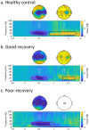
Results: Patients of poor recovery are characterised by a loss of significant beta rebound, beta-band connectivity, as well as impaired working memory speed and performances. Meanwhile, patients with good recovery have preserved these capacities to some extent. Our data further identified beta rebound to be closely associated with working memory speed and performances.
Conclusions: We provided novel findings that beta rebound and network connectivity as mechanistic evidence of impaired working memory in subacute stroke. These oscillatory features could potentially serve as a biomarker for brain stimulation technologies in stroke recovery.
Keywords: EEG; Oscillations; Prognosis; Stroke; Working memory.
© 2024. The Author(s).
Conflict of interest statement
Declarations. Ethics approval and consent to participate: This study was approved by the First Affiliated Hospital of Zhejiang University School of Medicine (Approval No. 2023 − 0694) and adhered to the Declaration of Helsinki. Consent for publication: All participants provided written informed consent. Competing interests: The authors declare no competing interests.
Figures


Similar articles
-
Cross-Frequency Coupling as a Biomarker for Early Stroke Recovery.Neurorehabil Neural Repair. 2024 Jul;38(7):506-517. doi: 10.1177/15459683241257523. Epub 2024 Jun 6. Neurorehabil Neural Repair. 2024. PMID: 38842027 Free PMC article.
-
Bidirectional Frontoparietal Oscillatory Systems Support Working Memory.Curr Biol. 2017 Jun 19;27(12):1829-1835.e4. doi: 10.1016/j.cub.2017.05.046. Epub 2017 Jun 9. Curr Biol. 2017. PMID: 28602658 Free PMC article.
-
Research on Differential Brain Networks before and after WM Training under Different Frequency Band Oscillations.Neural Plast. 2021 Mar 20;2021:6628021. doi: 10.1155/2021/6628021. eCollection 2021. Neural Plast. 2021. PMID: 33824657 Free PMC article.
-
Brain oscillatory correlates of working memory constraints.Brain Res. 2011 Feb 23;1375:93-102. doi: 10.1016/j.brainres.2010.12.048. Epub 2010 Dec 21. Brain Res. 2011. PMID: 21172316 Review.
-
Measurement and Modulation of Working Memory-Related Oscillatory Abnormalities.J Int Neuropsychol Soc. 2019 Nov;25(10):1076-1081. doi: 10.1017/S1355617719000845. Epub 2019 Jul 30. J Int Neuropsychol Soc. 2019. PMID: 31358081 Review.
Cited by
-
A bibliometric analysis of electroencephalogram research in stroke: current trends and future directions.Front Neurol. 2025 Apr 28;16:1539736. doi: 10.3389/fneur.2025.1539736. eCollection 2025. Front Neurol. 2025. PMID: 40356632 Free PMC article.
-
Exploring the Influence of Left Dorsolateral Prefrontal Cortex Intermittent Theta-Burst Stimulation on Working Memory Load: An EEG Study in Healthy Participants.CNS Neurosci Ther. 2025 Jun;31(6):e70486. doi: 10.1111/cns.70486. CNS Neurosci Ther. 2025. PMID: 40538226 Free PMC article.
References
-
- Lindeløv JK, Overgaard R, Overgaard M. Improving working memory performance in brain-injured patients using hypnotic suggestion. Brain. 2017;140(4):1100–6. - PubMed
-
- Synhaeve NE, Schaapsmeerders P, Arntz RM, Maaijwee NA, Rutten-Jacobs LC, Schoonderwaldt HC, et al. Cognitive performance and poor long-term functional outcome after young stroke. Neurology. 2015;85(9):776–82. - PubMed
MeSH terms
Grants and funding
LinkOut - more resources
Full Text Sources
Medical

