. 2024 Dec 4;12(1):187.
doi: 10.1186/s40478-024-01902-z.

CK1δ/ε-mediated TDP-43 phosphorylation contributes to early motor neuron disease toxicity in amyotrophic lateral sclerosis

Affiliations

CK1δ/ε-mediated TDP-43 phosphorylation contributes to early motor neuron disease toxicity in amyotrophic lateral sclerosis

Vivian I Ko et al. Acta Neuropathol Commun. .

Abstract

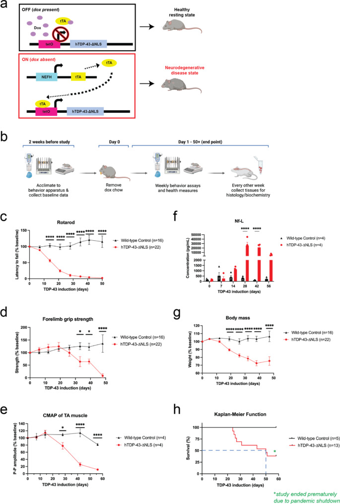
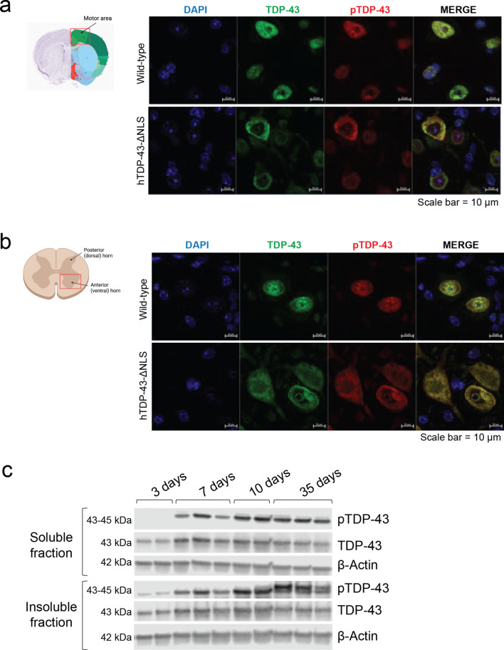
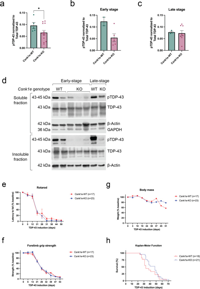
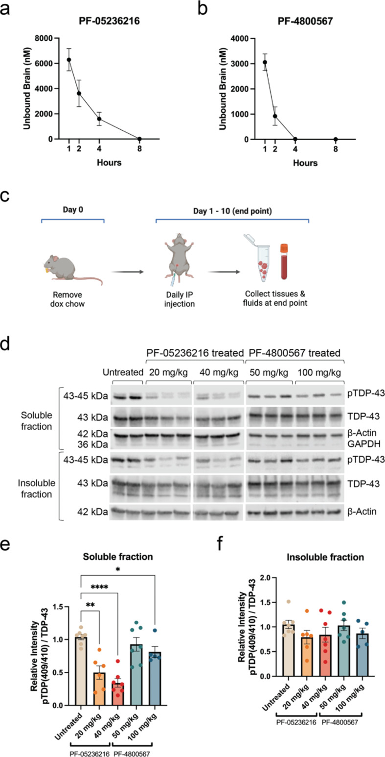
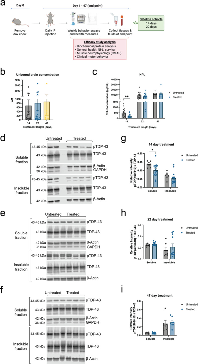
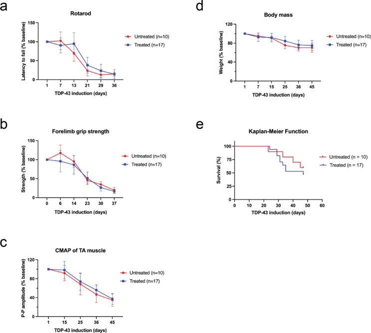
Hyperphosphorylated TDP-43 aggregates in the cytoplasm of motor neurons is a neuropathological signature of amyotrophic lateral sclerosis (ALS). These aggregates have been proposed to possess a toxic disease driving role in ALS pathogenesis and progression, however, the contribution of phosphorylation to TDP-43 aggregation and ALS disease mechanisms remains poorly understood. We've previously shown that CK1δ and CK1ε phosphorylate TDP-43 at disease relevant sites, and that genetic reduction and chemical inhibition could reduce phosphorylated TDP-43 (pTDP-43) levels in cellular models. In this study, we advanced our findings into the hTDP-43-ΔNLS in vivo mouse model of ALS and TDP-43 proteinopathy. This mouse model possesses robust disease-relevant features of ALS, including TDP-43 nuclear depletion, cytoplasmic pTDP-43 accumulation, motor behavior deficits, and shortened survival. We tested the effect of homozygous genetic deletion of Csnk1e in the hTDP-43-ΔNLS mouse model and observed a delay in the formation of pTDP-43 without significant ultimate rescue of TDP-43 proteinopathy or disease progression. Homozygous genetic deletion of Csnk1d is lethal in mice, and we were unable to test the role of CK1δ alone. We then targeted both CK1δ and CK1ε kinases by way of CK1δ/ε-selective PF-05236216 inhibitor in the hTDP-43-ΔNLS mouse model, reasoning that inhibiting CK1ε alone would be insufficient as shown by our Csnk1e knockout mouse model study. Treated mice demonstrated reduced TDP-43 phosphorylation, lowered Nf-L levels, and improved survival in the intermediate stages. The soluble TDP-43 may have been more amenable to the inhibitor treatment than insoluble TDP-43. However, the treatments did not result in improved functional measurements or in overall survival. Our results demonstrate that phosphorylation contributes to neuronal toxicity and suggest CK1δ/ε inhibition in combination with other therapies targeting TDP-43 pathology could potentially provide therapeutic benefit in ALS.

Keywords: Amyotrophic lateral sclerosis; Casein kinase 1 delta; Casein kinase 1 epsilon; Kinase inhibitors; Phosphorylation; TAR DNA-binding protein (TDP-43).


Conflict of interest statement

Declarations. Ethics approval and consent to participate: All procedures were formed in accordance with NIH Guide for the Care and Use of Experimental Animals and Studies were approved by the Institutional Animal Care and Use Committee (IACUC) of the University of California, San Diego. Consent for publication: N/A. Competing interests: The authors declare no competing interests.

Figures

Fig. 1
Fig. 1
hTDP-43-ΔNLS mouse model is a tet-off dox inducible system with significant motor deficits and neurodegenerative health. (a) When hTDP-43-ΔNLS mice were fed dox chow, the hTDP-43-ΔNLS transgene was suppressed, and mice grew normally into adulthood. The neurofilament heavy chain (NEFH) promoter line drove cell-specific expression in neurons with large caliber axons, which were primarily motor neurons of the brain and spinal cord. When dox chow was replaced with normal chow, the tetracycline activator protein (tTA) activated the transcription of the hTDP-43-ΔNLS transgene, which encoded the human TARDBP gene that contained a defective nuclear localization signal (ΔNLS). This initiated the progressive TDP-43 proteinopathy disease phenotypes and neurodegeneration in mice. (b) Experimental design used to characterize and validate the hTDP-43-ΔNLS mouse model. Mice were trained on behavior tasks in the weeks prior to the study start date, and the baseline data was collected before dox chow was changed to regular chow. Data on motor behavior and health measures was collected weekly. Satellite mice were run in parallel for tissue collection to have samples that represented different stages of disease progression. (c) The cohort for rotarod, grip strength test, and body mass included 16 wild-type control mice (9 male, 7 female) and 22 hTDP-43-ΔNLS mice (13 male, 9 female). Rotarod performance for hTDP-43-ΔNLS mice was significantly decreased by 14 days. (d) Forelimb grip test strength declined significantly by 32 days. (e) The cohort for CMAP and Nf-L included 4 wild-type control mice (2 male, 2 female) and 4 hTDP-43-ΔNLS mice (2 male, 2 female). CMAP recordings showed significant decline in the peak-to-peak amplitude in TA muscles by day 25 of induced TDP-43 expression. (f) hTDP-43-ΔNLS mice displayed elevated Nf-L levels by 14 days of induced TDP-43 expression, with significant increases by day 28. (g) Body mass quickly declined as disease progressed in hTDP-43-ΔNLS mice. (h) The cohort for survival included 5 wild-type control mice (3 male, 2 female) and 13 hTDP-43-ΔNLS mice (8 male, 5 female). The median survival was reached by day 50, as indicated by the blue dashed lines. The study ended prematurely in 2020 due to the COVID pandemic shutdown. Two-way ANOVA with repeated measures and Dunnett’s multiple comparisons, *p < 0.03, **p < 0.002, ***p < 0.0002, ****p < 0.0001. Error bars represent SEM
Fig. 2
Fig. 2
pTDP-43 accumulates in the cytoplasm of neurons and is detected before observable motor deficits in hTDP-43-ΔNLS mice. (a) Representative immunofluorescence images of neurons in cortex of wild-type control and hTDP-43-ΔNLS mice with staining for TDP-43 (green), pTDP-43 (red), and DAPI (blue). TDP-43 remained nuclear in wild-type cells while it became translocated and mislocalized to the cytoplasm in hTDP-43-ΔNLS samples. Much of the mislocalized TDP-43 was also found to be phosphorylated. Scale bars represent 10 μm. Overview of anatomical section was obtained from the Allen Mouse Brain Atlas, https://mouse.brain-map.org. (b) Similar observations of altered TDP-43 expression were also observed in lumbar spinal cord tissue sections. Scale bars represent 10 μm. (c) Representative immunoblots demonstrate that pTDP-43 was detected in soluble and insoluble protein fractions after 7 days of induced TDP-43 expression, with maximal signal intensity by day 10. pTDP-43 accumulated and was detected in the insoluble fraction before it could be detected in the soluble fraction. Later into the disease course, a higher molecular weight hyperphosphorylated TDP-43 species (top band) could be detected in the insoluble fraction
Fig. 3
Fig. 3
Deletion of Csnk1e delayed pTDP-43 formation but did not improve motor behavior on hTDP-43-ΔNLS mice. (a) Csnk1e-KO mice had generally reduced pTDP-43 levels in soluble protein fraction across all timepoints collected. (b-c) Data from panel 3a was grouped into early (less than 21 days) and late (more than 21 days) stages of disease progression. Only samples from early stage Csnk1e-KO mice demonstrated pTDP-43 reduction. Quantification of pTDP-43 reduction in soluble protein fraction was computed as a normalized ratio between pTDP-43 and total TDP-43 signals. The signal intensity for each pTDP-43 band was first normalized to β-Actin, then calculated as a ratio relative to total TDP-43 levels (which was also normalized to β-Actin). Mann-Whitney U test, *p < 0.03, **p < 0.002, ***p < 0.0002, ****p < 0.0001. Error bars represent SEM. (d) Fractionating the protein into soluble and insoluble compartments demonstrated a delay in soluble pTDP-43 formation in Csnk1e-KO mice from early to late stage. GAPDH signal was used to indicate the quality of fractionation between insoluble and soluble protein fractions. (e) The cohort for rotarod, forelimb grip strength test, and body mass included 17 Csnk1e-WT mice (5 male, 12 female) and 23 Csnk1e-KO mice (12 male, 11 female). Rotarod performance for Csnk1e-KO mice did not improve as compared to Csnk1e-WT control mice. (f) Forelimb grip strength test also did not display improvements. (g) Csnk1e deletion did not exacerbate the overall health of mice, and the body mass declined similarly in both groups. (h) The cohort for survival included 18 Csnk1e-WT mice (10 male, 8 female) and 21 Csnk1e-KO mice (10 male, 11 female). There was a modest extension of median survival temporarily for Csnk1e-KO mice. Two-way ANOVA with repeated measures and Dunnett’s multiple comparisons, *p < 0.03, **p < 0.002, ***p < 0.0002, ****p < 0.0001. Error bars represent SEM
Fig. 4
Fig. 4
Pharmacokinetic (PK) and pharmacodynamic (PD) characterization of CK1 inhibitors in vivo. (a) The PK profile for 40 mg/kg dose of CK1δ/ε-specific PF-05236216 inhibitor measured as unbound compound concentrations in the brain demonstrated a half-life approximately 4–6 h long. (b) The PK profile for 100 mg/kg dose of CK1ε-specific PF-4,800,567 inhibitor measured as unbound compound concentrations in the brain demonstrated a half-life of approximately 1–2 h long. Samples were collected 1-, 2-, 4-, 8-, and 24-hours after a single IP injection. Group sizes were n = 6, with 3 females and 3 males for each timepoint. (c) Schematic of the study design to test the target engagement for CK1δ/ε-specific PF-05236216 and CK1ε-specific PF-4,800,567 inhibitors in hTDP-43-ΔNLS mice. (d) Representative immunoblot images demonstrated reduced pTDP-43 levels in both soluble and insoluble protein fractions when mice were treated with either of the CK1 inhibitors at both doses tested. GAPDH signal was used to indicate the quality of fractionation between insoluble and soluble protein fractions. (e-f) Quantification of immunoblots showed pTDP-43 reduction in both soluble and insoluble protein fractions, with greater reduction observed in the soluble portion. Quantification of pTDP-43 reduction in soluble and insoluble protein fractions was computed as a normalized ratio between pTDP-43 and total TDP-43 signals. The signal intensity for each pTDP-43 band was first normalized to β-Actin, then calculated as a ratio relative to total TDP-43 levels (which was also normalized to β-Actin). Ratios were then compared to the untreated control group. This analysis incorporated normalization not only to an internal gene control within each sample using the housekeeping gene β-Actin, but also took into account the expression of the total TDP-43 (hTDP-43-ΔNLS transgene) since there may have been inherent genetic regulatory variability between samples. Two-way ANOVA, *p < 0.03, **p < 0.002, ***p < 0.0002, ****p < 0.0001. Error bars represent SEM
Fig. 5
Fig. 5
Mice treated with CK1δ/ε-selective inhibitor showed reduced TDP-43 phosphorylation and Nf-L levels. (a) Schematic of experimental design to test the efficacy of CK1δ/ε-specific inhibitor PF-05236216 in hTDP-43-ΔNLS mice. (b) Unbound inhibitor concentrations were detected and measured in the brain across 14, 22, and 47 days of inhibitor treatment, confirming the inhibitor could cross the blood brain barrier well. (c) Nf-L measurements from plasma samples across 14, 22, and 47 days of inhibitor treatment. Nf-L was significantly reduced by 2-fold in mice treated for 14 days. Mice treated for longer time frames did not show a difference compared to untreated mice. Mann-Whitney U test, *p < 0.03, **p < 0.002, ***p < 0.0002, ****p < 0.0001. Error bars represent SEM. (d) Representative immunoblot images demonstrated reduced pTDP-43 levels in both soluble and insoluble protein fractions when hTDP-43-ΔNLS mice were treated with CK1δ/ε-specific PF-05236216 inhibitor for 14 days. (e-f) 22-day and 47-day inhibitor treatment did not show a difference between treated and untreated groups. (g-i) Quantification of immunoblots showed greater pTDP-43 reduction in soluble protein fractions for 14-day long treated mice. No significant differences were detected in mice treated for either 22 or 47 days. Refer to Supplementary material file 2 for all immunoblots that were analyzed for this study. Quantification of pTDP-43 reduction in soluble and insoluble protein fractions was computed as a normalized ratio between pTDP-43 and total TDP-43 signals. The signal intensity for each pTDP-43 band was first normalized to β-Actin, then calculated as a ratio relative to total TDP-43 levels (which was also normalized to β-Actin). GAPDH signal was used to indicate the quality of fractionation between insoluble and soluble protein fractions. This analysis incorporated normalization not only to an internal gene control within each sample using the housekeeping gene β-Actin, but also took into account the expression of the total TDP-43 (hTDP-43-ΔNLS transgene) since there may have been inherent genetic regulatory variability between samples. Two-way ANOVA, *p < 0.03, **p < 0.002, ***p < 0.0002, ****p < 0.0001. Error bars represent SEM
Fig. 6
Fig. 6
Treating mice with CK1δ/ε-selective inhibitor was insufficient to rescue motor deficits for functional benefit. (a) The cohort for rotarod, forelimb grip strength test, CMAP, body mass, and survival included 10 untreated hTDP-43-ΔNLS mice (5 male, 5 female) and 17 treated hTDP-43-ΔNLS mice (9 male, 8 female). Rotarod performance for treated mice did not improve as compared to untreated control mice. (b) Forelimb grip strength test also did not display improvements. (c) Treated mice did not display changes in CMAP recordings of the TA muscles. (d) The overall health of mice treated with inhibitor was not exacerbated since the body mass declined similarly as untreated mice. (e) Treated mice reached median survival and end stage faster than untreated mice, likely due to an unintended adverse event with long-term treatment with this particular CK1δ/ε-selective inhibitor. Two-way ANOVA with repeated measures and Dunnett’s multiple comparisons, *p < 0.03, **p < 0.002, ***p < 0.0002, ****p < 0.0001. Error bars represent SEM

References

    1. Afroz T, Chevalier E, Audrain M, Dumayne C, Ziehm T, Moser R, Egesipe A-L, Mottier L, Ratnam M, Neumann M, Havas D, Ollier R, Piorkowska K, Chauhan M, Silva AB, Thapa S, Stöhr J, Bavdek A, Eligert V, Adolfsson O, Nelson PT, Porta S, Lee VM-Y, Pfeifer A, Kosco-Vilbois M, Seredenina T (2023) Immunotherapy targeting the C-terminal domain of TDP-43 decreases neuropathology and confers neuroprotection in mouse models of ALS/FTD. Neurobiol Dis 179:106050. 10.1016/j.nbd.2023.106050 - PubMed
    1. Alfieri JA, Pino NS, Igaz LM (2014) Reversible behavioral phenotypes in a conditional mouse model of TDP-43 proteinopathies. J Neurosci 34:15244–15259. 10.1523/JNEUROSCI.1918-14.2014 - PMC - PubMed
    1. Alquezar C, Salado IG, De La Encarnación A, Pérez DI, Moreno F, Gil C, De Munain AL, Martínez A, Martín-Requero Á (2016) Targeting TDP-43 phosphorylation by Casein Kinase-1δ inhibitors: a novel strategy for the treatment of frontotemporal dementia. Mol Neurodegener 11:36. 10.1186/s13024-016-0102-7 - PMC - PubMed
    1. Benatar M, Boylan K, Jeromin A, Rutkove SB, Berry J, Atassi N, Bruijn L (2016) ALS biomarkers for therapy development: state of the field and future directions: ALS biomarkers. Muscle Nerve 53:169–182. 10.1002/mus.24979 - PMC - PubMed
    1. Benatar M, Zhang L, Wang L, Granit V, Statland J, Barohn R, Swenson A, Ravits J, Jackson C, Burns TM, Trivedi J, Pioro EP, Caress J, Katz J, McCauley JL, Rademakers R, Malaspina A, Ostrow LW, Wuu J, Hussain S, Cooley A, Li Y, Wallace M, Steele J, Hernandez JP, Medina J, Paredes ME, Manso A, Ravelo N, Levy W, Whitehead P, Zuchner S, Pasnoor M, Jawdat O, Jabari D, Farmakidis C, Glenn M, Dimachkie MM, Herbelin L, Tanui H, Anderson S, Walker M, Liu T, McCally A, Heim A, Currence M, Harness Y, Sieren J, Gibson E, Gutierrez G, Bussey D, Previte R, Kittrell P, Joshi A, Conger A, Hastings D, Caristo I, Marandi M, Carty S, Taylor JP, Wu G, Rampersaud E, Schule R, Blitterswijk MV (2020) Validation of serum neurofilaments as prognostic and potential pharmacodynamic biomarkers for ALS. Neurology 95. 10.1212/WNL.0000000000009559 - PMC - PubMed

Publication types

MeSH terms