

Exploring the innovative application of cerium oxide nanoparticles for addressing oxidative stress in ovarian tissue regeneration
- PMID: 39633503
- PMCID: PMC11619646
- DOI: 10.1186/s13048-024-01566-2
Exploring the innovative application of cerium oxide nanoparticles for addressing oxidative stress in ovarian tissue regeneration
Abstract
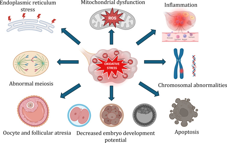
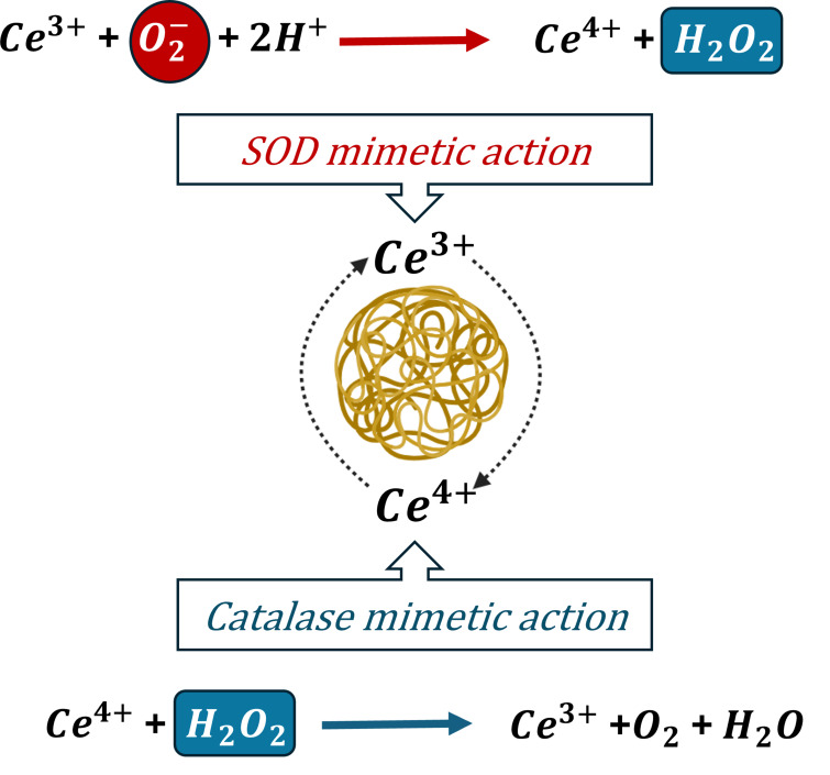
The female reproductive system dysfunction considerably affects the overall health of women and children on a global scale. Over the decade, the incidence of reproductive disorders has become a significant source of suffering for women. Infertility in women may be caused by a range of acquired and congenital abnormalities. Ovaries play a central role in the female reproductive function. Any defect in the normal functioning of these endocrine organs causes health issues and reproductive challenges extending beyond infertility, as the hormones interact with other tissues and biological processes in the body. The complex pathophysiology of ovarian disorders makes it a multifactorial disease. The key etiological factors associated with the diseases include genetic factors, hormonal imbalance, environmental and lifestyle factors, inflammatory conditions, oxidative stress, autoimmune diseases, metabolic factors, and age. Oxidative stress is a major contributor to disease development and progression affecting the oocyte quality, fertilization, embryo development, and implantation. The choice of treatment for ovarian disorders varies among individuals and has associated complications. Reproductive tissue engineering holds great promise for overcoming the challenges associated with the current therapeutic approach to tissue regeneration. Furthermore, incorporating nanotechnology into tissue engineering could offer an efficient treatment strategy. This review provides an overview of incorporating antioxidant nanomaterials for engineering ovarian tissue to address the disease recurrence and associated pathophysiology. Cerium oxide nanoparticles (CeO2 NPs) are prioritized for evaluation primarily due to their antioxidant properties. In conclusion, the review explores the potential applications of CeO2 NPs for effective and clinically significant ovarian tissue regeneration.
Keywords: Cerium oxide nanoparticles; Female infertility; Nanotechnology; Ovarian tissue engineering; Oxidative stress.
© 2024. The Author(s).
Conflict of interest statement
Declarations. Ethics approval and consent to participate: Not applicable. Competing interests: The authors declare no competing interests.
Figures
Similar articles
-
Cerium oxide nanoparticles: a promise for applications in therapy.J Exp Ther Oncol. 2011;9(1):47-51. J Exp Ther Oncol. 2011. PMID: 21275265 Review.
-
Cerium oxide nanoparticle elicits oxidative stress, endocrine imbalance and lowers sperm characteristics in testes of balb/c mice.Andrologia. 2018 Apr;50(3). doi: 10.1111/and.12920. Epub 2017 Nov 21. Andrologia. 2018. PMID: 29164652
-
Overview of Nrf2 as a target in ovary and ovarian dysfunctions focusing on its antioxidant properties.J Ovarian Res. 2025 Mar 22;18(1):60. doi: 10.1186/s13048-025-01639-w. J Ovarian Res. 2025. PMID: 40121445 Free PMC article. Review.
-
Hollow cerium nanoparticles synthesized by one-step method for multienzyme activity to reduce colitis in mice.World J Gastroenterol. 2025 Feb 7;31(5):98732. doi: 10.3748/wjg.v31.i5.98732. World J Gastroenterol. 2025. PMID: 39926211 Free PMC article.
-
Cerium oxide nanoparticles: the regenerative redox machine in bioenergetic imbalance.Nanomedicine (Lond). 2017 Feb;12(4):403-416. doi: 10.2217/nnm-2016-0342. Epub 2016 Dec 21. Nanomedicine (Lond). 2017. PMID: 28000542
Cited by
-
Reactive oxygen species in polycystic ovary syndrome: Mechanistic insights into pathogenesis and therapeutic opportunities.Redox Biol. 2025 Sep;85:103776. doi: 10.1016/j.redox.2025.103776. Epub 2025 Jul 17. Redox Biol. 2025. PMID: 40694958 Free PMC article. Review.
References
Publication types
MeSH terms
Substances
LinkOut - more resources
Full Text Sources
