. 2024 Dec 4;12(1):189.
doi: 10.1186/s40478-024-01883-z.

The neuropathological basis of elevated serum neurofilament light following experimental concussion

Affiliations

The neuropathological basis of elevated serum neurofilament light following experimental concussion

John D Arena et al. Acta Neuropathol Commun. .

Abstract

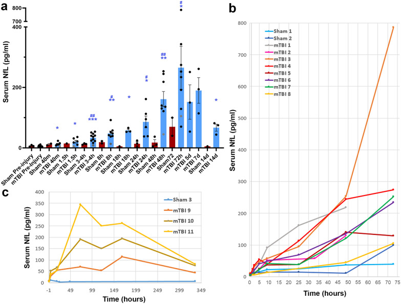
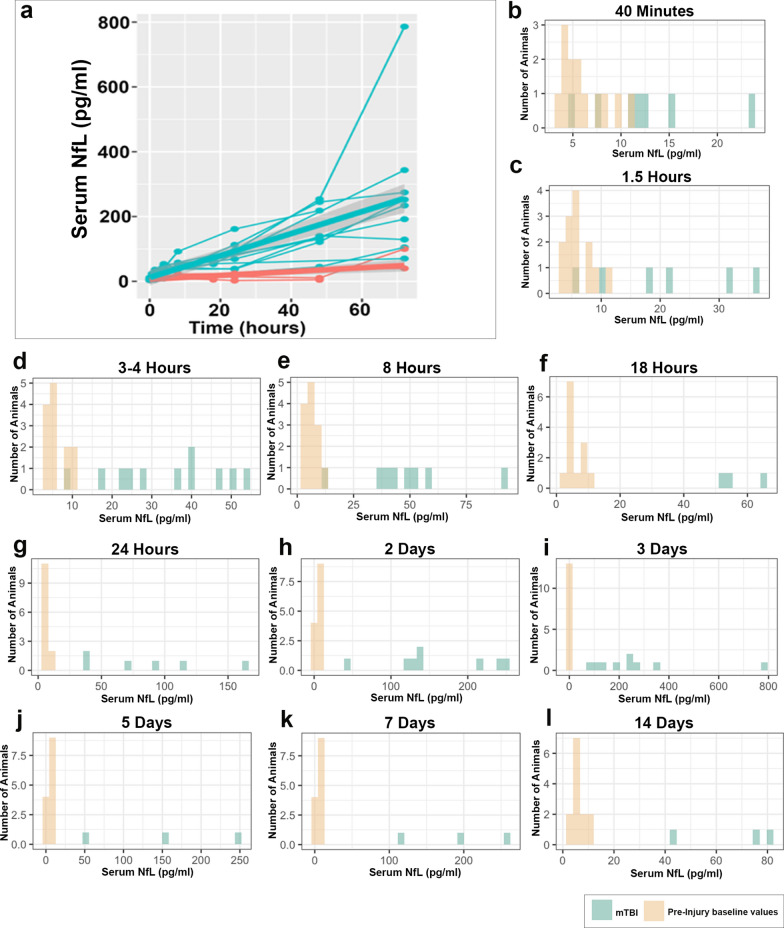
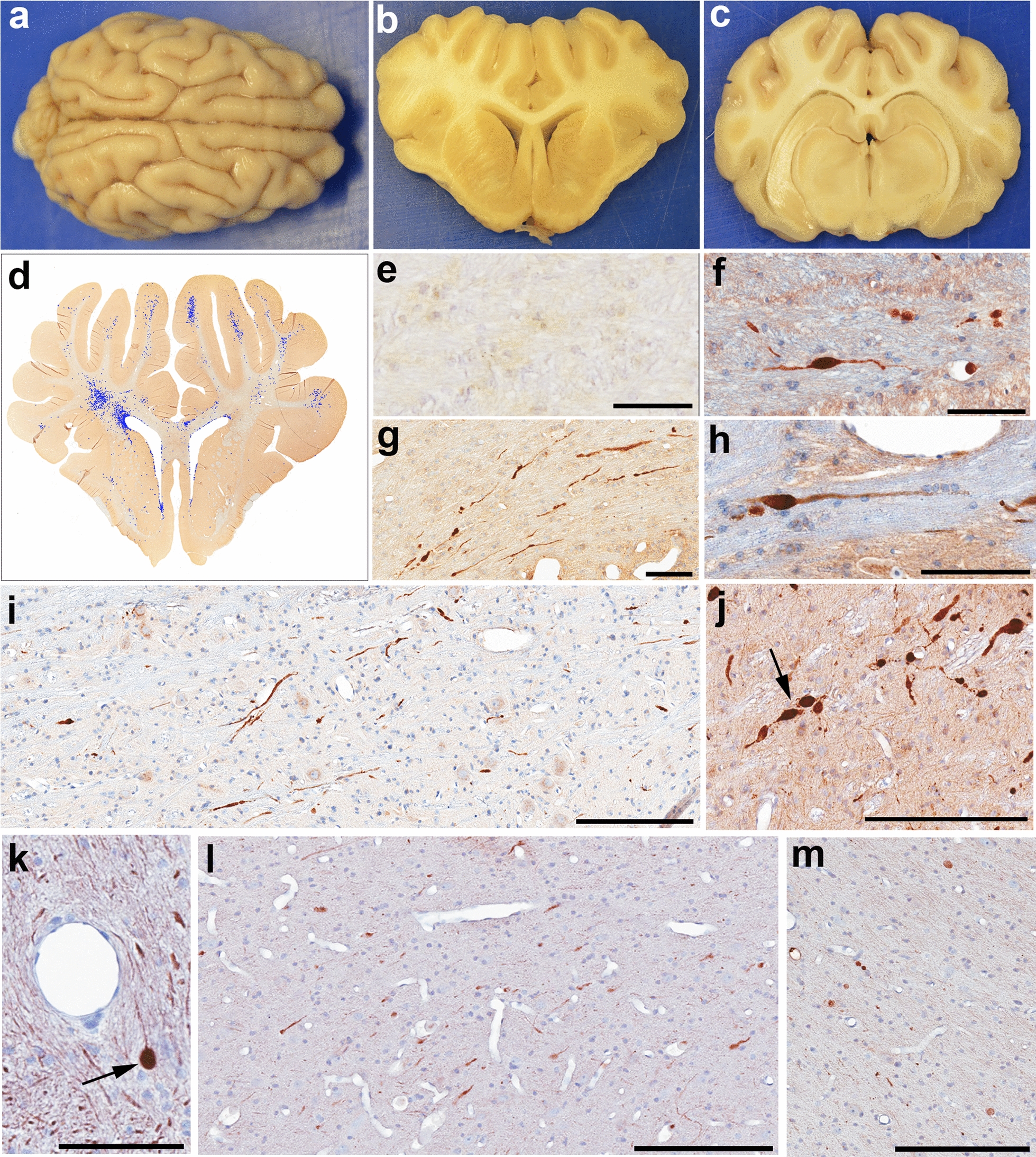
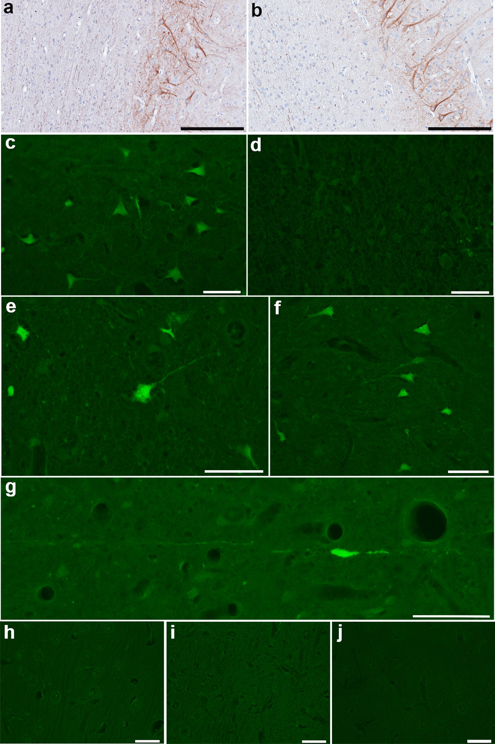
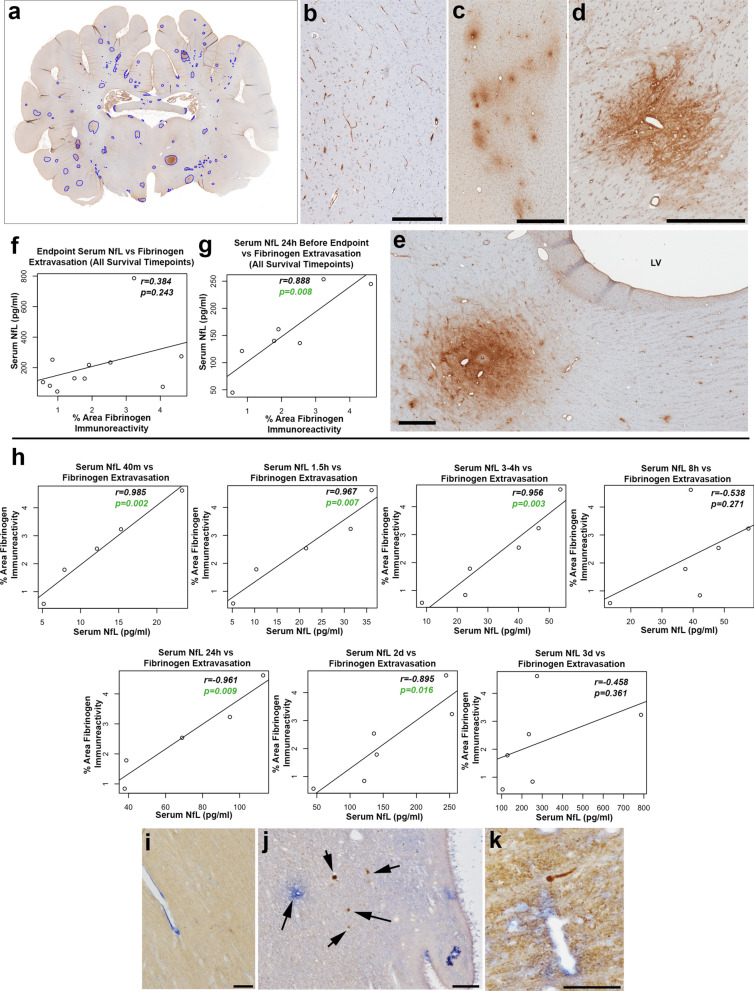
Mild traumatic brain injury (mTBI) or concussion is a substantial health problem globally, with up to 15% of patients experiencing persisting symptoms that can significantly impact quality of life. Currently, the diagnosis of mTBI relies on clinical presentation with ancillary neuroimaging to exclude more severe forms of injury. However, identifying patients at risk for a poor outcome or protracted recovery is challenging, in part due to the lack of early objective tests that reflect the relevant underlying pathology. While the pathophysiology of mTBI is poorly understood, axonal damage caused by rotational forces is now recognized as an important consequence of injury. Moreover, serum measurement of the neurofilament light (NfL) protein has emerged as a potentially promising biomarker of injury. Understanding the pathological processes that determine serum NfL dynamics over time, and the ability of NfL to reflect underlying pathology will be critical for future clinical research aimed at reducing the burden of disability after mild TBI. Using a gyrencephalic model of head rotational acceleration scaled to human concussion, we demonstrate significant elevations in serum NfL, with a peak at 3 days post-injury. Moreover, increased serum NfL was detectable out to 2 weeks post-injury, with some evidence it follows a biphasic course. Subsequent quantitative histological examinations demonstrate that axonal pathology, including in the absence of neuronal somatic degeneration, was the likely source of elevated serum NfL. However, the extent of axonal pathology quantified via multiple markers did not correlate strongly with the extent of serum NfL. Interestingly, the extent of blood-brain barrier (BBB) permeability offered more robust correlations with serum NfL measured at multiple time points, suggesting BBB disruption is an important determinant of serum biomarker dynamics after mTBI. These data provide novel insights to the temporal course and pathological basis of serum NfL measurements that inform its utility as a biomarker in mTBI.

Keywords: Blood brain barrier; Concussion; Diffuse axonal injury; Mild traumatic brain injury; Neurofilament light; Serum biomarkers.


Conflict of interest statement

Declarations. Ethics approval and consent to participate: All in vivo experiments were conducted in accordance with protocols approved by The University of Pennsylvania Institutional Animal Care and Use Committee. Consent for publication: Not applicable. Competing interests: Dr. Ramon Diaz-Arrastia has consulted with MesoScale Discoveries and BrainBox Solutions, Inc. Dr. Douglas Smith has consulted with Abbott Laboratories.

Figures

Fig. 1
Fig. 1
Temporal Serum NfL Measurements Following mTBI. a Serum NfL measurements for each time point measured in injured versus sham animals. *Denotes difference from baseline (*p < 0.05, **p < 0.01, ***p < 0.001) using T Test with adjustment for FDR. Note one animal did not have baseline serum NfL data and thus could not be included in this analysis. Individual data points for this animal are shown in gray. #Denotes difference from sham. (#p < 0.05, ##p < 0.01) using T Test with adjustment for FDR. b, c Serial NfL measurements in individual animals over time in those with survival of 48 or 72 h (b) and 2 weeks (c)
Fig. 2
Fig. 2
Range of Serum NfL Levels Over Time Following mTBI. a Serum NfL in individual injured animals to 3 days post-injury (blue) and shams (red) with linear mixed effects model with random slope demonstrating increasing differences between mTBI and sham over time. bl Range of serum NfL levels at each time point post-mTBI in comparison with all baseline measurements (mTBI and sham). It can be observed that serum NfL measurements completely separate from the baseline measurements after 8 h following mTBI
Fig. 3
Fig. 3
Gross Neuropathological Findings and Axonal Pathology. Representative example of the gross appearance of a the whole brain, and coronal tissue blocks at the level of b the lateral ventricles and c hippocampus 72 h following mTBI. The brain appears grossly normal and symmetrical, without evidence of significant brain swelling, hemorrhage or other focal pathologies. d Representative map showing the extent and distribution of APP + injured axonal profiles on a manually annotated tissue section following high-resolution digital scanning at 20X magnification. e An absence of APP immunoreactive swellings in the centrum semiovale of a sham animal at 72 h. fj Injured axons displaying APP immunoreactive swellings within the centrum semiovale at the level of the anterior hippocampus 48 h post-mTBI (f), adjacent to the lateral ventricle 48 h post-mTBI (g), at 72 h post-mTBI within the internal capsule (h) and the brainstem (i), and 2 weeks following mTBI within the brainstem (j). Note the arrow in (j) showing multiple serial swellings due to multiple points of partially interrupted transport. k Injured axons displaying NfL immunoreactive swellings within the centrum semiovale. Arrow denotes a large spheroid with apparent terminal disconnection. l NfL immunoreactive injured axons in the white matter of the superior frontal gyrus at 48 h post mTBI m NfL immunoreactive injured axons 2 weeks post-mTBI within the grey-white interface of the insular cortex. Scale Bars: F, G: 90 µm, E, H, K: 100 µm, I, J, L, M: 200 µm
Fig. 4
Fig. 4
NfL and FJC Staining of Neuronal Somata. a, b NfL immunoreactivity in neuronal somata and dendrites with normal morphologies within deeper layers of cortex as well as axons in regions of normal white matter in sham animal (a) and at 72 h post-mTBI (b). Fluoro-Jade C fluorescent staining observed in (c) positive control tissue from the cortex surrounding a focal contusion at 48 h post-injury. d Negative control section where the same region of tissue underwent identical treatment absent the application of FJC. e, f Isolated clusters of FJC positive neurons observed in just 2 of all animals examined following mTBI including in the perirhinal cortex at 72 h post-injury (e) and the somatosensory cortex at 48 h post-injury (f). g Representative example of an FJC + axon with varicose swelling in the thalamus at 2 weeks post-mTBI. hj Representative examples showing an absence of any FJC positive staining in the somatosensory cortex of a sham animal (h) and at 72 h post-mTBI (i), as well the motor cortex at 2 weeks post-mTBI (j). All scale bars 20 µm
Fig. 5
Fig. 5
Blood–Brain Barrier Permeability and Serum NfL. a Representative annotated map of fibrinogen extravasation at 72 h following mTBI. b Sham animals showing fibrinogen immunoreactivity only within the intravascular compartment. c Multiple foci of extravasated fibrinogen in the white matter close to the gray-white interface of the superior frontal gyrus 72 h following mTBI. d Large focus of extravasated fibrinogen in the midbrain at 72 h after mTBI. e Focus of extravasated fibrinogen adjacent to the lateral ventricle 2 weeks following mTBI. f, g Graphs showing correlations between extravasated fibrinogen determined via histological assessment and serum NfL at the endpoint (f) and 24 h prior to the endpoint (g) in all animals regardless of survival group. h Graphs showing correlations between serum NfL measured at different time points post-injury and extravasated fibrinogen determined via histological assessment in animals with a survival of 72 h. ik Multi-immunoenzymatic labelling for NfL (brown) and fibrinogen (blue). Note the absence of swollen NfL profiles and fibrinogen confined to the intravascular compartment in a sham animal (i). In contrast, representative examples show NfL immunoreactive swollen axonal profiles in close proximity to regions of extravasated fibrinogen within (j) the periventricular region of the lateral ventricle at 72 h following mTBI and k the corpus callosum at 48 h following mTBI. Scale Bars: B: 300 µm, C–E: 500 µm, I–K: 100 µm

References

    1. Adams JH, Doyle D, Ford I, Gennarelli TA, Graham DI, McLellan DR (1989) Diffuse axonal injury in head injury: definition, diagnosis and grading. Histopathology 15:49–59 - PubMed
    1. Adams JH, Graham DI, Gennarelli TA, Maxwell WL (1991) Diffuse axonal injury in non-missile head injury. J Neurol Neurosurg Psychiatry 54:481–483 - PMC - PubMed
    1. Adams JH, Graham DI, Murray LS, Scott G (1982) Diffuse axonal injury due to nonmissile head injury in humans: an analysis of 45 cases. Ann Neurol 12:557–563 - PubMed
    1. Al Nimer F, Thelin E, Nystrom H, Dring AM, Svenningsson A, Piehl F, Nelson DW, Bellander BM (2015) comparative assessment of the prognostic value of biomarkers in traumatic brain injury reveals an independent role for serum levels of neurofilament light. PLoS ONE 10:e0132177. 10.1371/journal.pone.0132177 - PMC - PubMed
    1. Alvarez JI, Saint-Laurent O, Godschalk A, Terouz S, Briels C, Larouche S, Bourbonniere L, Larochelle C, Prat A (2015) Focal disturbances in the blood-brain barrier are associated with formation of neuroinflammatory lesions. Neurobiol Dis 74:14–24. 10.1016/j.nbd.2014.09.016 - PubMed

Publication types

LinkOut - more resources