. 2025 Jan 7;151(1):8-30.
doi: 10.1161/CIRCULATIONAHA.123.066003. Epub 2024 Dec 5.

Sickle Trait and Alpha Thalassemia Increase NOS-Dependent Vasodilation of Human Arteries Through Disruption of Endothelial Hemoglobin-eNOS Interactions

Affiliations

Sickle Trait and Alpha Thalassemia Increase NOS-Dependent Vasodilation of Human Arteries Through Disruption of Endothelial Hemoglobin-eNOS Interactions

Steven D Brooks et al. Circulation. .

Abstract

Background: Severe malaria is associated with impaired nitric oxide (NO) synthase (NOS)-dependent vasodilation, and reversal of this deficit improves survival in murine models. Malaria might have selected for genetic polymorphisms that increase endothelial NO signaling and now contribute to heterogeneity in vascular function among humans. One protein potentially selected for is alpha globin, which, in mouse models, interacts with endothelial NOS (eNOS) to negatively regulate NO signaling. We sought to evaluate the impact of alpha globin gene deletions on NO signaling and unexpectedly found human arteries use not only alpha but also beta globin to regulate eNOS.

Methods: The eNOS-hemoglobin complex was characterized by multiphoton imaging, gene expression analysis, and coimmunoprecipitation studies of human resistance arteries. Novel contacts between eNOS and hemoglobin were mapped using molecular modeling and simulation. Pharmacological or genetic disruption of the eNOS-hemoglobin complex was evaluated using pressure myography. The association between alpha globin gene deletion and blood pressure was assessed in a population study.

Results: Alpha and beta globin transcripts were detected in the endothelial layer of the artery wall. Imaging colocalized alpha and beta globin proteins with eNOS at myoendothelial junctions. Immunoprecipitation demonstrated that alpha globin and beta globin form a complex with eNOS and cytochrome b5 reductase. Modeling predicted negatively charged glutamic acids at positions 6 and 7 of beta globin to interact with positively charged arginines at positions 97 and 98 of eNOS. Arteries from donors with a glutamic acid-to-valine substitution at beta globin position 6 (sickle trait) exhibited increased NOS-dependent vasodilation. Alpha globin gene deletions were associated with decreased arterial alpha globin expression, increased NOS-dependent vasodilation, and lower blood pressure. Mimetic peptides that targeted the interactions between hemoglobin and eNOS recapitulated the effects of these genetic variants on human arterial vasoreactivity.

Conclusions: Alpha and beta globin subunits of hemoglobin interact with eNOS to restrict NO signaling in human resistance arteries. Malaria-protective genetic variants that alter the expression of alpha globin or the structure of beta globin are associated with increased NOS-dependent vasodilation. Targeting the hemoglobin-eNOS interface could potentially improve NO signaling in diseases of endothelial dysfunction such as severe malaria or chronic cardiovascular conditions.

Keywords: alpha-Globins; alpha-Thalassemia; beta-Globins; hemoglobins; nitric oxide; nitric oxide synthase type III; sickle cell trait.


Conflict of interest statement

H.C.A., S.B., and P.C. are co-inventors on a patent held by the National Institute of Allergy and Infectious Diseases for structures and uses of beta globin mimetic peptides (PCT/US2023/065432). The other authors report no conflicts.

Figures
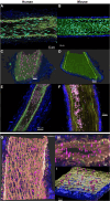
Figure 1.
Figure 1.
Alpha globin, beta globin, and eNOS comprise an autofluorescent complex that localizes to the internal elastic lamina layer near endothelial–smooth muscle cell junctions in human resistance arteries. Multiphoton (MP) imaging of intact human resistance arteries revealed autofluorescent puncta in artery wall that were not observed in murine resistance arteries. A, DAPI-stained human omental artery. Elastin narrow-band autofluorescence is shown in green, collagen second harmonic generation is shown in blue, DAPI-stained nuclei are shown in cyan, and the broad-spectrum autofluorescent puncta are shown in pink. B, DAPI-stained murine mesenteric artery exhibits similar structure and organization with elastin, collagen, and DAPI-stained nuclei present, but the broad-spectrum autofluorescent objects are absent. C, Three-dimensional (3-D) image of blood-free human mesenteric artery showing autofluorescent puncta in the internal elastic laminar layer of the artery wall. D, 3-D image of blood-free murine mesenteric artery showing the internal elastic laminar layer and collagen, but no autofluorescent puncta. Unstained, intact human omental artery (E) and murine mesenteric artery (F) imaged with red blood cells in the artery lumen for comparison with the autofluorescent puncta. G, In the wall of a DAPI-stained human resistance artery, the autofluorescent puncta (pink) localize to the internal elastic lamina (yellow) and tend to appear in gaps. H, 3-D image of the artery wall shows the autofluorescent objects (pink) are embedded in the internal elastic lamina (yellow). Collagen is shown in blue. I, 3-D image of artery wall with nuclei subtracted from the image reveals that the autofluorescent objects (pink) traverse the internal elastic lamina. DAPI indicates 4′,6-diamidino-2-phenylindole.
Figure 2.
Figure 2.
Fluorescent lifetime imaging microscopy (FLIM) identifies alpha globin, beta globin, and eNOS as constituents of an autofluorescent complex in the artery wall. Fluorescent lifetime was used to distinguish photons emitted from the antibody-conjugated fluorophores vs those emitted from the autofluorescent arterial wall object itself. A, The distribution of fluorescence lifetimes shifted after staining with a fluorophore-labeled antibody directed against alpha globin (HBA). B, Individual puncta exhibit mixed fluorescence lifetime signals after staining with HBA antibody, indicating that the antibody is bound to the autofluorescent object. C, Individual puncta have a shorter fluorescence lifetime when stained with antibody compared with unstained (P<0.0001 by t test). D through F, Fluorescence lifetime image analysis is shown for fluorophore-labeled antibodies against beta globin (HBB) and (G through I) against eNOS. J, The shift in fluorescence lifetime distribution for dual-labeled (HBA+eNOS) arteries is greater than the shift for single-labeled arteries (HBA only, eNOS only), indicating Förster resonance energy transfer (FRET), a process that can occur when fluorophores are within ≈10 nm of each other. K and L, Analysis of individual puncta reveals a significant shift in fluorescence lifetime of photons emitted from single-labeled arteries vs unstained (unstained, 4.48±0.04 ns; HBA antibody, 3.95±0.15 ns; eNOS antibody, 3.96±0.08 ns; P<0.0001 for each factor by 2-way ANOVA), whereas the costained sample had a mean lifetime of 3.29±0.19 ns (adjusted P value [Padj]<0.0001 vs singly or unstained arteries by Tukey Honestly Significant Difference test).
Figure 3.
Figure 3.
Constituents of a hemoglobin-eNOS-CYB5R3 complex are expressed in blood-free human resistance arteries. A, RNA was extracted from human omental resistance arteries that were perfused free of blood. Transcripts from alpha globin (HBA1 and HBA2) and beta globin (HBB) were quantified using RT-ddPCR and normalized to GAPDH. Endothelial nitric oxide synthase (NOS3) and myosin light chain kinase (MYLK), which are expected to be expressed in arterial endothelial or smooth muscle cells, respectively, served as comparators. The erythrocyte lineage specific gene Band 3 (SLC4A1) served as a sentinel for blood contamination. RNA processed in parallel without reverse transcriptase served as an additional control (no reverse transcriptase). B, The ratio of alpha globin transcripts originating from the HBA2 vs HBA1 genes in arterial tissue was compared with blood; the difference provides further evidence that alpha globin transcripts in arterial tissue are not attributable to blood contamination. RNA in situ hybridization on 5-μm sections of human omental artery with probes for HBA1 (C) and HBB (D); red arrows highlight positive signals for HBA1 and HBB in the endothelial cell layer. Additional histological studies are provided in Figure S4. E, Approximately 100 omental artery segments from 6 donors were divided into 3 pools (P1, P2, and P3) for immunoprecipitation studies. Proteins from pools P1 and P2 were subjected to immunoprecipitation using an antibody directed against alpha globin, whereas pool P3 was subjected to immunoprecipitation with an anti-IgG antibody. A lysate from HEK293 cells expressing Myc-eNOS provided a positive control for eNOS, whereas a red blood cell lysate provided positive controls for alpha and beta globin. Western blotting was performed for eNOS, CYB5R3, beta globin (HBB), and alpha globin (HBA). The image acquisition time was reduced for the bottom 2 panels (HBB and HBA) to prevent oversaturation of the image with the signals from the RBC lysates which had abundant hemoglobin. F, Protein from pools P1 and P2 was also tested for Band 3 through Western blot to assess for blood contamination and was not detected even at high exposure. G, Additional arteries were obtained from more donors and divided into pools P4, P5, and P6 to confirm the coimmunoprecipitation of eNOS with alpha globin and to test for HSP90. A HEK293 cell lysate expressing eNOS was used as a control and expressed HSP90. H, Artery pools P4 and P5 were immunoprecipitated with HBA antibody and then blotted for CYB5R3 using an antibody from a different manufacturer and more precise molecular weight markers to verify the sizes of CYB5R3 in arteries (34 kD) vs red blood cells (31 kD). The higher molecular weight of CYB5R3 in arterial tissues reflects the expression of the exon 1–encoded transmembrane domain and provides further evidence that the arterial lysates were not contaminated with blood. ddPCR indicates droplet digital polymerase chain reaction.
Figure 4.
Figure 4.
Structural predictions and functional studies of the interactions between the alpha subunit of hemoglobin and eNOS. A, A docking model of tetrameric hemoglobin (pink, green) interacting with an eNOS oxygenase domain dimer (yellow, orange). A model including CYB5R3 is provided in Figure S6. B, A close-up view of the amino acid residues of alpha globin that interact with eNOS, in which the sequence comprising the mimetic peptide is in red. The pink residues show the position of this sequence in the previous model of monomeric alpha globin with eNOS. C, Vasoconstriction of human omental resistance arteries in response to escalating doses of phenylephrine (PE; 10-9 to 10-3 M; black line) was measured using single-vessel ex vivo pressure myography (n=5 different donors). Phenylephrine responses were repeated after incubation with 5×10⁻⁶ M HbaX (orange) and after incubation with HbaX and a NOS inhibitor L-NAME at a concentration of 10-4 M (blue). HbaX-treated arteries constricted less to phenylephrine compared with phenylephrine alone (P<0.0001). NOS inhibition with L-NAME restored phenylephrine-induced vasoconstriction (P<0.0001 vs HbaX; P=0.09 vs phenylephrine alone). D, Vasoconstrictive responses to escalating doses of phenylephrine were conducted using a peptide with a scrambled sequence (ScrX) as a control. Differences between curves at individual doses of phenylephrine were assessed by repeated-measures 2-way ANOVA with post hoc Tukey test. *P< 0.05; **P<0.01; ***P<0.001.
Figure 5.
Figure 5.
Alpha globin gene expression and vasoreactivity of subcutaneous adipose resistance arteries from donors with HBA gene deletions. Gene expression levels were determined using droplet digital PCR on RNA extracted from adipose arteries after blood was perfused out with saline. A, The ratio of total alpha globin transcripts (HBA1 and HBA2) relative to HBB transcripts. B through D, HBA2, HBA1, and HBB gene transcript levels relative to GAPDH. E, HBA2 to HBA1 transcript ratio. F through H, Expression levels of an endothelial (NOS3) and smooth muscle gene (MYLK) serve as positive controls, and an erythrocyte gene (SLC4A1) is a control for blood contamination. Gene expression levels in arteries from αα/αα, -α/αα, and -α/-α donors were compared using the Kruskal-Wallis test; P values <0.05 are indicated on the figure. I, Vasoconstrictive responses of adipose resistance arteries from 7 αα/αα donors (blue) and 8 -α/αα donors (orange) were characterized in response to escalating doses of phenylephrine. Arteries from -α/αα donors constricted less to phenylephrine compared with arteries from αα/αα donors (P<0.0001 by global F test; statistical comparisons between -α/αα and αα/αα at individual PE doses are provided by repeated-measures 2-way ANOVA with post hoc Tukey test; *P<0.05; **P<0.01; ***P<0.001). Myographic studies of arteries from the 2 −α/−α donors were excluded from analysis according to prespecified criteria, one because of age and the other because of vessel nonreactivity to KCl or PE. J and K, Genotype-specific responses to phenylephrine alone (solid lines), after exposure to the alpha globin mimetic peptide HbaX (dashed lines), and after exposure to HbaX plus the NOS inhibitor L-NAME (dotted lines). L, Responses from both genotype groups are superimposed for comparison. Vessel diameters were expressed as percentage of baseline diameters. HbaX inhibited the maximal vasoconstrictive response to phenylephrine within each genotype group (αα/αα group: Padj<0.0001; -α/αα group: Padj=0.0425; by repeated-measures 2-way ANOVA with post hoc Tukey test). The effect of HbaX on maximal phenylephrine responses was less in the -α/αα group compared with the αα/αα group (P=0.0003 by Mann-Whitney test). L-NAME restored the maximal vasoconstrictive response to phenylephrine in the presence of HbaX within each genotype group (αα/αα group: Padj<0.0001 vs PE+HbaX, Padj=0.8206 vs PE alone; -α/αα group: Padj=0.1109 vs PE+HbaX, Padj=0.9109 vs PE alone; by repeated-measures 2-way ANOVA with post hoc Šidák test). *P<0.05; **P<0.01; ***P<0.001; ****P<0.0001. PCR indicates polymerase chain reaction.
Figure 6.
Figure 6.
Association of HBA copy number with blood pressure among healthy Cambodian children. Left, Systolic and diastolic blood pressures stratified by age and alpha globin genotype. Color-filled boxes indicate the interquartile range, solid horizontal lines indicate the medians, vertical lines indicate the range, and values that are >1.5*IQR away from the median are presented as individual symbols (ie, Tukey box-and-whisker plot). Right, The linear trends of blood pressure rising with age are provided for each alpha globin genotype group represented as lines, and the SE of each linear model is represented with shading. Total sample size was n=704, with 529 αα/αα, 160 −α/αα, and 15 −α/−α genotypes. Multivariable linear regression models for factors associated with systolic and diastolic blood pressure are presented in the Table. A decrease in HBA copy number by 1 gene copy was associated with a decrease in systolic blood pressure of 1.97±0.71 mm Hg (P=0.0057) and a decrease in diastolic blood pressure of 2.25±0.72 mm Hg (P=0.0020) in analyses adjusted for age and height. Sex was not associated with blood pressure, and therefore male and female children were analyzed together.
Figure 7.
Figure 7.
The beta subunit of endothelial hemoglobin interacts with eNOS to regulate human arterial vasoreactivity. A, The complex of heterotetrameric hemoglobin A with the eNOS oxygenase domain (eNOSox) dimer after molecular dynamic simulation. B, As in A, but for hemoglobin S. See also animations of molecular dynamic simulations in supplementary materials. C, Detailed view of beta globin–eNOS interactions involving the beta globin E6 and E7 residues. D, These interactions are absent when valine replaces glutamic acid at position 6 (E6V), leading to reconfiguration of the interface. E, Subcutaneous adipose resistance artery vasoconstrictive responses to phenylephrine from HbAS (heterozygous for hemoglobin A and S) donors (n=4) vs HbAA (homozygous for hemoglobin A) donors (n=7) (P<0.0001; *P<0.05, **P<0.01 at each dose of PE). L-NAME partially restored phenylephrine-induced vasoconstriction of arteries from HbAS donors (P=0.0367 vs baseline). F, Docking model of tetrameric hemoglobin with eNOS oxygenase domain dimer. Beta globin residues predicted to interact with eNOS are shown in shades of pink. G, Detail of beta globin residues 4 to 17 that comprise the mimetic peptide Hbb-1. H, Human omental artery vasoconstrictive responses to phenylephrine (PE), after incubation with Hbb-1 (PE+Hbb-1), and after addition of NOS inhibitor L-NAME (PE+Hbb-1+L-NAME; n=7 donors). Hbb-1 inhibited phenylephrine-induced vasoconstriction (P<0.0001). L-NAME restored phenylephrine-induced vasoconstriction in Hbb-1 treated arteries (P<0.0001). *P<0.05, **P<0.01 at each dose of PE. I, Human omental artery vasoconstrictive responses to phenylephrine alone and after treatment with a scrambled version of Hbb-1 (Hbb-1–Scr) alone or with L-NAME (n=5 different donors).

References

    1. Palmer R, Ferrige A, Moncada S. Nitric oxide release accounts for the biological activity of endothelium-derived relaxing factor. Nature. 1987;327:524–526. doi: 10.1038/327524a0 - PubMed
    1. Lancaster JR. Simulation of the diffusion and reaction of endogenously produced nitric oxide. Proc Natl Acad Sci U S A. 1994;91:8137–8141. doi: 10.1073/pnas.91.17.8137 - PMC - PubMed
    1. Butler AR, Megson IL, Wright PG. Diffusion of nitric oxide and scavenging by blood in the vasculature. Biochim Biophys Acta. 1998;1425:168–176. doi: 10.1016/s0304-4165(98)00065-8 - PubMed
    1. Vaughn MW, Kuo L, Liao JC. Effective diffusion distance of nitric oxide in the microcirculation. Am J Physiol. 1998;274:H1705–H1714. doi: 10.1152/ajpheart.1998.274.5.H1705 - PubMed
    1. Rhodin JA. The ultrastructure of mammalian arterioles and precapillary sphincters. J Ultrastruct Res. 1967;18:181–223. doi: 10.1016/s0022-5320(67)80239-9 - PubMed

MeSH terms

LinkOut - more resources