Review
. 2022 Jan 24;11(9):1783-1808.
doi: 10.1515/nanoph-2021-0711. eCollection 2022 Apr.

The perspectives of broadband metasurfaces and photo-electric tweezer applications

Affiliations
Review

The perspectives of broadband metasurfaces and photo-electric tweezer applications

Geon Lee et al. Nanophotonics. .

Abstract

With strong demands of real-time monitoring of biomolecules or environmental pollutants, overcoming technical hurdles on control and detection of freely diffusive nanoscale objects become a question of issue to solve in a variety of research fields. Most existing optical techniques inevitably require labeling to the target material, which sometimes denature the measuring biomaterials. For highly efficient real-time monitoring without complicated pretreatment or labeling, many successes in development of label-free or non-destructive detection techniques via increased sensitivity were accomplished by the additional structures. Metasurface-based two-dimensional photonic/electric devices have recently represented extraordinary performances in both manipulation and sensing for various small particles and biochemical species, repeatedly overcoming the limit of detection achieved right before. In parallel, various metasurface-based devices were also introduced promoting transportation of targets into optical hotspot sites, overcoming diffusion limits. We noted this point, therefore, reviewed two major research fields such as metasurface-assisted material sensing and transportation technologies that have contributed to present prospective sensing technologies, then showed perspective views on how great synergy can be created when two technologies are cleverly integrated. Recently, a trend of conceptual merging of optical detection and transporting schemes beyond both diffraction limit and diffusion limit leads to a creation of exceptional performance in molecular detections. In this review, the trends of the latest technologies accomplishing this purpose by hybridization of various composite materials and functional metasurfaces will be introduced.

Keywords: biosensor; dielectrophoresis; metasurface; optical sensing; plasmonic trapping; tweezer.


Figures

Figure 1:
Figure 1:
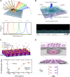
Conceptual illustration of biochemical substance monitoring over the metasurface-based structures at the ultrabroad band of electromagnetic waves. Significantly improved light-molecule interactions by the metasurfaces and increased absorption cross-sections of the molecules provide improved sensing performances even in extremely low density of the molecules as described.
Figure 2:
Figure 2:
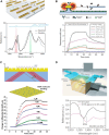
All-dielectric metasurface enhanced sensing. (A) A broadband on-demand resonance behavior for every incidence angle (bottom) from high-Q germanium-based metasurfaces (top). Reproduced with permission from ref. [1], copyright 2019, AAAS (American Association for the Advancement of Science). (B) Polydimethylsiloxane (PDMS) microfluidic chamber bonded to the PhCM (top). The air cylindrical holes arranged in a silicon nitride film (bottom). Adapted with permission from ref. [2], copyright 2018, Optical Society of America. (C) CEA molecule detection by dielectric metasurface (top). FL-intensity plot versus CEA concentration (orange closed circle) with fitted curve (dashed curve) using the Hill equation (bottom). Adapted with permission from ref. [3], copyright 2020, American Chemical Society. (D) Chiral molecule detection using dye/DNA functionalization with periodic silicon disk-arrays. Reproduced with permission from ref. [4], copyright 2020, American Chemical Society.
Figure 3:
Figure 3:
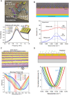
Plasmonic metasurface for high sensitive molecule sensing. (A) Multi-resonant mid-IR metasurface (top) and experimental reflectance spectra (bottom). Reproduced with permission from ref. [5], copyright 2018, Springer Nature. (B) Pillar-supported disk structure increases the effective sensing volume and performance (top). Real-time sensing of interaction of two types of IgGs as antibody concentration (bottom). Adapted with permission from ref. [6], copyright 2017, Springer Nature. (C) The diagram of nanogroove hyperbolic metasurface (top) and plasmonic sensing of binding interaction of bovine serum albumin molecule by the change in differential GH shift (bottom). Reproduced with permission from ref. [7], copyright 2017, John Wiley and Sons. (D) A graphical representation of the GC-HMM sensor device with a fluid flow channel (top). Measured time-varying wavelength shifts while 10 pM biotin injection (bottom). Adapted with permission from ref. [8], copyright 2016, Springer Nature.
Figure 4:
Figure 4:
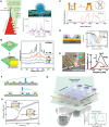
2D material combined metasurface sensor. (A) DNA adsorption on the graphene-combined nanoslot metasurface (top). Change in THz transmission spectra due to ssDNA on a graphene covered nanoslot metasurface (bottom). Adapted with permission from Ref. [18], copyright 2020, Elsevier. (B) Illustration of acoustic plasmon resonator architecture (top) and measured absorption spectra about resonators with and without a silk film (bottom). Lorentzian fitting curves are shown as well (dashed lines). Adapted with permission from Ref. [19], copyright 2019, Springer Nature. (C) Active metasurface sensors for protein fingerprinting (top) and measured reflectance spectra of monolayer protein A/G and controlled Fermi level of graphene via a gate voltage (bottom). Reproduced with permission from Ref. [20], copyright 2019, American Chemical Society. (D) Graphene-metallic hybrid metasurface sensor (top) and spectral responses of various concentrations of glucose on the hybrid metasurface (bottom). Reproduced with permission from Ref. [21], copyright 2018, Springer Nature.
Figure 5:
Figure 5:
SEIRA based optical sensor. (A) Truncated view of the nanoscale antennas (NAs) 2D-array (top). Measured reflectance of the NAs-array is shown: the black and red curve are referred to bare NAs-arrays and NAs-array covered with a 50 nm PMMA layer respectively (bottom). Adapted with permission from ref. [24], copyright 2019, Elsevier. (B) Illustration of SEIRA based gas sensor (top) and measured transmission of two types of photonic crystal slab (PCS) (bottom). Adapted with permission from ref. [25], copyright 2018, American Chemical Society.
Figure 6:
Figure 6:
Highly selective THz metasensor. (A) Schematic of steroid sensing using THz nanoslot sensing (left). THz absorption spectra of steroid hormones in pellet types and transmission spectra of nanoslot-arrays (right). Reproduced with permission from ref. [26], copyright 2019, American Chemical Society. (B) Schematic diagram of THz nanogap metasurface sensing of dsDNA and single-strand RNA viruses. Reproduced with permission from ref. [27], copyright 2017, Optical Society of America. (C) Diagram of metasurface-assisted THz sensing of kanamycin sulfate. Reproduced with permission from ref. [28], copyright 2015, Springer Nature. (D) Illustration of the nanofluidic THz metasensor and its cross-sectional device structure. Reproduced with permission from ref. [29], copyright 2018, AIP Publishing.
Figure 7:
Figure 7:
Schematic illustration of photo-electric tweezing platform of nanoparticles and biochemical substances using plasmonic metasurfaces. The critical role of the photo-electric tweezing platform is based on the large-scale plasmonic structures to trap the target particle/molecules and detect the targets simultaneously.
Figure 8:
Figure 8:
Principles and applications of plasmonic particle tweezing. (A) Diffraction limit on optical trapping by far-field focusing. (B) Plasmonic trapping on gold nanorods and its potential energy landscape. Reproduced with permission from ref. [38], copyright 2011, Springer Nature. (C) Migration of particle by excited SPP evanescent field. Reproduced with permission from ref. [39], copyright 1996, Optical Society of America. (D) SPP plasmofluidic ordering of nanoparticles. Adapted with permission from ref. [41], copyright 2014, Springer Nature. (E) Plasmonic trapping of Eschetichia coli bacteria on metallic nanorods. Reproduced with permission from ref. [42], copyright 2009, American Chemical Society. (F) Effect of SIBA restoring force on augmenting plasmonic trapping potential depth on metallic nanoaperture. Adapted with permission from ref. [38], copyright 2011, Springer Nature. (G) Enantio-selective chiral plasmonic trapping on annular aperture. Reproduced with permission from ref. [43], copyright 2016, American Chemical Society.
Figure 9:
Figure 9:
Principles and applications of AC electrokinetic particle tweezing. (A) DEP force acting on a particle under uniform- (left) and non-uniform E-field (right). (B) ACEO fluid motion on surface of potential applied electrodes [60]. (C) Particle trapping at stagnation zone of ACEO and thermophoresis on nanhole-array. Adapted with permission from ref. [63], copyright 2020, Springer Nature. (D) Dielectrophoretic particle trapping on nanogap and fluorescent microscopic image of trapped nanodiamond. Reproduced with permission from ref. [64], copyright 2016, American Chemical Society. (E) Simulations of particle dynamics on vertical nanogap electrode for combined DEP/ACEO for relocation of particles on the metasurface. Adapted with permission from ref. [61], copyright 2020, Springer Nature. (F) Dielectrophoretic nano-biopsy and fluorescent microscopic images of DNA trapping and releasing. Adapted with permission from ref. [65], copyright 2019, Springer Nature.
Figure 10:
Figure 10:
Surface-enhanced sensing on metasurface combined with self-guided passive transport. (A) Evaporation-induced molecule concentration over the plasmonic focusing nanocone and Raman spectra for detection of 1 fM lysosome. Adapted with permission from ref. [84], copyright 2011, Springer Nature. (B) Evaporation-induced molecule concentration and Raman spectra for detection of rhodamine 6G on hydrophobic SERS platform. Adapted with permission from ref. [85], copyright 2016, National Academy of Sciences. (C) Capillary force-driven nanostructure clustering for achievement of enhanced optical signals. Raman spectra for detection of rhodamine 6G on mushrooms before and after clustering. Adapted with permission from ref. [87], copyright 2010, American Chemical Society. (D) Evaporation-assisted molecule concentration at the trench of ribbon-resonator. Adapted with permission from ref. [90], copyright 2021, Springer Nature. (E) Mechanical sweeping for concentration of gold nanoparticles into THz nanoslot. Adapted with permission from ref. [91], copyright 2017, Optical Society of America. (F) Molecular depletion zone and diffusion on plasmonic sensor with uniform- (upper) and orthogonal functionalization (lower). Adapted with permission from ref. [92], copyright 2019, Royal Society of Chemistry. (G) In-flow imaging platform with diatomic all-dielectric metasurface for extracellular vesicle detection. Reproduced with permission from ref. [97], copyright 2021, Springer Nature. (H) Time-lapse spectral shift for detection of isopropyl alcohol on nanoholes by flow-over and flow-through strategies. Reproduced with permission from ref. [101], copyright 2010, AIP Publishing.
Figure 11:
Figure 11:
Surface-enhanced sensing on metasurface combined with field-induced active transport. (A) BSA protein trapping on double-nanohole and time-lapse monitoring of optical transmission intensity for detection of protein folding. Adapted with permission from ref. [104], copyright 2012, American Chemical Society. (B) Plasmonic nanopore bowtie for trapping and Raman sequencing of DNA molecule. Adapted with permission from ref. [112], copyright 2015, American Chemical Society. (C) Gold nanoparticle trapping at nanohole and real-time monitoring of Raman spectra for detection of DNA bases. Adapted with permission from ref. [119], copyright 2019, Springer Nature. (D) ACEO-enhanced gold nanoparticle gathering for improved scattering intensity for protein detection. Adapted with permission from ref. [123], copyright 2017, American Chemical Society. (E) Nanoparticle trapping on split-trench resonator and time-lapse monitoring of transmission spectral shift for detection of nanoparticle. Adapted with permission from ref. [126], copyright 2021, American Chemical Society. (F) Particle concentration into nanoslot by combined DEP/ACEO and real-time monitoring of terahertz reflectance for detection of nano-vesicles. Adapted with permission from ref. [62], copyright 2021, John Wiley and Sons.
Figure 12:
Figure 12:
Conceptual illustration of photo-electric tweezers for biochemical substances detection on the metasurface by physical matching of particle capturing sites to the optical sensing hotspots.

References

    1. Leitis A., Tittl A., Liu M., et al. Angle-multiplexed all-dielectric metasurfaces for broadband molecular fingerprint retrieval. Sci. Adv. . 2019;5:eaaw2871. doi: 10.1126/sciadv.aaw2871. - DOI - PMC - PubMed
    1. Romano S., Zito G., Torino S., et al. Label-free sensing of ultralow-weight molecules with all-dielectric metasurfaces supporting bound states in the continuum. Photon. Res. . 2018;6:726. doi: 10.1364/prj.6.000726. - DOI
    1. Iwanaga M. All-dielectric metasurface fluorescence biosensors for high-sensitivity antibody/antigen detection. ACS Nano . 2020;14:17458–17467. doi: 10.1021/acsnano.0c07722. - DOI - PubMed
    1. Solomon M. L., Abendroth J. M., Poulikakos L. V., Hu J., Dionne J. A. Fluorescence-detected circular dichroism of a chiral molecular monolayer with dielectric metasurfaces. J. Am. Chem. Soc. . 2020;142:18304–18309. doi: 10.1021/jacs.0c07140. - DOI - PubMed
    1. Rodrigo D., Tittl A., Ait-Bouziad N., et al. Resolving molecule-specific information in dynamic lipid membrane processes with multi-resonant infrared metasurfaces. Nat. Commun. . 2018;9:2160. doi: 10.1038/s41467-018-04594-x. - DOI - PMC - PubMed

LinkOut - more resources