Review
. 2024 Nov 1;27(12):111309.
doi: 10.1016/j.isci.2024.111309. eCollection 2024 Dec 20.

Histatins, proangiogenic molecules with therapeutic implications in regenerative medicine

Affiliations
Review

Histatins, proangiogenic molecules with therapeutic implications in regenerative medicine

Héctor Tapia et al. iScience. .

Abstract

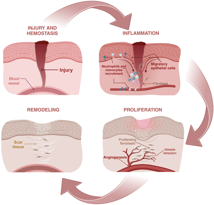
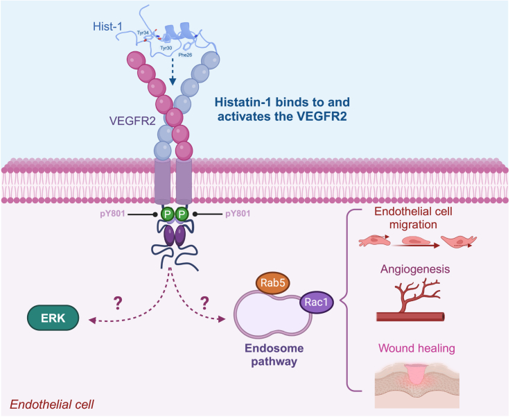
Recent studies show that a group of salivary peptides, collectively known as histatins, are potent inducers of wound healing in both soft and hard tissues. Among these molecules, histatin-1 stands out for its ability to stimulate the repair of skin, oral mucosal, and osseous tissue. Remarkably, all these effects are associated with the capacity of histatin-1 to promote angiogenesis via inducing endothelial cell adhesion, migration, and signaling. These findings have opened new opportunities in the field of regenerative medicine, leading to an increasing number of articles and patents proposing therapeutic uses of histatin-1. However, this scenario raises a relevant concern regarding the appropriate use of these molecules, since, unlike the mode of action, little is known about the molecular mechanism by which they promote angiogenesis and wound healing. Recent studies shed light on the pharmacodynamics of histatin-1, by identifying the endothelial receptor that it binds and downstream signaling. This perspective will discuss current evidence on the role of histatins in wound healing and angiogenesis, emphasizing their impact on regenerative medicine.

Keywords: Biological sciences; Health sciences; Medicine; Natural sciences; Physiology.


Conflict of interest statement

The authors declare no competing interest.

Figures

Figure 1
Figure 1
Wound healing The different phases of epithelial wound healing are described. These include hemostasis, inflammation, proliferation, and remodeling (see the main text for details).
Figure 2
Figure 2
Human histatins (A) Amino acid sequence of main human histatins. The two histatin genes, HTN1 and HTN3, are indicated with their protein products, histatin-1 and histatin-3, respectively. Derivative fragments of these histatins (histatin-2 and histatin-5) are also shown. (B) The 3D structure of histatin-1 is shown, highlighting residues Phe26, Tyr30, and Y34, which are critical for biological activity in endothelial cells. The 3D model was previously proposed by Mateluna et al., in molecular modeling studies. The 3D structure of full-length histatin-3 is shown and it was obtained from AlphaFold Protein Structure Database (repository code: AF_AFP15516F1).,
Figure 3
Figure 3
Timeline: retrospective analysis of histatin-dependent effects in tissue repair The timeline shows the main findings related to the roles histatins in cell and tissue repair. These include the effects at the cellular level, namely cell adhesion, spreading, and migration. Also, the effects of histatins in soft and mineralized tissue repair are shown, along with the different therapeutic strategies that have been proposed in animal models. The impact of histatins on angiogenesis and the consequences in tissue repair are highlighted.
Figure 4
Figure 4
Pharmacodynamics of histatin-1 The image summarizes the findings that histatin-1 is a potent angiogenic factor, which binds to the VEGFR2, leading to the activation of downstream signaling. While ERK and Rab5/Rac-dependent signaling have been described downstream histatin-1, their sequential activation and requirement of VEGFR2 remain to be explored.

Similar articles

Cited by

References

    1. Melino S., Santone C., Di Nardo P., Sarkar B. Histatins: salivary peptides with copper(II)- and zinc(II)-binding motifs: perspectives for biomedical applications. FEBS J. 2014;281:657–672. doi: 10.1111/febs.12612. - DOI - PubMed
    1. Pan L., Zhang X., Gao Q. Effects and mechanisms of histatins as novel skin wound-healing agents. J. Tissue Viability. 2021;30:190–195. doi: 10.1016/j.jtv.2021.01.005. - DOI - PubMed
    1. Torres P., Castro M., Reyes M., Torres V.A. Histatins, wound healing, and cell migration. Oral Dis. 2018;24:1150–1160. doi: 10.1111/odi.12816. - DOI - PubMed
    1. van Dijk I.A., Veerman E.C.I., Reits E.A.J., Bolscher J.G.M., Stap J. Salivary peptide histatin 1 mediated cell adhesion: a possible role in mesenchymal-epithelial transition and in pathologies. Biol. Chem. 2018;399:1409–1419. doi: 10.1515/hsz-2018-0246. - DOI - PubMed
    1. Mateluna C., Torres P., Rodriguez-Peña M., Silva P., Matthies D.J., Criollo A., Bikker F.J., Bolscher J.G.M., Wilson C.A.M., Zapata-Torres G., Torres V.A. Identification of VEGFR2 as the Histatin-1 receptor in endothelial cells. Biochem. Pharmacol. 2022;201 doi: 10.1016/j.bcp.2022.115079. - DOI - PubMed

LinkOut - more resources