. 2024 Oct 17;4(6):669-678.
doi: 10.1021/acsphyschemau.4c00052. eCollection 2024 Nov 27.

Atomistic Multiscale Modeling of Colloidal Plasmonic Nanoparticles

Affiliations

Atomistic Multiscale Modeling of Colloidal Plasmonic Nanoparticles

Luca Nicoli et al. ACS Phys Chem Au. .

Abstract

A novel fully atomistic multiscale classical approach to model the optical response of solvated real-size plasmonic nanoparticles (NPs) is presented. The model is based on the coupling of the Frequency Dependent Fluctuating Charges and Fluctuating Dipoles (ωFQFμ), specifically designed to describe plasmonic substrates, and the polarizable Fluctuating Charges (FQ) classical force field to model the solvating environment. The resulting ωFQFμ/FQ approach accounts for the interactions between the radiation and the NP, as well as with the surrounding solvent molecules, by incorporating mutual interactions between the plasmonic substrate and solvent. ωFQFμ/FQ is validated against reference TD-DFTB/FQ calculations, demonstrating remarkable accuracy, particularly in reproducing plasmon resonance frequency shifts for structures below the quantum-size limit. The flexibility and reliability of the approach are also demonstrated by simulating the optical response of homogeneous and bimetallic NPs dissolved in pure solvents and solvent mixtures.


Conflict of interest statement

The authors declare no competing financial interest.

Figures

Figure 1
Figure 1
Pictorial view of the multiscale scheme employed to develop ωFQFμ/FQ.
Figure 2
Figure 2
Graphical scheme of the computational protocol employed to compute ωFQFμ/FQ absorption spectra of colloidal plasmonic NPs (see also Sections S2.1, S2.2, S2.4, and S2.3 in the Supporting Information).
Figure 3
Figure 3
Vacuo-to-water PRF shifts (in eV) computed by using DFTB/FQ, ωFQFμ/FQ, and DFT/DFTBWAT/FQ levels of theory. A graphical depiction of the structure used for each calculation is presented and the elements are colored according to the level of theory used (orange-DFTB, gray-ωFQFμ, blue-FQ). In the green inset a zoom of the DFTB/DFTBWAT/FQ structure is reported to highlight the presence of water molecules treated at the DFTB level of theory (orange).
Figure 4
Figure 4
ωFQFμ/FQ absorption spectra of (A) Ag3851 and (B) Au3851 in vacuo (VAC) and water (WAT1). Δλ is the vacuo-to-water solvatochromic shift in nm. All spectra are normalized to the corresponding maximum in vacuo.
Figure 5
Figure 5
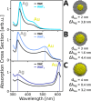
ωFQFμ/FQ absorption spectra of core–shell Au@Ag spherical NPs in vacuo (VAC) and in aqueous solution (WAT1) as a function of the diameter of the Au core (dAu: (A) 2.0 nm, (B) 3.0 nm and (C) 4.0 nm). The solvatochromic shifts of “Ag” (ΔλAg) and “Au” (ΔλAu) peaks are reported in nm. All spectra are normalized to the maximum in vacuo.
Figure 6
Figure 6
ωFQFμ/FQ absorption cross section (σabs) of core–shell Au@Ag spherical NP with an Au core diameter of 3 nm and featuring an alloyed (50% Au/Ag) external layer of 1 nm (see right panel) in vacuo (VAC) and aqueous solution (WAT1). Δλ indicates the solvatochromic shift (in nm). All spectra are normalized to the maximum in vacuo.
Figure 7
Figure 7
ωFQFμ/FQ absorption cross section (σabs) of core–shell Au@Ag spherical NP with an Au core diameter of 3 nm and featuring an alloyed (50% Au/Ag) external layer of 1 nm (see right panel) in vacuo (VAC) and a 1:1 water–ethanol mixture (WAT-ETH). The vacuo-to-mixture solvatochromic shift is given in nm. All spectra are normalized to the maximum in vacuo.

References

    1. Mayer K. M.; Hafner J. H. Localized surface plasmon resonance sensors. Chem. Rev. 2011, 111, 3828–3857. 10.1021/cr100313v. - DOI - PubMed
    1. McNamara K.; Tofail S. A. Nanoparticles in biomedical applications. Adv. Phys. X 2017, 2, 54–88. 10.1080/23746149.2016.1254570. - DOI - PubMed
    1. Dutta A.; Medda A.; Patra A. Recent advances and perspectives on colloidal semiconductor nanoplatelets for optoelectronic applications. J. Phys. Chem. C 2021, 125, 20–30. 10.1021/acs.jpcc.0c09416. - DOI
    1. Hu X.; Li G.; Yu J. C. Design, fabrication, and modification of nanostructured semiconductor materials for environmental and energy applications. Langmuir 2010, 26, 3031–3039. 10.1021/la902142b. - DOI - PubMed
    1. Yang P.; Zheng J.; Xu Y.; Zhang Q.; Jiang L. Colloidal synthesis and applications of plasmonic metal nanoparticles. Adv. Mater. 2016, 28, 10508–10517. 10.1002/adma.201601739. - DOI - PubMed

LinkOut - more resources