. 2024 Nov 20:15:1443940.
doi: 10.3389/fimmu.2024.1443940. eCollection 2024.

The FGFR inhibitor Rogaratinib reduces microglia reactivity and synaptic loss in TBI

Affiliations

The FGFR inhibitor Rogaratinib reduces microglia reactivity and synaptic loss in TBI

Rida Rehman et al. Front Immunol. .

Abstract

Background: Traumatic brain injury (TBI) induces an acute reactive state of microglia, which contribute to secondary injury processes through phagocytic activity and release of cytokines. Several receptor tyrosine kinases (RTK) are activated in microglia upon TBI, and their blockade may reduce the acute inflammation and decrease the secondary loss of neurons; thus, RTKs are potential therapeutic targets. We have previously demonstrated that several members of the Fibroblast Growth Factor Receptor (FGFR) family are transiently phosporylated upon TBI; the availability for drug repurposing of FGFR inhibitors makes worthwhile the elucidation of the role of FGFR in the acute phases of the response to TBI and the effect of FGFR inhibition.

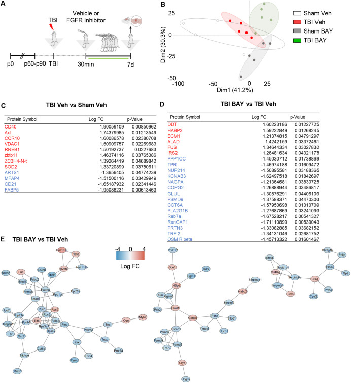
Methods: A closed, blunt, weight-drop mild TBI protocol was employed. The pan-FGFR inhibitor Rogaratinib was administered to mice 30min after the TBI and daily up to 7 days post injury. Phosphor-RTK Arrays and proteomic antibody arrays were used to determine target engagement and large-scale impact of the FGFR inhibitor. pFGFR1 and pFGFR3 immunostaining were employed for validation. As outcome parameters of the TBI injury immunostainings for NeuN, VGLUT1, VGAT at 7dpi were considered.

Results: Inhibition of FGFR during TBI restricted phosphorylation of FGFR1, FGFR3, FGFR4 and ErbB4. Phosphorylation of FGFR1 and FGFR3 during TBI was traced back to Iba1+ microglia. Rogaratinib substantially dowregulated the proteomic signature of the neuroimmunological response to trauma, including the expression of CD40L, CXCR3, CCL4, CCR4, ILR6, MMP3 and OPG. Prolonged Rogaratinib treatment reduced neuronal loss upon TBI and prevented the loss of excitatory (vGLUT+) synapses.

Conclusion: The FGFR family is involved in the early induction of reactive microglia in TBI. FGFR inhibition selectively prevented FGFR phosphorylation in the microglia, dampened the overall neuroimmunological response and enhanced the preservation of neuronal and synaptic integrity. Thus, FGFR inhibitors may be relevant targets for drug repurposing aimed at modulating microglial reactivity in TBI.

Keywords: proteomics; reactive microglia; receptor tyrosine kinase; synapses; traumatic brain injury.


Conflict of interest statement

The authors declare that the research was conducted in the absence of any commercial or financial relationships that could be construed as a potential conflict of interest. The author(s) declared that they were an editorial board member of Frontiers, at the time of submission. This had no impact on the peer review process and the final decision.

Figures

Figure 1
Figure 1
FGFR Inhibitor selectively alters RTK phosphorylation pattern at 3h post injury. (A) Outline of the experimental design. BAY121 (Rogaratinib) was administered by oral gavage at the dose of 25 mg/kg 30 min after TBI and samples were obtained 3h after TBI. (B) Principal component analysis (PCA) plot displayed group wise distribution of samples, highlighting the separation between TBI-Veh samples (red) and the BAY121-treated samples, which overlap with Sham-Veh samples. The group specific ellipses indicate 95% confidence interval. (C-J) Antibody phospho-array focused proteomic analysis of RTK phosphorylation patterns upon TBI with or withouth BAY121 treatment. Differential phosphorylation analysis showed the inhibitor significantly decreased the phosphorylated levels of (D) pFGFR3, (E), pFGFR4 (the array does not include pFGFR1) and (F) ErbB4 after TBI. However, the inhibitor showed no effect on phosphorylation levels of (C) FGFR2, (G) HGFR, (H) cRET, (I) EphA3, and (J) EphB2. Significance for differentially phosphorylated proteins was set at p<0.05 (FDR adjusted). [(B-J): n=5-6/group; *p<0.05, **p<0.01, ***p<0.001].
Figure 2
Figure 2
Upregulation of pFGFR1 and pFGFR3 in microglia 3h post injury. (A) Outline of the experimental design. BAY121 (Rogaratinib) was administered by oral gavage at the dose of 25 mg/kg 30 min after TBI and samples for immunohistology were obtained at 3h after TBI. (B-D) Immunostainings for pFGFR1 (green) and Iba1 (red) for Sham- Veh, TBI Veh, Sham- BAY121 and TBI BAY121 treated mice. Quantification of pFGFR1 immunostaining intensity in Iba1+ cells (C) and fraction of pFGFR1+ cells (D) display a significant increase upon TBI, which is negated by the treatment with BAY121. (D-F) Immunostaining for pFGFR3 (green) and Iba1 (red) shows the upregulation of pFGFR immunoreactivity in Iba1+ cells and the increase in the fraction of pFGFR3 upon TBI. Both indexes are decreased by treatment with BAY121. n=4/group; >300 cells per animal n.s., not significant; **p<0.01; ***p<0.001, ****p<0.0001. Overview Scale bar: 50µm. Inset scale bar: 20µm.
Figure 3
Figure 3
FGFR inhibitor suppresses immune-related proteome signature at 3d post injury. (A) Outline of the experimental design; BAY121 was administered 30 mins after trauma and continued for 3d (25 mg/Kg once daily by oral gavage, vehicle alone as control). Samples were collected 3d post injury. (B) PCA plot of proteomic data shows the separation of TBI-Veh from BAY121-treated samples. (C, D) After modified differential protein expression analysis (FDR <0.05), subsets of upregulated (Red) and downregulated (Blue) proteins with log fold change and individual significance for (D) TBI-Veh compared to Sham–Veh and (D) TBI-BAY121 compared to TBI-Veh (n=5/group). (E) Protein-protein-interaction analysis revealed distinct clustering of downregulated and upregulated proteins in TBI-BAY121 vs TBI Veh; the cluster of downregulated proteins is enriched with immune- and inflammation-related proteins.
Figure 4
Figure 4
Prolonged FGFR inhibitor treatment significantly alters the TBI-related proteome profiles at 7d post injury. (A) Outline of the experimental design; BAY121 was administered 30 mins after trauma and continued for 7d (25 mg/Kg/day). Samples were collected 7d post injury. (B) PCA plot shows the substantial separation between TBI groups and groups treated with BAY121. (C, D) After modified differential protein expression analysis (FDR <0.05), subset of significantly upregulated (Red) and downregulated (Blue) proteins with log fold change and individual significance for (C) TBI-Veh vs Sham–Veh and (D) TBI-BAY121 vs TBI-Veh. (E) Protein-protein interaction analysis by STRING reveals three distinct networks affected by prolonged BAY121 treatment, related to protein synthesis proteasomal degradation and inflammation (n=5/group).
Figure 5
Figure 5
Prolonged FGFR inhibitor administration preserves neuronal density at 7 dpi. (A) Outline of the experimental design; BAY121 was administered 30 mins after trauma and continued for 7d (25 mg/Kg daily by oral gavage). Samples were collected 7d post injury. (B, C) Immunostaining for NeuN showed a significantly decreased number of neurons in the ijury site of TBI-Veh group compared to Sham–Veh; a significantly higher number of neurons was seen in the TBI-BAY121 group. Note that microglial density was also increased in TBI-Veh but not in TBI-BAY121. (n=4/group; **p<0.01, ***p<0.001, ****p<0.0001; Scalebar 70µm) (D, E) Loss of VGLUT1 density after TBI is dependent on FGFR signaling 7d after TBI. (n=4/group; ***p<0.001; Scale bar: overview = 70μm; insert = 5μm) (F, G) VGAT density in the TBI core is significantly reduced at 7dpi. (n=4/group; **p<0.01; Scale bar: overview = 70μm; insert = 5μm).

References

    1. Morganti-Kossmann MC, Semple BD, Hellewell SC, Bye N, Ziebell JM. The complexity of neuroinflammation consequent to traumatic brain injury: from research evidence to potential treatments. Acta Neuropathol. (2019) 137:731–55. doi: 10.1007/s00401-018-1944-6 - DOI - PubMed
    1. Jassam YN, Izzy S, Whalen M, McGavern DB, El Khoury J. Neuroimmunology of traumatic brain injury: time for a paradigm shift. Neuron. (2017) 95:1246–65. doi: 10.1016/j.neuron.2017.07.010 - DOI - PMC - PubMed
    1. Davalos D, Grutzendler J, Yang G, Kim JV, Zuo Y, Jung S, et al. . ATP mediates rapid microglial response to local brain injury in vivo . Nat Neurosci. (2005) 8:752–8. doi: 10.1038/nn1472 - DOI - PubMed
    1. Zanier ER, Fumagalli S, Perego C, Pischiutta F, De Simoni MG. Shape descriptors of the "never resting" microglia in three different acute brain injury models in mice. Intensive Care Med Exp. (2015) 3:39. doi: 10.1186/s40635-015-0039-0 - DOI - PMC - PubMed
    1. Somebang K, Rudolph J, Imhof I, Li L, Niemi EC, Shigenaga J, et al. . CCR2 deficiency alters activation of microglia subsets in traumatic brain injury. Cell Rep. (2021) 36:109727. doi: 10.1016/j.celrep.2021.109727 - DOI - PMC - PubMed

MeSH terms

Substances