Review
. 2024 Nov 20;3(12):101411.
doi: 10.1016/j.jacadv.2024.101411. eCollection 2024 Dec.

Right Ventricular Remodeling and Function in Hypoplastic Left Heart Syndrome

Affiliations
Review

Right Ventricular Remodeling and Function in Hypoplastic Left Heart Syndrome

Thomas Salaets et al. JACC Adv. .

Abstract

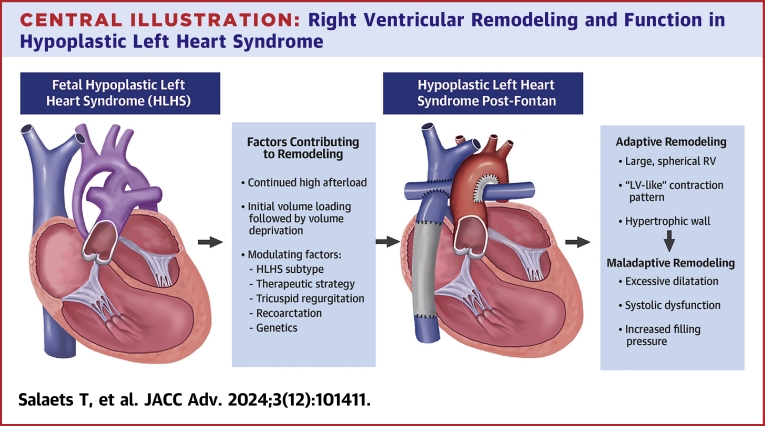
The right ventricle (RV) in hypoplastic left heart syndrome (HLHS) becomes the systemic ventricle pumping against systemic afterload. It also has to adapt to an initially increased volume load followed by a decrease in volume load after Fontan completion. Anatomical HLHS subtype, therapeutic strategy, tricuspid valve regurgitation, recoarctation, and genetics influence RV size and function. The resulting remodeling process can be maladaptive and lead to ventricular systolic and diastolic dysfunction. While systolic dysfunction is a strong predictor for mortality before Fontan, there is increasing evidence for the impact of progressive diastolic dysfunction after Fontan. This comprehensive review summarizes the (recent) empirical observations that increased understanding of RV remodeling and function in HLHS. It aims at clinicians and researchers wishing to increase their understanding of the physiology of this disease. It highlights the potential for future scientific work on the assessment and preservation of myocardial health throughout the palliation.

Keywords: Fontan; congenital cardiology; hypoplastic left heart syndrome; single ventricle disease; ventricular remodeling.


Conflict of interest statement

Dr Salaets was supported for this work by an international ‘long stay’ grant from the 10.13039/501100003130Research Foundation Flanders (FWO V401622N), by the Frans Van de Werf Fund for Clinical Cardiovascular Research (2022 awardee) and by ‘VZW De Kleine Hartjes’. All other authors have reported that they have no relationships relevant to the contents of this paper to disclose.

Figures

None
Graphical abstract
Figure 1
Figure 1
Fetal HLHS (A) Anatomical diagram. (B) Hypothetical PV loop. Full figure legend and justification of PV loop can be found in the Supplemental Appendix.
Figure 2
Figure 2
HLHS After Norwood (A) Anatomical diagram. (B) Hypothetical PV loop. Full figure legend and justification of PV loop can be found in the Supplemental Appendix.
Figure 3
Figure 3
HLHS After BCPC (A). Anatomical diagram. (B) Hypothetical PV loop early and late after BCPC. Full figure legend and justification of PV loop can be found in the Supplemental Appendix.
Figure 4
Figure 4
HLHS Late After TCPC (A) Anatomical diagram. (B) Hypothetical PV loop, including PV loops after Fontan failure with preserved EF (pEF) and reduced EF (rEF). Full figure legend and justification of PV loop can be found in the Supplemental Appendix.
Central illustration
Right Ventricular Remodeling and Function in Hypoplastic Left Heart Syndrome From fetal life onwards, the RV in patients with HLHS remodels in response to the specific hemodynamic conditions and patient specific modulators. This results in extraordinary geometrical, mechanical, and biological adaptations that might become maladaptive at various stages of the Fontan palliation.

Similar articles

References

    1. Norwood W.I., Lang P., Hansen D.D. Physiologic repair of aortic atresia-hypoplastic left heart syndrome. N Engl J Med. 1983;308(1):23–26. doi: 10.1056/NEJM198301063080106. - DOI - PubMed
    1. Sanz J., Sánchez-Quintana D., Bossone E., Bogaard H.J., Naeije R. Anatomy, function, and dysfunction of the right ventricle: JACC state-of-the-art review. J Am Coll Cardiol. 2019;73(12):1463–1482. doi: 10.1016/J.JACC.2018.12.076. - DOI - PubMed
    1. Friedberg M.K., Redington A.N. Right versus left ventricular failure. Circulation. 2014;129(9):1033–1044. doi: 10.1161/CIRCULATIONAHA.113.001375. - DOI - PubMed
    1. Sun L., van Amerom J.F.P., Marini D., et al. MRI characterization of hemodynamic patterns of human fetuses with cyanotic congenital heart disease. Ultrasound Obstet Gynecol. 2021;58(6):824–836. doi: 10.1002/UOG.23707. - DOI - PubMed
    1. Brooks P.A., Khoo N.S., Mackie A.S., Hornberger L.K. Right ventricular function in fetal hypoplastic left heart syndrome. J Am Soc Echocardiogr. 2012;25(10):1068–1074. doi: 10.1016/J.ECHO.2012.06.005. - DOI - PubMed

LinkOut - more resources