. 2024 Jan-Dec;16(1):2437253.
doi: 10.1080/19490976.2024.2437253. Epub 2024 Dec 5.

Gut microbiota CLA and IL-35 induction in macrophages through Gαq/11-mediated STAT1/4 pathway: an animal-based study

Affiliations

Gut microbiota CLA and IL-35 induction in macrophages through Gαq/11-mediated STAT1/4 pathway: an animal-based study

Xiaomin Su et al. Gut Microbes. 2024 Jan-Dec.

Abstract

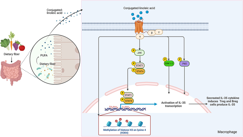
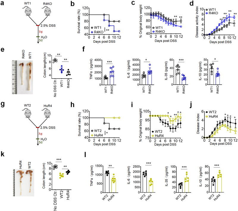
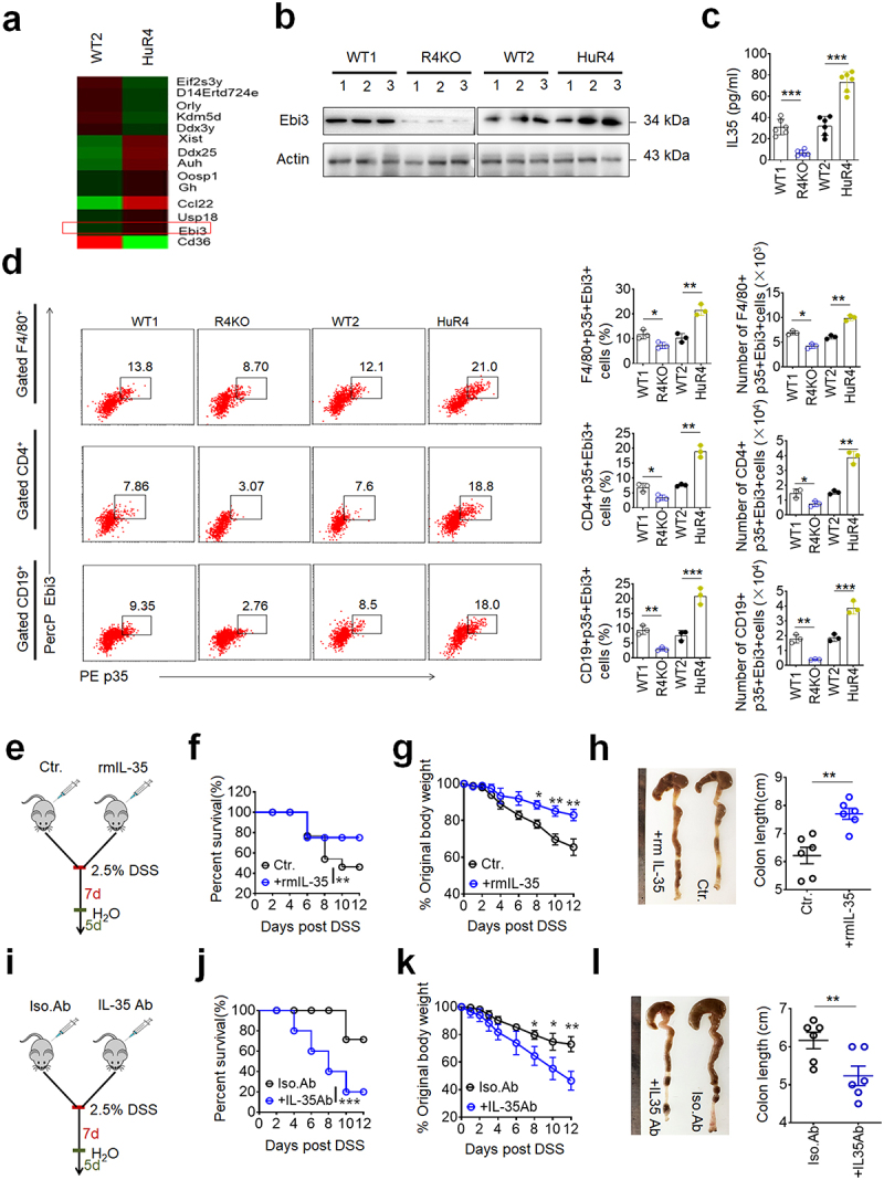
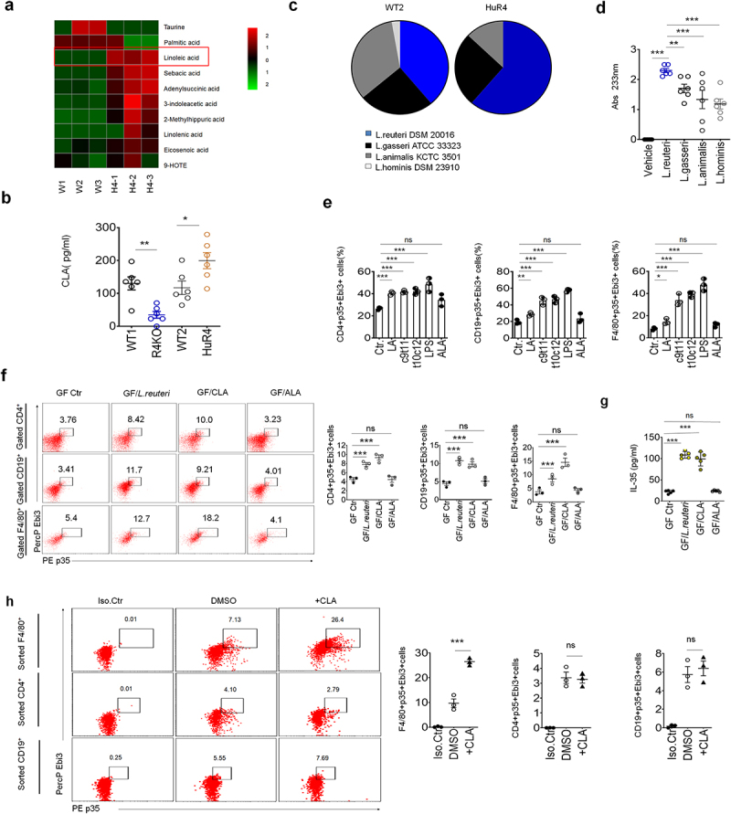
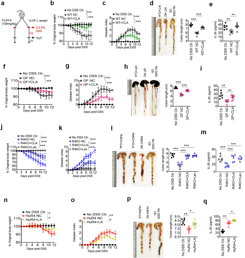
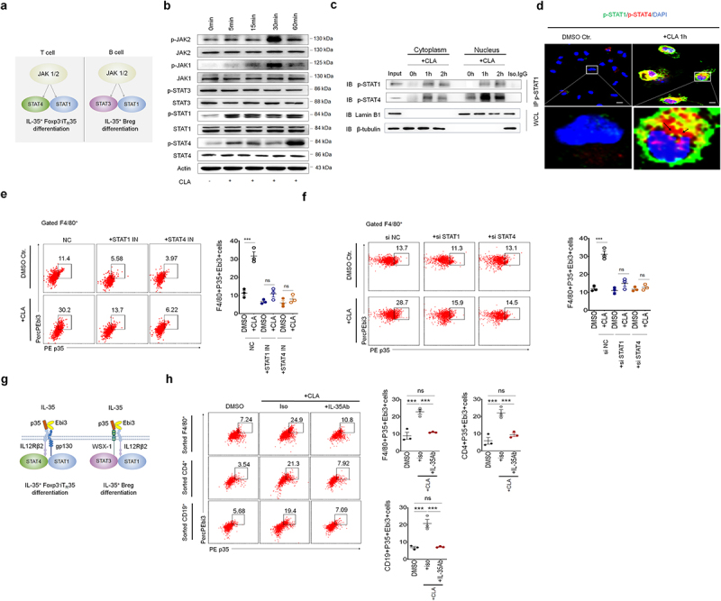
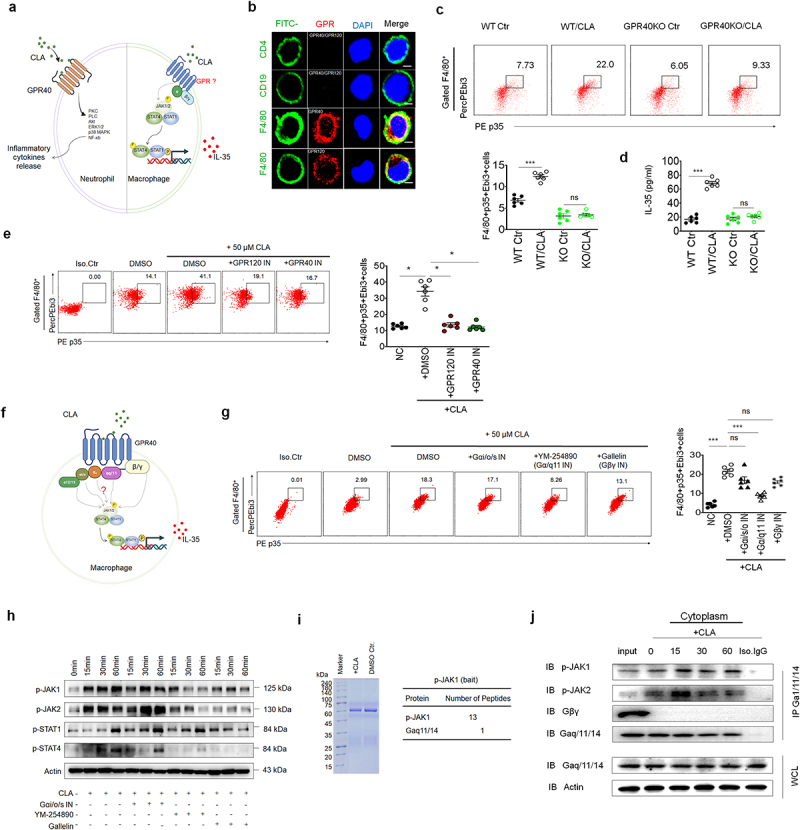
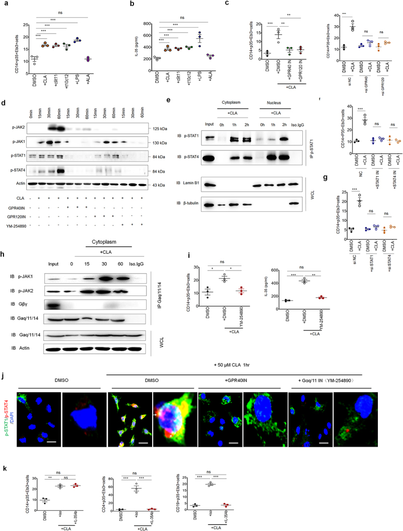
Gut microbiota/metabolites not only participate in the food and energy metabolism but also contribute to the host immune response and homeostasis. The alternation of gut microbiota/metabolites has been widely related to intestinal and extra-intestinal disorders such as intestinal bowel diseases (IBDs). Bactericidal substances from gut epithelial cells can regulate the composition of gut microbiota. However, the effects of regenerating protein 4 (REG4) (human)/(Reg4) (mice), a potentially bactericidal substance from gut epithelial cells, on the gut immune homeostasis maintain elusive. Here, we found that REG4/Reg4 is essential in maintaining gut immune homeostasis through REG4/Reg4 associated gut microbiota. Reg4 knockout (KO) mice were highly sensitive to DSS-mediated colitis, whereas human REG4 intestine epithelial cell transgenic (huREG4IECtg) mice exhibited more resistance to DSS-mediated colitis. Mechanistically, sequencing of gut microbiota and liquid chromatography-mass spectrometry showed that REG4/Reg4 could affect the composition of gut microbiota. REG4/Reg4 associated gut microbiota such as Lactobacillus could metabolize linoleic acid (LA) into conjugated linoleic acid (CLA). Immunoprecipitation and immunoblot showed that CLA could effectively promote the expression of IL-35 in macrophages through Gαq/11 mediated activation STAT1/4. Thus, our results demonstrate that REG4/Reg4 plays a critical role in maintaining gut immune homeostasis through CLA-mediated IL-35+ macrophages.

Keywords: Conjugated linoleic acid; IL-35; Reg4; gut microbiota; macrophages.


Conflict of interest statement

No potential conflict of interest was reported by the author(s).

Figures

None
Graphical abstract
Figure 1.
Figure 1.
REG4/Reg4 resists to dss-mediated colitis. (a) The design for the dss-induced colon inflammation model for b-f. Reg4 knockout (R4KO) and their littermate control mice (WT1) were exposed to 2.5 % DSS for 7 d, and then changed with normal water for 5 d in b, c and d; (b) survival rate of R4KO and WT1 mice; n = 18; (c) body weight of R4KO and WT1 mice; n = 6; (d) disease activity index (DAI) of R4KO and WT1 mice; n = 6; (e) colon length of R4KO and WT1 mice; n = 6; (f) ELISA of tnf-α, IL-6, IL-10 and IL-35 in the sera of R4KO and WT1 mice; (g) the design for the dss-induced colon inflammation model for h-l. huREG4IECtg (HuR4) and littermate control mice (WT2) were exposed to 2.5 % DSS for 7 d, then and changed with normal water for 5 d in h, I and j or 2 d in k and l; (h) survival rate of HuR4 and WT2 mice; n = 18; (i) body weight of HuR4 and WT2 mice; n=6; (j) disease activity index (DAI) of HuR4 and WT2 mice; n = 6; (k) colon length of HuR4 and WT2 mice; n = 6; (l) ELISA of tnf-α, IL-6, IL-10 and IL-35 in the sera of HuR4 and WT2 mice. Wilcoxon’s test in b and h; one-way ANOVA test in c, d, e, I, j and k; Student’s t-test in f and l. *p < 0.05, **p < 0.05, ***p < 0.05. data is a representative of at least three experiments.
Figure 2.
Figure 2.
REG4/Reg4 mediated resistance to dss-mediated colitis is dependent on IL-35. (a) RNA-seq of colon lamina propria (CLP) tissues in huREG4IECtg (HuR4) and their littermate control mice (WT2); (b) Western blotting of Ebi3 expression in colon lamina propria (CLP) tissues of R4KO, HuR4 and control mice. (n = 3); (c) ELISA of IL-35 in the sera of R4KO, HuR4 and control mice; (d) flow cytometry of F4/80+p35+Ebi3+, CD4+p35+Ebi3+ and CD19+p35+Ebi3+ cells in the CLP of R4KO, HuR4 and control mice. The proportion and absolute number F4/80+p35+Ebi3+, CD4+p35+Ebi3+ and CD19+p35+Ebi3+ cells in the colon were compared (n = 3); (e) the design for the effects of recommendation mouse IL-35 (rmIL-35) on R4KO mice on dss-mediated colitis for f-h; (f) survival rate of R4KO mice with (+rmIL-35) or without (ctr.) rmIL-35; n = 18; (g) body weight changes of R4KO mice with (+ rmIL-35) or without (ctr.) rmIL-35; n = 6; (h) colon length in of R4KO mice with (+rmIL-35) or without (ctr.) rmIL-35; n = 6; (i) the design for the effects of mouse IL-35 blocking antibody (IL-35Ab) on huREG4IECtg mice on DSS mediated colitis for j-l; (j) survival rate of HuR4 mice with (+IL-35 ab) or without (isotypic control, Iso.Ab) IL-35 blocking antibody; n = 18; (k) body weight changes of HuR4 mice with (+IL-35 ab) or without (Iso.Ab) IL-35 blocking antibody; n = 6; (l) colon length of HuR4 mice with (+IL-35 ab) or without (Iso. Ab) IL-35 blocking antibody; n = 6. Wilcoxon’s test in f and j; one-way ANOVA test in g and k; Student’s t-test in c, d, h and l. *p < 0.05, **p < 0.05, ***p < 0.05. a representative of at least three experiments.
Figure 3.
Figure 3.
Gut microbiota derived CLA induces IL-35 production in macrophages (a) LC-MS of the sera from huREG4IECtg (H4–1, H4–2 and H4–3) and their littermate control mice (W1, W2 and W3); (b) analyses of conjugated linoleic acid (CLA) in the sera of R4KO, HuR4 and their control mice; (c) proportion of different Lactobacilli in the colon contents of WT2 and HuR4 mice; (d) the levels of conjugated linoleic acid (cis-9 and trans-11 isomes) in the supernatants of different Lactobacillus species. L. reuteri, Lactobacillus Reuteri isolated from HuR4 mice; L.Animalis, Lactobacillus animalis (BNCC, China); L. gasserri, Lactobacillus gasserri (BNCC, China); L.Hominis, Lactobacillus hominis (BNCC, China); (e) flow cytometry of CD4+p35+Ebi3+, CD19+ p35+Ebi3+ and F4/80+ p35+Ebi3+ cells after exposure to vehicle, LA, CLA1 (c9t11, cis-9 CLA), CLA2 (t10c12, trans-10 CLA) and LPS, ALA (adenylsuccinic acid); (f) flow cytometry of CD19+p35+Ebi3+, CD4+p35+Ebi3+ and F4/80+p35+Ebi3+ cells in the colon lamina propria (CLP) tissues from GF mice with L. reuteri (GF/L.Reuteri), or CLA (GF/CLA), or ALA(GF/ALA) gavage or without (GF ctr) treatment; (g) ELISA of IL-35 in the colon tissues of GF mice with L.Reuteri (GF/L.Reuteri), CLA (GF/CLA), ALA(GF/ALA) gavage or without (GF ctr) treatment in vivo; (h) flow cytometry of p35+Ebi3+ cells in sorted CD4+ T cells, CD19+ B cells or F4/80+ macrophages with CLA(+CLA, cis-9 LA) or without (DMSO). Iso.Ctr, isotype IgG control. One-way ANOVA test; *p < 0.05, **p < 0.05, ***p < 0.05. a representative of at least three experiments.
Figure 4.
Figure 4.
CLAs reduce sensitivity to DSS mediated colitis. (a) A schematic illustration showing the design for CLA/L.Reuteri gavage studies. (b) Body weight changes; n = 6; (c) disease index; n = 6; (d) colon length; n = 6 (e) ELISA of IL-35 in sera; wild type (WT) mice were treated with 100mg/kg CLA (WT+CLA) gavage then given 2.5% DSS for 7 d, and normal drinking water for another 5 d; (f) body weight changes; n = 6; (g) disease index; n = 6; (h) colon length; n = 6; (i) ELISA of IL-35 in sera; germ-free (GF) mice were treated with 100mg/kg CLA (GF+CLA) gavage, and then with 2% DSS; (j) body weight changes; n = 6; (k) disease index; n=6; (l) colon length; n = 6; (m) ELISA of IL-35 in sera; Reg4 knockout (R4KO) mice and littermate control mice were treated with 100mg/kg CLA (R4KO+CLA) or with 1 × 10⁹ L.Reuteri (R4KO+Lac) gavage, and then with 2.5% DSS; (n) body weight changes; n = 6; (o) disease index; n = 6; (p) colon length; n = 6; (q) ELISA of IL-35 in sera; huREG4IECtg (HuR4). Mice were treated with 100mg/kg LA (R4KO+LA) gavage, and then given with 2.5% DSS. NC, PBS control group under DSS colitis model. No DSS ctr., no DSS and no other treatment group. One-way ANOVA test; *p < 0.05, **p < 0.05, ***p < 0.05. a representative of at least two experiments.
Figure 5.
Figure 5.
CLA mediated IL-35 production is through activating STAT1/4 in macrophages. (a) JAK-stat signal pathway is involved in inducing IL-35+ T and B cells; (b) immunoblotting of phosphor (p)-JAK2/JAK1/STAT1/STAT3/STAT4 and total JAK2/JAK1/STAT1/STAT3/STAT4) in the bone marrow derived macrophages (BMDMs) upon exposure to cis-9 CLA at the indicated time. Actin, loading control; (c) immunoblotting of p-STAT1 and p-STAT4 in the BMDMs after immunoprecipitated with anti-p-STAT1. WCL, whole cell lysis. LaminB1 and β-tubulin, loading control in nucleus and cytoplasm respectively; (d) immunostaining of p-STAT1(green) and p-STAT4(red) in the BMDMs with (+ cis-9 CLA 1h) or without (DMSO ctr.) CLA stimulation. Macrophages were observed by confocal microscopy with an oil immersion objective of 40 × and a digital zoom of 3. Nucleus was stained with DAPI (blue). Arrows indicated binding sites; scale bar=10 μM; (e) flow cytometry of F4/80+p35+Ebi3+cells in the BMDMs. BMDMs were pre-exposed to STAT1 inhibitor (+STAT1IN) or STAT4 inhibitor (+STAT4IN) for 6 h, and then cells were stimulated with (+ cis-9 CLA) or without (DMSO. Ctr) CLA for 24hrs; (f) flow cytometry of F4/80+p35+Ebi3+cells in BMDMs after transfected with STAT1 siRNA (+siSTAT1) or STAT4 siRNA (+siSTAT4) for 24hrs, and then stimulated with (+cis-9 CLA) or without (DMSO. Ctr) CLA for 24hrs; (g) classical signal molecules which is involved in IL-35 mediated IL-35+ Foxp3-iTR35 differentiation and IL-35+ Breg differentiation; (h) flow cytometry of p35+Ebi3+cells in the co-culture of CD4+ and CD19+ cells with F4/80+cells upon exposure to CLA (+CLA) with IL-35 antibody (+IL-35Ab) or isotypic antibody (+iso) for 24hrs. One-way ANOVA test; *p < 0.05, **p < 0.05, ***p < 0.05. a representative of at least two experiments.
Figure 6.
Figure 6.
CLA mediated STAT1/4 activation is through GPR Gαq/11. (a) Conjugated linoleic acid (CLA) stimulates signaling pathways via the GPR40 receptor in neutrophil but it is unclear in macrophages; (b) immunostaining of GPR40/GPR120 (red) in CD4+ T cells, CD19+B cells and F4/80+ macrophages (green). Macrophages were observed by confocal microscopy with an oil immersion objective of 40 × and a digital zoom of 3. DAPI (blue) for nuclear staining; scale bar =10 μM; (c) flow cytometry of F4/80+p35+Ebi3+ cells of colon lamina propria (CLP) tissues from GPR40 -/- mice (GPR40KO) or control mice(wt) with CLA (CLA) or without (Ctr) CLA gavage; (d) ELISA of IL-35 in sera from GPR40 -/- mice(ko) or control mice(wt) with CLA (cis-9 CLA) or without (Ctr) CLA gavage; (e) flow cytometry of F4/80+p35+Ebi3+cells in BMDMs after exposure to CLA (+50μM CLA, DMSO) with GPR40 inhibitor (50μM CLA+GPR40IN) or GPR120 inhibitor (50μM CLA+GPR120IN) for 24hrs. Iso. Ctr, isotype control. DMSO, unstimulated control; (f) G-protein subunits including α12/13、αi/o、αs、αq/11 and βγ, which were potentially involved in the production of IL-35 by CLA in macrophages. (g) Flow cytometry of F4/80+p35+Ebi3+cells in the BMDMs after exposed to CLA (50μM CLA, DMSO) with Gαi/o/s inhibitor (50μM CLA+Gαi/o/sIN) or Gα/q11 inhibitor YM-254890 (50μM CLA + Ga/q11IN) or Gβγ inhibitor gallelin (50μM CLA + GβγIN) for 24hrs. Iso. Ctr, isotype control. DMSO, unstimulated control; (h) immunoblotting of p-JAK1/JAK2/STAT1/STAT4 in BMDMs exposed to CLA with Gαi/o/s inhibitor (Gαi/o/sIN +), Gα/q11 inhibitor (YM-254890+) or Gβγ inhibitor (galletin+) at indicated time. (-), no stimulated control. Actin, a loading control; (i) interaction of Gα/q11(also known as Gα/q11/14) with phospho-JAK1 after CLA stimulation. BMDMs were exposed to CLA (+CLA) or DMSO control (Dmso.Ctr), and then immunoprecipitated with anti-phospho-JAK1; (j) immunoblotting of p-JAK1/JAK2, Gβγ and Ga/q11/14 in the BMDM lyses immunoprecipitated with anti-Ga/q11/14. WCL, whole cell lysis; IP, immunoprecipitation; IB, immunoblotting; Iso. IgG, isotype IgG control. One-way ANOVA test; *p < 0.05, **p < 0.05, ***p < 0.05. a representative of at least two experiments.
Figure 7.
Figure 7.
CLA induces IL-35 expression in human monocytes/macrophages. (a)Flow cytometry of CD14+p35+Ebi3+cells in the human peripheral blood monocyte cells derived macrophages (BMDMs) upon exposure to CLA (+CLA), c9t11 (cis-9 CLA), t10c12 (trans-10 CLA), ALA(+ALA) or LPS for 24hrs. DMSO, unstimulated control; (b) ELISA of IL-35 in the supernatants of BMDMs upon exposure to CLA (+CLA), c9t11 (cis-9 CLA), t10c12 (trans-10 CLA), ALA(+ALA) or LPS for 24hrs. DMSO, unstimulated control; (c) flow cytometry of CD14+p35+Ebi3+ in the BMDMs upon exposure to CLA(+CLA) with GPR40 inhibitor (CLA+GPR40IN) or GPR120 inhibitor (CLA+GPR120IN) for 24 h or GPR40 (si GPR40) or GPR120 (si GPR120) siRNAs or control (si NC) for 3 d. DMSO, unstimulated control; (d) immunoblotting of p-JAK2/JAK1/STAT1/STAT4 in the BMDMs upon exposure to CLA with GPR40 inhibitor (GPR40IN), GPR120 inhibitor (GPR120IN) or Gα/q11 inhibitor (YM-254890) at the indicated time. Actin as the loading control; (e) immunoblotting of p-STAT1 and p-STAT4 in the BMDM after immunoprecipitation with anti-p-STAT1. WCL: whole cell lysis, Lamin B1 and β-tubulin, loading control in nucleus and cytoplasm respectively; (f) flow cytometry of CD14+p35+Ebi3+cells in the BMDMs. Cells were pre-exposure to STAT1 inhibitor (+STAT1IN) or STAT4 inhibitor (+STAT4IN) for 6 h and then stimulated with (+CLA) or without (DMSO. Ctr) CLA for 24hrs; (g) flow cytometry of CD14+p35+Ebi3+cells in the BMDMs. Cells were transfected with STAT1 siRNA (+siSTAT1) or STAT4 siRNA (+siSTAT4) for 24hrs, and then stimulated with (+CLA) or without (DMSO. Ctr) CLA for 24hrs; (h) immunoblotting of phospho-JAK1/JAK2, Gβγ and Gαq/11 in the BMDM after immunoprecipitation with anti- Gαq/11. WCL, whole cell lysis; IP, immunoprecipitation; IB, immunoblotting; Iso.IgG, isotype IgG control; (i) flow cytometry of CD14+p35+Ebi3+cells in BMDMs and ELISA of IL-35 in the supernatant of BMDMs upon exposure to CLA (+CLA) with or without Gαq/11 (DMSO) inhibitor for 24hrs; (j) immunostaining of p-STAT1(green) and p-STAT4(red) in the BMDMs with (50μM CLA 1h) or without (DMSO) CLA stimulation. Nuclear was stained with DAPI (blue). Arrows indicated binding sites. (k) Flow cytometry of CD14+p35+Ebi3+cells, CD4+p35+Ebi3+cells or CD19+p35+Ebi3+cells in the coculture of the BMDMs with CD4+ and CD19+cells upon exposure to CLA (+CLA) with (+IL-35Ab) or without (isotypic antibody, iso) IL-35 antibody for 24hrs; one-way ANOVA test; *p < 0.05, **p < 0.05, ***p < 0.05. a representative of at least two experiments.

Similar articles

Cited by

References

    1. Foppa C, Rizkala T, Repici A, Hassan C, Spinelli A.. Microbiota and IBD: current knowledge and future perspectives. Dig Liver Dis. 2023;56(6):911–21. PMID: 38008696. doi:10.1016/j.dld.2023.11.015. - DOI - PubMed
    1. Sato Y, Tsujinaka S, Miura T, Kitamura Y, Suzuki H, Shibata C. Inflammatory bowel disease and colorectal cancer: epidemiology, etiology, surveillance, and management. Cancers (Basel). 2023;15(16):4154. PMID: 37627182. doi:10.3390/cancers15164154. - DOI - PMC - PubMed
    1. Klag T, Stange EF, Wehkamp J. Defective antibacterial barrier in inflammatory bowel disease. Dig Dis. 2013;31(3–4):310–316. PMID: 24246980. doi:10.1159/000354858. - DOI - PubMed
    1. Nicholson JK, Holmes E, Kinross J, Burcelin R, Gibson G, Jia W, Pettersson S. Host-gut microbiota metabolic interactions. Science. 2012;336(6086):1262–1267. PMID: 22674330. doi:10.1126/science.1223813. - DOI - PubMed
    1. Rooks MG, Garrett WS. Gut microbiota, metabolites and host immunity. Nat Rev Immunol. 2016;16(6):341–352. PMID: 27231050. doi:10.1038/nri.2016.42. - DOI - PMC - PubMed

MeSH terms

LinkOut - more resources