

Evaluation of indices for the assessment and classification of keratoconus based on optical coherence tomography and Scheimpflug technology
- PMID: 39636209
- PMCID: PMC11823291
- DOI: 10.1111/opo.13425
Evaluation of indices for the assessment and classification of keratoconus based on optical coherence tomography and Scheimpflug technology
Abstract
Purpose: To compare the parameters and indices of a novel swept-source optical coherence tomography device (SS-OCT, ANTERION) with those of a rotating Scheimpflug camera (RSC)-based tomograph (Pentacam) in normal and keratoconic (KC) eyes.
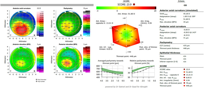
Methods: This prospective, monocentric, cross-sectional study included individuals with unoperated normal and KC eyes, selecting one eye per subject. Ectasia-specific parameters analysed with the SS-OCT were difference in mean keratometry (Kmean) in the inferior and superior meridians, maximum keratometry value (Kmax), elevation of the posterior surface at the thinnest point, screening corneal objective risk of ectasia (SCORE) and thinnest point thickness. With the RSC, parameters determined were Belin/Ambrosio total deviation value (BAD-D), index of height decentration and index of vertical asymmetry. KC classification with the SS-OCT was based on the anterior and posterior radii of curvature and thinnest point thickness according to the ABCD classification of the RSC system.
Results: This study included 117 individuals with healthy eyes and 335 eyes with KC. The indices with the highest diagnostic discriminatory ability between the two cohorts were SCORE, difference of Kmean in the inferior and superior meridians and posterior elevation of the thinnest point (SS-OCT), as well as the index of height decentration, index of vertical asymmetry and BAD-D (RSC). The classifications using SS-OCT defined mild-stage KC as Kmax, posterior elevation of the thinnest point and thinnest point thickness as ≤50.9 D, ≤30 and ≥472 μm, respectively. Moderate stage values were 51-55.9 D, 31-69 and 471-438 μm, respectively, while respective advanced stage were ≥56 D, ≥70 and ≤437 μm.
Conclusion: The diagnostic capabilities for both devices were found to be comparable. KC classification using SS-OCT can be independently based on the anterior surface, posterior surface and corneal thickness.
Keywords: ANTERION; Pentacam; corneal tomography; epithelium thickness; keratoconus; swept‐source optical coherence tomography.
© 2024 The Author(s). Ophthalmic and Physiological Optics published by John Wiley & Sons Ltd on behalf of College of Optometrists.
Conflict of interest statement
All authors declare no conflicts of interest. RH has received fees as a speaker for Heidelberg Engineering GmbH.
Figures


References
-
- Ambrosio R Jr, Belin MW. Imaging of the cornea: topography vs tomography. J Refract Surg. 2010;26:847–849. - PubMed
-
- Dave T. Current developments in measurement of corneal topography. Cont Lens Anterior Eye. 1998;21(Suppl 1):S13–S30. - PubMed
-
- Ambrosio R Jr, Lopes BT, Faria‐Correia F, Salomao MQ, Buhren J, Roberts CJ, et al. Integration of Scheimpflug‐based corneal tomography and biomechanical assessments for enhancing ectasia detection. J Refract Surg. 2017;33:434–443. - PubMed
-
- Herber R, Hasanli A, Lenk J, Vinciguerra R, Terai N, Pillunat LE, et al. Evaluation of corneal biomechanical indices in distinguishing between normal, very asymmetric, and bilateral keratoconic eyes. J Refract Surg. 2022;38:364–372. - PubMed
-
- Vinciguerra R, Ambrosio R Jr, Elsheikh A, Roberts CJ, Lopes B, Morenghi E, et al. Detection of keratoconus with a new biomechanical index. J Refract Surg. 2016;32:803–810. - PubMed
MeSH terms
LinkOut - more resources
Full Text Sources
Research Materials
