

ESR Essentials: how to get to valuable radiology AI: the role of early health technology assessment-practice recommendations by the European Society of Medical Imaging Informatics
- PMID: 39636421
- PMCID: PMC12081502
- DOI: 10.1007/s00330-024-11188-3
ESR Essentials: how to get to valuable radiology AI: the role of early health technology assessment-practice recommendations by the European Society of Medical Imaging Informatics
Abstract
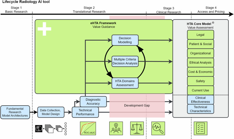
AI tools in radiology are revolutionising the diagnosis, evaluation, and management of patients. However, there is a major gap between the large number of developed AI tools and those translated into daily clinical practice, which can be primarily attributed to limited usefulness and trust in current AI tools. Instead of technically driven development, little effort has been put into value-based development to ensure AI tools will have a clinically relevant impact on patient care. An iterative comprehensive value evaluation process covering the complete AI tool lifecycle should be part of radiology AI development. For value assessment of health technologies, health technology assessment (HTA) is an extensively used and comprehensive method. While most aspects of value covered by HTA apply to radiology AI, additional aspects, including transparency, explainability, and robustness, are unique to radiology AI and crucial in its value assessment. Additionally, value assessment should already be included early in the design stage to determine the potential impact and subsequent requirements of the AI tool. Such early assessment should be systematic, transparent, and practical to ensure all stakeholders and value aspects are considered. Hence, early value-based development by incorporating early HTA will lead to more valuable AI tools and thus facilitate translation to clinical practice. CLINICAL RELEVANCE STATEMENT: This paper advocates for the use of early value-based assessments. These assessments promote a comprehensive evaluation on how an AI tool in development can provide value in clinical practice and thus help improve the quality of these tools and the clinical process they support. KEY POINTS: Value in radiology AI should be perceived as a comprehensive term including health technology assessment domains and AI-specific domains. Incorporation of an early health technology assessment for radiology AI during development will lead to more valuable radiology AI tools. Comprehensive and transparent value assessment of radiology AI tools is essential for their widespread adoption.
Keywords: Artificial intelligence; Radiology; Stakeholder participation; Technology assessment (Biomedical); Value-based healthcare.
© 2024. The Author(s).
Conflict of interest statement
Compliance with ethical standards. Guarantor: The scientific guarantor of this publication is Jacob J. Visser. Conflict of interest: M.H. is speakers honoraria from industry; support for attending meetings and/or travel from scientific societies; EuSoMII Board member, ESR eHealth & Informatics Subcommittee member, ECR Imaging Informatics/Artificial Intelligence and Machine Learning Chairperson 2025, committee member with FMS (Dutch), and Radiology: Artificial Intelligence associate editor and trainee editorial board advisory panel (all unpaid). M.E.K. is a Scientific Editorial Board member of European Radiology and has not taken part in the review and decision process of this paper. J.J.V.: Grant to the institution from Qure.ai/Enlitic; consulting fees from Tegus; payment to an institution for lectures from Roche; travel grant from Qure.ai; participation on advisory board from Contextflow, Noaber Foundation, and NLC Ventures; leadership role on the steering committee of the PINPOINT Project (payment to institution from AstraZeneca) and RSNA Common Data Elements Steering Committee (unpaid); chair scientific committee EuSoMII (unpaid); chair ESR value-based radiology subcommittee (unpaid); phantom shares in Contextflow and Quibim; member editorial Board European Journal of Radiology (unpaid). The other authors of this manuscript declare no relationships with any companies, whose products or services may be related to the subject matter of the article. Statistics and biometry: No complex statistical methods were necessary. Informed consent: Written informed consent was not required. Ethical approval: Institutional Review Board approval was not required. Study subjects or cohorts overlap: Not applicable. Methodology: Practice recommendations
Figures
Comment in
-
Avoiding Boeing 737 Max moments: ideal versus real radiology AI assessment.Eur Radiol. 2025 Jun;35(6):3429-3431. doi: 10.1007/s00330-024-11226-0. Epub 2024 Dec 5. Eur Radiol. 2025. PMID: 39636424 No abstract available.
References
-
- Mehrizi MHR, Gerritsen SH, de Klerk WM et al (2022) How do providers of artificial intelligence (AI) solutions propose and legitimize the values of their solutions for supporting diagnostic radiology workflow? A technography study in 2021. Eur Radiol 33:915–924. 10.1007/s00330-022-09090-x - DOI - PMC - PubMed
Publication types
MeSH terms
LinkOut - more resources
Full Text Sources
Medical
Miscellaneous
