. 2024 Dec 5;50(1):44.
doi: 10.1007/s11064-024-04290-x.

Filbertone-Induced Nrf2 Activation Ameliorates Neuronal Damage via Increasing BDNF Expression

Affiliations

Filbertone-Induced Nrf2 Activation Ameliorates Neuronal Damage via Increasing BDNF Expression

Jeong Heon Gong et al. Neurochem Res. .

Abstract

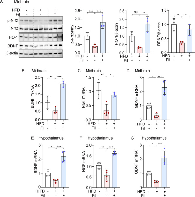
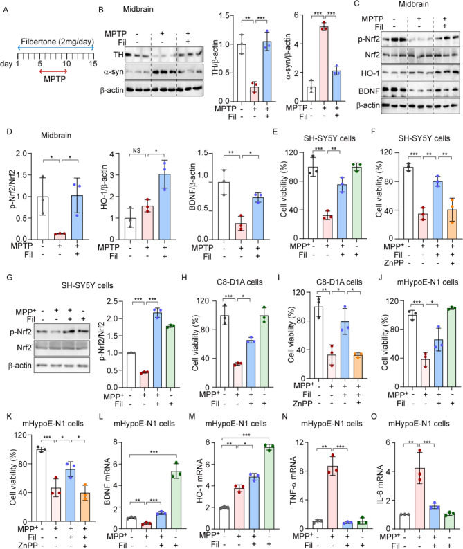
Neurotrophic factors are endogenous proteins that promote the survival of various neuronal cells. Increasing evidence has suggested a key role for brain-derived neurotrophic factor (BDNF) in the dopaminergic neurotoxicity associated with Parkinson's Disease (PD). This study explores the therapeutic potential of filbertone, a bioactive compound found in hazelnuts, in neurodegeneration, focusing on its effects on neurotrophic factors and the nuclear factor erythroid 2-related factor 2 (Nrf2) signaling pathway. In our study, filbertone markedly elevated the expression of neurotrophic factors, including BDNF, Glial cell line-Derived Neurotrophic Factor (GDNF), and Nerve Growth Factor (NGF), in human neuroblastoma SH-SY5Y cells, mouse astrocyte C8-D1A cells, and mouse hypothalamus mHypoE-N1 cells. Moreover, filbertone effectively countered neuroinflammation and reversed the decline in neurotrophic factors and Nrf2 activation induced by a high-fat diet (HFD) in neurodegeneration models. The neuroprotective effects of filbertone were further validated in models of neurotoxicity induced by palmitic acid (PA) and the neurotoxin MPTP/MPP+, where it was observed to counteract PA and MPTP/MPP+-induced decreases in cell viability and neuroinflammation, primarily through the activation of Nrf2 and the subsequent upregulation of BDNF and heme oxygenase-1 expression. Nrf2 deficiency negated the neuroprotective effects of filbertone in MPTP-treated mice. Consequently, our finding suggests that filbertone is a novel therapeutic agent for neurodegenerative diseases, enhancing neuronal resilience through the Nrf2 signaling pathway and upregulation of neurotrophic factors.

Keywords: Filbertone; Heme oxygenase-1; Neuroinflammation; Neurotrophic factors; Nuclear factor erythroid 2-related factor 2; Parkinson’s disease.


Conflict of interest statement

Declarations. Ethical Approval: All experimental procedures were approved by the Ethics Committee of the University of Ulsan University (HTC-22-010) in accordance with the National Institutes of Health guidelines. Efforts were made to minimize animal suffering, and all sample sizes for the assessment parameters were calculated to minimize the number of animals used. Consent to Paricipate: Not applicable. Consent for Publication: Not applicable. Competing Interests: The authors declare no competing interests.

Figures

Fig. 1
Fig. 1
Filbertone enhances neurotrophic factors in human neuroblastoma SH-SY5Y cells and in mouse astrocyte C8-D1A cells and mHypoE-N1 cells. (A and B) SH-SY5Y cells (A), C8-D1A cells (B) were treated with filbertone (0, 5, 10, 20, 40, 80, 160 and 320 µM) for 24 h. Cell viability was determined by WST-8 assay. (C-F) SH-SY5Y cells (C) were incubated with filbertone (0, 2, 10, and 50 µM) for 24 h. mRNA levels of BDNF were measured by qRT-PCR. (D-F) C8-D1A cells were incubated with filbertone (0, 2, 10, and 50 µM) for 24 h. mRNA levels of BDNF, GDNF, and NGF were measured by qRT-PCR. Data are mean ± SD (n = 3); *p < 0.05, **p < 0.01, and ***p < 0.001, calculated by one-way ANOVA followed by Tukey’s test
Fig. 2
Fig. 2
Filbertone counteracts the decrease in neurotrophic factors and the activation of Nrf2 caused by HFD in the brain (A-G) Six-week-old male C57BL/6 were fed a normal chow diet (NCD) or high fat diet (HFD) for 16 weeks with filbertone (Fil). (A) p-Nrf2, Nrf2, HO-1, and BDNF in midbrain were measured by western blotting. Quantification of protein expression is shown in the right panel. (B-D) The expression of BDNF (B), NGF (C), GDNF (D) mRNA in midbrain were determined by qRT-PCR. (E-G) In hypothalamus, the expression levels of neurotrophic factors, BDNF (E), NGF (F), and GDNF (G) were detected by qRT-PCR. Data represent mean ± SD; *p < 0.05, **p < 0.01 and ***p < 0.001, calculated by one-way ANOVA followed by Tukey’s test
Fig. 3
Fig. 3
Filbertone attenuates neuronal inflammation and the activation of glial cells in the brain (A and B) In midbrain, expression levels of inflammatory cytokines, TNF-a (A) and IL-1b (B) were detected by qRT-PCR. (C-E) In hypothalamus, the expression levels of inflammatory cytokines, TNF-a (C), IL-1b (D), and MCP-1 (E) were detected by qRT-PCR. (F-H) In hypothalamus, the expression levels of microglia activation markers, CD68 (F), CD11b (G), and Iba-1 (H) were detected by qRT-PCR. (I and J) The expression levels of astrocyte activation marker GFAP (I) and neuron damage marker HSP72 (J) were detected by qRT-PCR. Data represent mean ± SD; *p < 0.05, **p < 0.01 and ***p < 0.001, calculated by one-way ANOVA followed by Tukey’s test
Fig. 4
Fig. 4
Filbertone-mediated Nrf2 activation protects against PA-induced neurotoxicity through the increase of BDNF and HO-1. To determine effects of filbertone (Fil) on palmitic acid (PA)-induced neurotoxicity in SH-SY5Y cells, the cells were incubated with filbertone (20 µM, 1 h) followed by PA (250 µM, 24 h). (A-D) Expression levels of BDNF (A), HO-1 (B), TNF-a (C), and IL-6 (D) were measured by qRT-PCR. (E) Viability of SH-SY5Y cells, the cells treated with filbertone and PA. The viability was detected by WST-8 assay. (F) Viability of SH-SY5Y cells. The cells were incubated with ZnPP (100 nM) in the presence of filbertone, followed by PA. (G) Viability of C8-D1A cells treated with filbertone and PA. (H) Viability of C8-D1A cells. The cells were incubated with ZnPP in the presence of filbertone, followed by PA. (I) Viability of mHypoE-N1cells, the cells treated with filbertone and PA. (J) Viability of mHypoE-N1 cells. The cells were incubated with ZnPP in the presence of filbertone, followed by PA. (K) Representative immunoblots for SH-SY5Y cells treated with filbertone and PA. Quantification of protein expression is shown in the right panel. Data represent mean ± SD; *p < 0.05, **p < 0.01, ***p < 0.001, and NS; not significant, calculated by one-way ANOVA followed by Tukey’s test
Fig. 5
Fig. 5
Filbertone-mediated activation of Nrf2 protects against neurotoxicity induced by MPTP/MPP+through the induction of BDNF and HO-1 (A-D) Six-week-old male C57BL/6 were intraperitoneally (i.p.) injected with MPTP (25 mg/kg) or vehicle. Five days before treatment with MPTP, mice received 0.2% filbertone treatment (A). (B) tyrosine hydroxylase (TH) and a-syn midbrain were measured by western blotting and densitometric analysis. (C-D) p-Nrf2, Nrf2, HO-1, and BDNF were measured by western blotting (C). Quantification of protein expression is shown in (D). (E) Viabilities of SH-SY5Y cells were detected by WST-8 assay. The cells were incubated with filbertone, followed by MPP+. (F) Viabilities of SH-SY5Y cells. The cells treated with MPP+ in the presence of filbertone and ZnPP (100 nM). (G) Representative immunoblots for SH-SY5Y cells treated with filbertone and MPP+. Quantification of protein expression is shown in the right panel. (H) Viabilities of C8-D1A cells were detected by WST-8 assay. The cells were incubated with filbertone, followed by MPP+. (I) Viabilities of C8-D1A cells. The cells treated with MPP+ in the presence of filbertone and ZnPP (100 nM). (J) Viabilities of mHypoE-N1 cells were detected by WST-8 assay. The cells were incubated with filbertone, followed by MPP+. (K) Viabilities of mHypoE-N1 cells. The cells treated with MPP+ in the presence of filbertone and ZnPP (100 nM). (L-O) The cells incubated with filbertone (20 µM, 1 h), followed by MPP+ (500 µM, 24 h) in mHypoE-N1 cells. Expression levels of BDNF (L), HO-1 (M), TNF-a (N), and IL-6 (O) were measured by qRT-PCR. Data represent mean ± SD; *p < 0.05, **p < 0.01, ***p < 0.001, calculated by one-way ANOVA followed by Tukey’s test
Fig. 6
Fig. 6
The deficiency of Nrf2 negates the protective effects of filbertone against neurotoxicity in SH-SY5Y and MPTP-treated mice (A) SH-SY5Y cells were incubated with filbertone (0, 5, 10, 20, and 40 µM) for 24 h. p-Nrf2, Nrf2, and HO-1 were measured by western blotting. Quantification of protein expression is shown in the right panel. (B) C8-D1A cells were incubated with filbertone (0, 5, 10, 20, and 40 µM) for 24 h. The mRNA levels of HO-1 were detected by RT-PCR. (C) mHypoE-N1 cells were incubated with filbertone (0, 5, 10, 20, and 40 µM) for 24 h. The mRNA levels of HO-1 were detected by RT-PCR. (D) Protein expression of BDNF in SH-SY5Y cells. BDNF were measured by western blotting. Quantification of protein expression is shown in the right panel. (E) The protein expression of BDNF, HO-1, p-Nrf2, and Nrf2 in SH-SY5Y cells were detected by western blotting. Quantification of protein expression is shown in the right panel. (F) The mRNA levels of BDNF, HO-1, and Nrf2 in SH-SY5Y cells were measured by RT-PCR. (G) BDNF and tyrosine hydroxylase (TH) expression in the mouse midbrain were detected by western blotting. Quantification of protein expression is shown in the right panel. Data represent mean ± SD; *p < 0.05, **p < 0.01, ***p < 0.001, NS; not significant, calculated by one-way (A-D) and two- way (E-G) ANOVA followed by Tukey’s test

References

    1. Feigin VL, Vos T, Nichols E, Owolabi MO, Carroll WM, Dichgans M, Deuschl G, Parmar P, Brainin M, Murray C (2020) The global burden of neurological disorders: translating evidence into policy. Lancet Neurol 19:255–265 - PMC - PubMed
    1. Temple S (2023) Advancing cell therapy for neurodegenerative diseases. Cell Stem Cell 30:512–529 - PMC - PubMed
    1. Nussbaum RL, Ellis CE (2003) Alzheimer’s disease and Parkinson’s disease. N Engl J Med 348:1356–1364 - PubMed
    1. Chopade P, Chopade N, Zhao Z, Mitragotri S, Liao R, Chandran Suja V (2023) Alzheimer’s and Parkinson’s disease therapies in the clinic. Bioeng Transl Med 8:e10367 - PMC - PubMed
    1. Agnello L, Ciaccio M (2022) Neurodegenerative diseases: from molecular basis to Therapy. Int J Mol Sci 23 - PMC - PubMed

LinkOut - more resources