. 2025 Feb;66(2):471-481.
doi: 10.1111/epi.18192. Epub 2024 Dec 5.

The Imaging Database for Epilepsy And Surgery (IDEAS)

Affiliations

The Imaging Database for Epilepsy And Surgery (IDEAS)

Peter N Taylor et al. Epilepsia. 2025 Feb.

Abstract

Objective: Magnetic resonance imaging (MRI) is a crucial tool for identifying brain abnormalities in a wide range of neurological disorders. In focal epilepsy, MRI is used to identify structural cerebral abnormalities. For covert lesions, machine learning and artificial intelligence (AI) algorithms may improve lesion detection if abnormalities are not evident on visual inspection. The success of this approach depends on the volume and quality of training data.

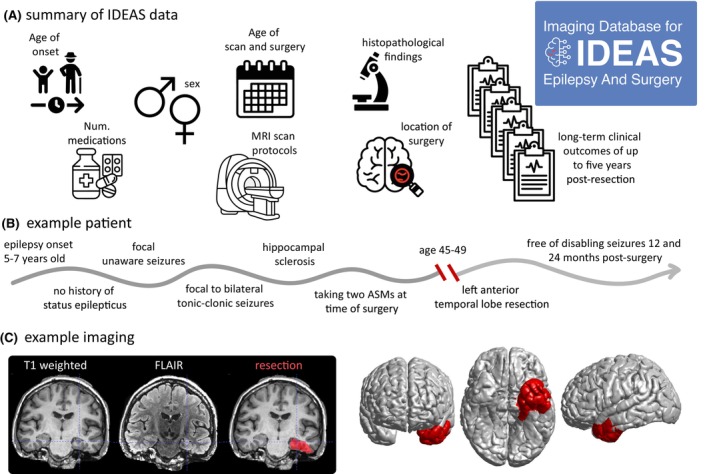
Methods: Herein, we release an open-source data set of pre-processed MRI scans from 442 individuals with drug-refractory focal epilepsy who had neurosurgical resections and detailed demographic information. We also share scans from 100 healthy controls acquired on the same scanners. The MRI scan data include the preoperative three-dimensional (3D) T1 and, where available, 3D fluid-attenuated inversion recovery (FLAIR), as well as a manually inspected complete surface reconstruction and volumetric parcellations. Demographic information includes age, sex, age a onset of epilepsy, location of surgery, histopathology of resected specimen, occurrence and frequency of focal seizures with and without impairment of awareness, focal to bilateral tonic-clonic seizures, number of anti-seizure medications (ASMs) at time of surgery, and a total of 1764 patient years of post-surgical followup. Crucially, we also include resection masks delineated from post-surgical imaging.

Results: To demonstrate the veracity of our data, we successfully replicated previous studies showing long-term outcomes of seizure freedom in the range of ~50%. Our imaging data replicate findings of group-level atrophy in patients compared to controls. Resection locations in the cohort were predominantly in the temporal and frontal lobes.

Significance: We envisage that our data set, shared openly with the community, will catalyze the development and application of computational methods in clinical neurology.

Keywords: MRI; data; epilepsy; prediction; surgery.


Conflict of interest statement

The authors have no conflict of interest to disclose.

Figures

FIGURE 1
FIGURE 1
Summary of the Imaging Database for Epilepsy And Surgery (IDEAS) data set. (A) Various anonymized patient data are included in the release. (B) Clinical demographic information for an example patient (subject 5 in the database). (C) Example imaging including pre‐operative T1‐weighted magnetic resonance imaging (MRI), fluid‐attenuated inversion recovery (FLAIR) MRI, and resection mask, all aligned in the same space and orientation. Right panels show a surface visualization from the shared FreeSurfer data.
FIGURE 2
FIGURE 2
Seizure outcome up to 5 years after epilepsy surgery. (A) Survival plot of proportion of patients remaining seizure‐free following epilepsy surgery and with available follow‐up at yearly intervals, showing time to first seizure. Each colored line represents a different surgical procedure. ATLRx, anterior temporal resection; ETLesx, extratemporal lesionectomy; ETLx, extratemporal resection; Hx, hemispherectomy; and TLesx, temporal lesionectomy. (B) Survival plot of proportion of patients who did (blue), and did not (red), have focal aware seizures (FASs) following surgery in years 1, 2, remaining free of seizures with impaired awareness in subsequent years. (C–E) Twelve‐month seizure outcome did not differ by age at onset of epilepsy, (C), age at surgery (D), or duration of epilepsy (E). All results broadly replicate those described previously in more extensive cohorts from 1990 to 2012.,
FIGURE 3
FIGURE 3
Widespread reductions in volume and thickness in mesial temporal lobe epilepsy (mTLE). Reduced volumes are shown in left and right mTLE ipsilaterally in the hippocampus and thalamus, with widespread bilateral thickness reductions in the neocortex. These results are concordant with those of other studies.
FIGURE 4
FIGURE 4
Post‐operative resection mask densities for 433 individuals.

Similar articles

Cited by

References

    1. Markiewicz CJ, Gorgolewski KJ, Feingold F, Blair R, Halchenko YO, Miller E, et al. The OpenNeuro resource for sharing of neuroscience data. elife. 2021;10:e71774. - PMC - PubMed
    1. Biswal BB, Mennes M, Zuo XN, Gohel S, Kelly C, Smith SM, et al. Toward discovery science of human brain function. Proc Natl Acad Sci. 2010;107(10):4734–4739. - PMC - PubMed
    1. Mueller SG, Weiner MW, Thal LJ, Petersen RC, Jack CR, Jagust W, et al. Ways toward an early diagnosis in Alzheimer's disease: the Alzheimer's Disease Neuroimaging Initiative (ADNI). Alzheimers Dement. 2005;1(1):55–66. - PMC - PubMed
    1. Di Martino A, Yan CG, Li Q, et al. The autism brain imaging data exchange: towards a large‐scale evaluation of the intrinsic brain architecture in autism. Mol Psychiatry. 2014;19(6):659–667. - PMC - PubMed
    1. ADHD‐200Consortium . The ADHD‐200 consortium: a model to advance the translational potential of neuroimaging in clinical neuroscience. Front Syst Neurosci. 2012;6:62. - PMC - PubMed

MeSH terms