. 2025 Feb 10;64(7):e202419996.
doi: 10.1002/anie.202419996. Epub 2024 Dec 17.

Self-Assembly and Biological Properties of Highly Fluorinated Oligonucleotide Amphiphiles

Affiliations

Self-Assembly and Biological Properties of Highly Fluorinated Oligonucleotide Amphiphiles

Quentin Laurent et al. Angew Chem Int Ed Engl. .

Abstract

Nucleic acids, used as therapeutics to silence disease-related genes, offer significant advantages over small molecule drugs: they provide high specificity, the ability to target "undruggable" molecules, and adaptability to a wide range of disease phenotypes. However, their instability in biological media, as well their rapid clearance from the organism limit their applicability, necessitating the use of nanocarriers to overcome these challenges. Among these strategies, spherical nucleic acids (SNA)-composed of a densely packed corona of oligonucleotides around a nanoparticle-have emerged as a powerful tool, in particular when self-assembled from DNA amphiphiles. This non-covalent strategy however has caveats, especially when it comes to stability in complex biological media, where these SNAs disassemble in contact to serum proteins. Here, we developed highly fluorinated DNA amphiphiles that readily self-assemble into SNAs and have tunable stability profiles in biological media. They are made of branched fluorinated moieties with potentially improved biodegradability as compared to their linear counterparts. Depending on the number of fluorophilic interactions, the self-assembled SNAs can have excellent serum stabilities-up to days-and readily deliver nucleic acid therapeutics for gene silencing applications. These systems show great potential as promising candidates for nucleic acid-based therapies.

Keywords: delivery; fluorine; gene silencing; oligonucleotides; self-assembly.


Conflict of interest statement

The authors declare no conflict of interest.

Figures

Figure 1
Figure 1
A) Synthesis and self‐assembly of F27‐oligonucleotide conjugates by strain‐promoted alkyne‐azide cycloaddition (SPAAC). The oligonucleotide is a 2’‐fluoroarabino nucleic acid (FANA) antisense oligonucleotide (FASO) against firefly luciferase. B) Native AGE (2 %, native) showing the self‐assembly of 1 b and 2 b into higher order structures with low mobility (>100 bp for 18‐mer oligonucleotides). C) Representative AFM images of 1 b and 2 b deposited on a mica surface in air (see Figure S10–S11). D) Synthesis and self‐assembly of HE12 micelles terminated (4 b) or not (4 a) with an F27 moiety to strengthen the hydrophobic core.
Figure 2
Figure 2
A) Number weighted size distributions obtained by DLS characterization of 1 b and 2 b in 1x TAMg buffer (solid lines) and in pure water (dashed lines). B) CMC measurements of 1 b and 2 b monitored by fluorescence emission of encapsulated Nile Red dye. Error bars represent SD from two independent experiments. C) 19F NMR of 1 b and 2 b in pure water showing either mainly disassembled (1 b) or partly assembled (2 b) amphiphiles.
Figure 3
Figure 3
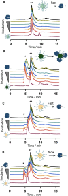
Size exclusion chromatography chromatograms of 1 b (A), 2 b (B), 4 a (C) and 4 b (D) after incubation in buffer+10 % FBS at 37 °C for 1 h (red), 2 h (orange), 8 h (blue), 24 h (teal) and 48 h (green). Purple line=SNA alone; black line=10 % FBS alone; *=SNA with protein corona; **=serum albumin and albumin‐bound conjugates.
Figure 4
Figure 4
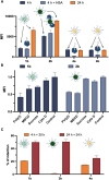
A) Cellular uptake of SNAs in HeLa cells after 4 h or 24 h of incubation, or after 4 h of incubation with 8 h of pre‐incubation with HSA, measured by flow cytometry. B) Cellular uptake of SNAs in HeLa cells after 4 h, with cells pre‐incubated for 30 min with different endocytosis inhibitors, measured by flow cytometry. Error bars represent SD from two different experiments. For statistical significance see Figure S19. C) Retention of SNAs in HeLa cells after 4 and 24 h of incubation, with or without an additional 20 or 24 h delay after media exchange to allow for cellular export, measured by flow cytometry.
Figure 5
Figure 5
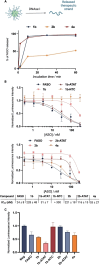
A) Percentage of release of the FASO therapeutic strand from the constructs measured by analyzing denaturing PAGE gels (Figure S23). B) Firefly luciferase knockdown activity after incubation for 24 h in HeLa cells with varying concentrations of the different SNAs transfected with LipofectamineTM, normalized to negative control and cell viability. This data was used to extrapolate the IC50 concentrations of the different constructs. Error bars represent standard deviation of three replicates for each sample. C) Firefly luciferase knockdown activity after incubation for 72 h in HeLa cells with the different SNAs without the help of transfection agent, normalized to negative control and cell viability. Error bars represent SD of three replicates for each sample. For statistical significance, see Figure S21.

References

    1. Roberts T. C., Langer R., Wood M. J. A., Nat. Rev. Drug Discovery 2020, 19, 673–694. - PMC - PubMed
    1. Kulkarni J. A., Witzigmann D., Thomson S. B., Chen S., Leavitt B. R., Cullis P. R., van der Meel R., Nat. Nanotechnol. 2021, 16, 630–643. - PubMed
    1. Bauhuber S., Hozsa C., Breunig M., Göpferich A., Adv. Mater. 2009, 21, 3286–3306. - PubMed
    1. Juliano R. L., Nucleic Acids Res. 2016, 44, 6518–6548. - PMC - PubMed
    1. Kulkarni J. A., Witzigmann D., Chen S., Cullis P. R., van der Meel R., Acc. Chem. Res. 2019, 52, 2435–2444. - PubMed

LinkOut - more resources