. 2024 Dec 10;121(50):e2401899121.
doi: 10.1073/pnas.2401899121. Epub 2024 Dec 5.

Leukemia inhibitory factor (LIF) receptor amplifies pathogenic activation of fibroblasts in lung fibrosis

Affiliations

Leukemia inhibitory factor (LIF) receptor amplifies pathogenic activation of fibroblasts in lung fibrosis

Hung N Nguyen et al. Proc Natl Acad Sci U S A. .

Abstract

Fibrosis drives end-organ damage in many diseases. However, clinical trials targeting individual upstream activators of fibroblasts, such as TGFβ, have largely failed. Here, we target the leukemia inhibitory factor receptor (LIFR) as an "autocrine master amplifier" of multiple upstream activators of lung fibroblasts. In idiopathic pulmonary fibrosis (IPF), the most common fibrotic lung disease, we found that lung myofibroblasts had high LIF expression, and the fibroblasts in fibroblastic foci coexpressed LIF and LIFR. In IPF, fibroblastic foci are the "leading edge" of fibrosis and a key site of disease pathogenesis. TGFβ1, one of the principal drivers of fibrosis, up-regulated LIF expression in IPF fibroblasts. We found that TGFβ1, IL-4, and IL-13 stimulations of fibroblasts require the LIF-LIFR axis to evoke a strong fibrogenic effector response in fibroblasts. In vitro antibody blockade of LIFR on IPF lung fibroblasts reduced the induction of profibrotic genes after TGFβ1 stimulation. Silencing LIF and LIFR reduced profibrotic fibroblast activation following TGFβ1, IL-4, and IL-13 stimulations. We also demonstrated that LIFR amplified profibrotic stimuli in precision-cut lung slices from IPF patients. These LIFR signals were transduced via JAK2, and STAT1 in IPF lung fibroblasts. Together, we find that LIFR drives an autocrine circuit that amplifies and sustains pathogenic activation of IPF fibroblasts. Targeting a single, downstream master amplifier on fibroblasts, like LIFR, is an alternative therapeutic strategy that simultaneously attenuates the profibrotic effects of multiple upstream stimuli.

Keywords: autocrine communication; fibroblasts; leukemia inhibitory factor; pulmonary fibrosis; receptors OSM-LIF.


Conflict of interest statement

Competing interests statement:C.P.H. reports consulting fees from AstraZeneca, Sanofi, and Takeda, outside of the submitted work. L.M.S. reports consulting: AstraZeneca, Lilly, Genentech. L.P.H. reports consulting: Boehringer Ingelheim, Pliant Therapeutics, Clario, and Abbvie Therapeutics. J.H.Y. reports consulting fees from Bridge Biotherapeutics and Genentech outside the submitted work. M.B.B. is a founder of Mestag Therapeutics and a consultant to GSK, Moderna, Third Rock Ventures, and 4F0 Ventures. J.W. and I.C. are employed by Abpro Corporation. H.N.N., Y.J., E.Y.K., and M.B.B. (lead) are co-inventors for PCT patent application (US2022/075673) concerning a method to treat fibrosis by targeting LIFR, the subject of this manuscript.

Figures

Fig. 1.
Fig. 1.
LIF+LIFR+ fibroblasts are in fibroblast foci of the IPF lung. (A) Gene expression in bulk RNA-seq of lung tissue from IPF patients (n = 231) compared to control (n = 267). Genes in the IL6 family with significantly different expression between IPF and healthy cohorts are shown (adj. P < 0.05). (BE) Analysis of the sc RNA-seq dataset of IPF lung tissue from Adams et al. (22) (B) Dot plot of LIF expression in cell lineages in the human IPF lung. (CF) Fine clustering analysis of stromal cells. The dotted blue line denotes myofibroblasts. (C) UMAP visualization of (Left) stromal cell clusters and expression of (Middle) COL1A1 and (Right) ACTA2. (D) UMAP visualization of LIF expression in (Left) control lung and (Right) IPF lung. (E) Violin plot of LIF expression in lung myofibroblasts from control or IPF patients. (F) UMAP visualization of LIFR expression in (Left) control lung and (Right) IPF lung. (G) Expression of LIF and LIFR in fibroblasts located in fibroblastic foci in IPF lung tissue. Representative fibroblastic foci are shown. (Left) Immunofluorescence staining against LIFR and LIF, DAPI counterstaining, and Hematoxylin & eosin staining; (Top Right) merged image of immunofluorescence staining using isotype control and DAPI; (Bottom Right) percentage of fibroblastic foci containing LIF+LIFR+ fibroblasts in IPF lung tissue (N = 6). The number inside each bar indicates the number of fibroblastic foci observed in one lung tissue section per patient.
Fig. 2.
Fig. 2.
LIF and LIFR amplify the induction of profibrotic genes by TGFβ, IL-4, and IL-13 in IPF lung fibroblasts. Early passage, human lung fibroblasts were derived from control or IPF donors. Each dotted line represents a different patient. (A) Basal (unstimulated) expression of LIF in fibroblasts derived from control and IPF donors. (B) IPF lung fibroblasts were incubated with media only (basal) or TGFβ1 (5 ng/mL) for 24 h and (Left) the expression of LIF in fibroblasts was measured by qPCR and (Right) LIF level in culture supernatant was measured by ELISA. (C) IPF lung fibroblasts were stimulated with TGFβ1 (5 ng/mL) in the presence of an antibody against LIFR (LIFR mAb) or its isotype control (Ctrl IgG) for 24 h and the expression of COL1A1, COL1A2, POSTN, and ACTA2 was analyzed with qPCR. (D) IPF lung fibroblasts were transfected with siRNA against LIFR or a control siRNA. 48 h after transfection with siRNA, cells were stimulated with TGFβ1 (5 ng/mL) for 24 h. Expression of LIFR, COL1A1, COL1A2, POSTN, and ACTA2 was analyzed with qPCR. (E) IPF lung fibroblasts were transfected with siRNA against LIF or a control siRNA. 48 h after transfection with siRNA, cells were stimulated with TGFβ1 (5 ng/mL) for 24 h. Expression of LIFR, COL1A1, COL1A2, POSTN, and ACTA2 was analyzed with qPCR. (F) Primary IPF lung fibroblasts were transfected with siRNAs against LIFR and LIF (Anti-LIFR + Anti-LIF) or a control siRNA. 48 h after transfection with siRNA, cells were stimulated with IL-4 (50 ng/mL), IL-13 (50 ng/mL), TGFβ1 (5 ng/mL), or left unstimulated (basal). The protein levels were measured by ELISA in cell lysates at 72 h poststimulation. (G) Primary IPF lung fibroblasts were stimulated with TGFβ1 (5 ng/mL), IL-4 (50 ng/mL), or IL-13 (50 ng/mL), or left unstimulated (basal) for 72 h. The expression of LIF, LIFR, and IL6ST was analyzed with qPCR. Data shown are fold-change over the basal (unstimulated) condition. (AE) Quantitative PCR data were normalized with GAPDH. (A and DG) Mean and SD are shown. (A) Unpaired t test, (B) paired t test, (C) paired t test, (D: LIFR qPCR) One-way ANOVA test, Dunnett’s multiple comparison, (D: COL1A1, COL1A2, POSTN, and ACTA2 qPCR) paired t test, (E: LIF qPCR) unpaired t test, Dunnett’s multiple comparison, (E: COL1A1, COL1A2, POSTN, and ACTA2 qPCR) paired t test, (F) One-way ANOVA, Sidak’s multiple comparison test, and (G) One-way ANOVA, Tukey's multiple comparison test. *, **, ***, ****, P < 0.05, 0.01, 0.001, 0.0001. ns: not significant.
Fig. 3.
Fig. 3.
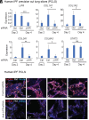
Targeting LIFR in human PCLS reduces expression of fibrotic genes. Human IPF PCLS were incubated with control (Ctrl) siRNA or siRNA against LIFR on day 0. (A) On days 2 and 4, gene expression (normalized to HPRT) was measured by qPCR. (B) On day 4, PCLS were imaged by immunofluorescence with mAb specific for COL1A1 and FN1. (A) Mean + SD shown from 5 slices per condition. Unpaired Student’s t test. *P < 0.05, **P < 0.01, ***P < 0.001, ns = nonsignificant.
Fig. 4.
Fig. 4.
LIFR amplifies the expression of profibrotic genes via JAK2 signaling in IPF lung fibroblasts. JAK2 mediates the induction of profibrotic genes by TGFβ1, IL-4, and IL-13 in lung fibroblasts. (A) Human IPF lung fibroblasts were transfected with control siRNA (Ctrl), with siRNA against LIFR and LIF (LIFR + LIF), or with siRNA against IL11R and IL11 (IL11R + IL11). Cell lysates 72 h after siRNA transfection were run on SDS-PAGE gel with immunoblotting with antibodies specific for phosphorylated (p) JAK1 (Tyr1034/1035), JAK1, pJAK2 (Tyr1007/1008), JAK2, and beta-tubulin (loading control). (B and C) Human IPF lung fibroblasts were transfected with a control siRNA or anti-JAK2 siRNA. 48 h after siRNA transfection, fibroblasts were stimulated with TGFβ1 (5 ng/mL). At 24 h after stimulation by TGFβ1: (B) cell lysates were run on SDS-PAGE gel with immunoblotting with antibodies specific to JAK2 and tubulin (loading control). (C) Gene expression (normalized to GAPDH) was measured by qPCR. (D) Human IPF lung fibroblasts were stimulated with TGFβ1 (5 ng/mL); at 0, 2, and 4 h after stimulation, cell lysates were run on SDS-PAGE gel with immunoblotting with antibodies specific for phosphorylated pJAK2 (Tyr1007/1008 and Tyr221), JAK2, phosphorylated STAT1, STAT1, phosphorylated STAT3, STAT3, and beta-tubulin (loading control). Protein quantification was done using ImageJ. (E) Human IPF lung fibroblasts were treated with vehicle (DMSO) or JAK inhibitors at 8 or 24 μM. 0.5 h later, cells were left unstimulated (basal) or stimulated with TGFβ1 (5 ng/mL), IL-4 (50 ng/mL), or IL-13 (50 ng/mL). 48 h after stimulation, COL1A1 and ACTA2 expression (normalized to GAPDH) were measured by qPCR. (CE) Mean and SD shown. (A and D) The immunoblot is representative of two IPF fibroblast cell lines tested. In the quantification of (D), the dots represent two IPF fibroblast cell lines, with two technical replicates each. (C and D) Each dot represents a different IPF lung fibroblast cell line (n = 4). (C) Paired Student’s t test. (D) One-way ANOVA, Dunnett's multiple comparison test. *, **, ***, ****, P < 0.05, 0.01, 0.001, 0.0001.
Fig. 5.
Fig. 5.
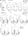
LIFR mediates the increased response to TGFβ1 in IPF lung tissue. (A) Human control and IPF lung fibroblasts (n = 4 patients) were transfected with siRNA against LIFR or a control siRNA. 48 h after transfection with siRNA: (A) LIFR expression was measured by qPCR (normalized to GAPDH). The relative expression after treatment with anti-LIFR siRNA compared to after treatment with control siRNA (normalized as 1.0) is shown. (B) Cells were left at basal (unstimulated) condition or stimulated with TGFβ1 (5 ng/mL) for 24 h. Expression of LIFR, COL1A1, COL1A2, POSTN, and ACTA2 was analyzed with qPCR (normalized to GAPDH). The relative expression after TGFβ1 stimulation compared to basal condition (normalized as 1) is shown. (CE) Human PCLS from control or IPF patients were tested in vitro. (CD) Human PCLS were transfected with siRNA against LIFR or a control siRNA. 48 h after transfection with siRNA: (C) LIFR expression was measured by qPCR (normalized to HPRT). (D) Human PCLS were left at basal (unstimulated) condition or stimulated with TGFβ1 (5 ng/mL) for 24 h. Expression of LIFR, COL1A1, COL1A2, POSTN, and ACTA2 was analyzed with qPCR (normalized to HPRT). (E) Human PCLS were treated with vehicle (DMSO) or baricitinib at 30 μM. 0.5 h later, PCLS were left unstimulated (basal) or stimulated with TGFβ1 (5 ng/mL) for 96 h. Expression of COL1A1 and COL1A2 was analyzed with qPCR (normalized to HPRT). (A and B) Each dot represents a different primary lung fibroblast cell line. (CE) Each dot represents a different, nonserial, replicate lung slice from a single control subject or IPF patient. (AE) Mean and SD shown. (A and C) Unpaired Student’s t test. (B and D) One-way ANOVA, Sidak's multiple comparison test. *, **, ***, ****, P < 0.05, 0.01, 0.001, 0.0001. ns, nonsignificant (P > 0.05).

Update of

References

    1. Jeganathan N., Sathananthan M., The prevalence and burden of interstitial lung diseases in the USA. ERJ Open Res. 8, 00630–02021 (2022). - PMC - PubMed
    1. Ley B., Collard H. R., King T. E., Clinical course and prediction of survival in idiopathic pulmonary fibrosis. Am. J. Respir. Crit. Care Med. 183, 431–440 (2011). - PubMed
    1. Maher T. M., Strek M. E., Antifibrotic therapy for idiopathic pulmonary fibrosis: Time to treat. Respir. Res. 20, 205 (2019). - PMC - PubMed
    1. Frangogiannis N., Transforming growth factor-β in tissue fibrosis. J. Exp. Med. 217, e20190103 (2020). - PMC - PubMed
    1. Allen J. E., IL-4 and IL-13: Regulators and effectors of wound repair. Annu. Rev. Immunol. 41, 229–254 (2023). - PubMed

MeSH terms

LinkOut - more resources