. 2024 Dec 6;386(6726):1154-1161.
doi: 10.1126/science.adp1779. Epub 2024 Dec 5.

Target-conditioned diffusion generates potent TNFR superfamily antagonists and agonists

Affiliations

Target-conditioned diffusion generates potent TNFR superfamily antagonists and agonists

Matthias Glögl et al. Science. .

Abstract

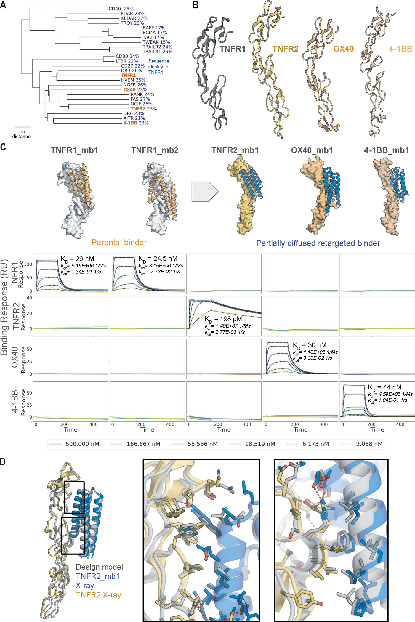
Despite progress in designing protein-binding proteins, the shape matching of designs to targets is lower than in many native protein complexes, and design efforts have failed for the tumor necrosis factor receptor 1 (TNFR1) and other protein targets with relatively flat and polar surfaces. We hypothesized that free diffusion from random noise could generate shape-matched binders for challenging targets and tested this approach on TNFR1. We obtain designs with low picomolar affinity whose specificity can be completely switched to other family members using partial diffusion. Designs function as antagonists or as superagonists when presented at higher valency for OX40 and 4-1BB. The ability to design high-affinity and high-specificity antagonists and agonists for pharmacologically important targets in silico presages a coming era in protein design in which binders are made by computation rather than immunization or random screening approaches.


Conflict of interest statement

Competing interests:

M.G., A.Kr., R.R. and D.B. are co-inventors on a provisional patent application 63/641,829 submitted by the University of Washington for the design, composition, and function of the proteins created in this study.

Figures

Fig. 1.
Fig. 1.. Diffusion of shape complementary binding proteins.
A. Previous de novo designed binders (green) generated by docking pregenerated scaffolds bury less surface area (Å2) against their target than many native complexes (orange); the approach developed here enables the design of very large interfaces (blue). B. TNFR1 is a challenging target, with a flat surface and few surface hydrophobic residues which are shown in red on the structure (PDB ID: 6KP8). Residues selected as target hotspots for RFdiffusion are shown as sticks. C. Representative RF_Diffusion trajectory against TNFR1 starting from a random residue distribution placed against the target (top left). At each denoising step (Xt, top), the network generates a predicted structure (^X0, bottom), and interpolates towards this structure to generate the next step (Xt-1). D. Comparison with previous TNFR1 design efforts. The RFdiffusion TNFR1 binder designs generated here (orange) have substantially higher buried SASA and contact molecular surface (CMS) than designs against TNFR1 generated previously using the Rosetta RIF dock method (red) that failed to bind. Designs generated against multiple targets using RIFdock (3)(green) and short chain RFdiffusion (7) (blue) are also shown for comparison. E. (top) Design models of binders TNFR1_mb1 (orange) and TNFR1_mb2 (blue) in complex with TNFR1 (gray). (bottom) SPR measurements of binding to TNFR1. F. Site saturation mutagenesis (SSM) results confirm design models of TNFR1 binders and associated entropy. All 2014 and 2033 single amino acid substitutions for TNFR1_mb1 and TNFR1_mb2 were expressed on yeast surface and probed using FACS with biotinylated TNFR1 followed by deep sequencing. Positions that were strongly conserved (low entropy, blue) were in the core and at the binding interface, while most surface residues away from the interface had high entropy (yellow). Entropy was calculated based on the overall change of affinity. Lower entropy (blue) is related to conserved interactions in the interface or core. Zoom-ins show dense interaction networks of low entropy residues (TNFR1 is in gray).
Fig. 2.
Fig. 2.. Partial diffusion generates picomolar binders.
A. Schematic of partial diffusion process for TNFR1_mb2. The backbone of the input structure is represented as a collection of independent residues (first panel), noise is added (second panel), and then RFdiffusion is used to remove the noise, which results in a similar but better fitting model (orange, right) compared to the input structure (purple). B. Partial diffusion increases interface contacts. Contact molecular surface and interface buried solvent accessible surface area is depicted for input designs (orange) and the respective partial diffused variants in blue squares (TNFR1_mb1), triangles (TNFR1_mb2), and circles (TNFR1_mb3). C. Partial diffusion increases interface interaction density and binding affinity. For TNFR1_mb2 an additional interface forms (left panel), while an existing interface remains largely unchanged (right). For TNFR1_mb3, improved shape matching leads to additional interactions (bottom middle insets). The corresponding TNFR1 SPR traces are on the right (6 steps of 5x dilutions from 500 nM).
Fig. 3.
Fig. 3.. Partial diffusion generates high-specificity TNFR2, OX40, and 4–1BB binders.
A. Phylogenetic tree of TNFT superfamily receptors. The tree was constructed using the UPGMA method based on a multiple sequence alignment performed with ClustalW. TNFR1, TNFR2, OX40, and 4–1BB investigated in this study are highlighted in orange. The scale bar represents a distance of 0.1 substitutions per site. Pairwise sequence identity to TNFR1 is indicated in blue. B. Comparison of structures of TNFR1, TNFR2, OX40, and 4–1BB (PDB IDs: 7KP8 (17), 3ALQ (17, 18), 2HEV (19), 6BWV (20)). C. (Top) Original design models and partial diffusion generated model for the highest affinity TNFR2, OX40 and 4–1BB binders (also see Fig. S6). The two TNFR1 binders (orange, left) were superimposed on the new targets (right) and partially diffused to yield target-matched backbones (blue). (Bottom) SPR measurements show that the binders are highly specific for the targets they were diffused against. D. Crystal structure (colors) of TNFR2_mb1 (blue) in complex with TNFR2 (yellow) superimposed on the computationally design model (grey). Boxed interface regions on the backbone superposition on the left are shown with interface sidechains in the zoom ins on the right.
Fig. 4.
Fig. 4.. Design of soluble oligomeric 4–1BB and OX40 superagonists.
A. Designed binders antagonize TNFɑ signaling. HEK293-blue cells were incubated with 100 pM TNF-α and serial dilutions of designed binders and NFkB-dependent activation were measured. Curves are fit to data from two independent replicates. B. Schematic overview of 4–1BB signaling. Clustering of trimeric 4–1BB Ligand (4–1BB-L, green) with three copies of 4–1BB receptor (gray) leads to intracellular formation of TRAF1/2 trimers (orange) and intracellular hexamers of zinc-RING finger domains leading to downstream signaling. C. Multivalent presentation of 4–1BB binder design on cyclic oligomers activates 4–1BB signaling on luciferase reporter cell lines, compared to native 4–1BB ligand (4–1BB-L) alone and complexed with an anti-his antibody. 4–1BB_mb2 was fused to various oligomerization domains, examples for different cyclic oligomeric states with a valency of one for binder alone (C1_1) to valency of eight (C8_N2) are shown. For details on oligomers see also Table S4 and Figure S10. D. Multivalent presentation of OX40 binder design on cyclic oligomers activates OX40 signaling on luciferase reporter cell lines. OX40_mb1 was fused to various oligomerization domains, examples for different cyclic oligomeric states with a valency of one for binder alone (C1_1) to valency of eight (C8_N2). For details on oligomers see also Table S4 and Figure S10. E. Model of 4–1BB (gray) upon binding of 4–1BB_mb_2 (blue) fused to the N-terminus of a cyclic hexamer (orange) compared to native complex with 4–1BB-L (green) (right, PDB ID: 6BWV). Distance between receptor (M101) to center is indicated (R). F. Geometry and oligomerization state dependence of designed 4–1BB and OX40 agonists. Binders 4–1BB_mb2 and OX40_mb1 were fused N- or C-terminal to 24 oligomerization domains each (see also Table 2). Central panel shows maximum recorded signal at 200 pM, with each dot representing one oligomer-binder fusion, separated in groups depending on oligomeric state for C2-C8 oligomers. Each Dotted line indicates signal of the native ligand (OX40) or native ligand plus antibody (4–1BB). Examples of design models of oligomerization domains that show high or low signal in specific groups for 4–1BB are shown in the surrounding panels, with numbers indicating their signal in the central plot. Fusion sites of binders are indicated by a red sphere and distance to center (R) labeled. Chains are colored in a gradient from N-terminus (yellow) to C-terminus (blue).

References

    1. Kortemme T, De novo protein design-From new structures to programmable functions. Cell 187, 526–544 (2024). - PMC - PubMed
    1. Marchand A, Van Hall-Beauvais AK, Correia BE, Computational design of novel protein-protein interactions - An overview on methodological approaches and applications. Curr. Opin. Struct. Biol. 74, 102370 (2022). - PubMed
    1. Cao L, Coventry B, Goreshnik I, Huang B, Sheffler W, Park JS, Jude KM, Marković I, Kadam RU, Verschueren KHG, Verstraete K, Walsh STR, Bennett N, Phal A, Yang A, Kozodoy L, DeWitt M, Picton L, Miller L, Strauch E-M, DeBouver ND, Pires A, Bera AK, Halabiya S, Hammerson B, Yang W, Bernard S, Stewart L, Wilson IA, Ruohola-Baker H, Schlessinger J, Lee S, Savvides SN, Garcia KC, Baker D, Design of protein-binding proteins from the target structure alone. Nature 605, 551–560 (2022). - PMC - PubMed
    1. Chevalier A, Silva D-A, Rocklin GJ, Hicks DR, Vergara R, Murapa P, Bernard SM, Zhang L, Lam K-H, Yao G, Bahl CD, Miyashita S-I, Goreshnik I, Fuller JT, Koday MT, Jenkins CM, Colvin T, Carter L, Bohn A, Bryan CM, Fernández-Velasco DA, Stewart L, Dong M, Huang X, Jin R, Wilson IA, Fuller DH, Baker D, Massively parallel de novo protein design for targeted therapeutics. Nature 550, 74–79 (2017). - PMC - PubMed
    1. Gainza P, Wehrle S, Van Hall-Beauvais A, Marchand A, Scheck A, Harteveld Z, Buckley S, Ni D, Tan S, Sverrisson F, Goverde C, Turelli P, Raclot C, Teslenko A, Pacesa M, Rosset S, Georgeon S, Marsden J, Petruzzella A, Liu K, Xu Z, Chai Y, Han P, Gao GF, Oricchio E, Fierz B, Trono D, Stahlberg H, Bronstein M, Correia BE, De novo design of protein interactions with learned surface fingerprints. Nature 617, 176–184 (2023). - PMC - PubMed

Publication types

MeSH terms

Substances