. 2024 Dec 6;386(6726):eadl4237.
doi: 10.1126/science.adl4237. Epub 2024 Dec 6.

Programming tissue-sensing T cells that deliver therapies to the brain

Affiliations

Programming tissue-sensing T cells that deliver therapies to the brain

Milos S Simic et al. Science. .

Abstract

To engineer cells that can specifically target the central nervous system (CNS), we identified extracellular CNS-specific antigens, including components of the CNS extracellular matrix and surface molecules expressed on neurons or glial cells. Synthetic Notch receptors engineered to detect these antigens were used to program T cells to induce the expression of diverse payloads only in the brain. CNS-targeted T cells that induced chimeric antigen receptor expression efficiently cleared primary and secondary brain tumors without harming cross-reactive cells outside of the brain. Conversely, CNS-targeted cells that locally delivered the immunosuppressive cytokine interleukin-10 ameliorated symptoms in a mouse model of neuroinflammation. Tissue-sensing cells represent a strategy for addressing diverse disorders in an anatomically targeted manner.


Conflict of interest statement

Competing interests: Several patents have been filed related to this work. These include, but are not limited to, US APP # 63/464,497, 17/042,032, 17/040,476, 17/069,717, 15/831,194, 15/829,370, 15/583,658, 15/096,971, and 15/543,220. W.A.L. is shareholder of Gilead Sciences, Allogene, and Intellia Therapeutics, and previously consulted for Cell Design Labs, Gilead, Allogene, and SciFi Foods. H.O. is on the on the scientific advisory boards for Neuvogen and Eureka Therapeutics. S.L.H. currently serves on the scientific advisory boards of Accure, Alector, and Annexon; has previously consulted for BD, Moderna, NGM Bio, and Pheno Therapeutics; and previously served on the board of directors of Neurona. S.L.H. has received travel reimbursement and writing support from F. Hoffmann-La Roche and Novartis AG for anti-CD20 therapy–related meetings and presentations.

The remaining authors declare no competing interests.

Figures
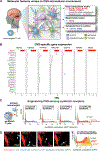
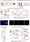
Fig. 1.
Fig. 1.. Identification of CNS-specific extra-cellular antigens that can be recognized by synNotch receptors.
(A) Conceptual rationale for the design of therapeutic T cells that can recognize CNS-specific antigens to trigger a therapeutic response locally. Specific extracellular molecular features of the CNS that could be used for recognition are highlighted. (B) Box plots showing tissue-specific expression of candidate CNS-specific genes BCAN, CSPG5, PTPRZ1, NrCAM, CDH10, GRM3, and MOG across a subset of tissue samples in GTEx v7. Units shown are normalized RNA-seq counts (transcripts per million) taken from GTEx portal v7. Brain expression is shown in red. See fig. S1 for the pipeline used to identify CNS-specific targets. (C) Validation of synNotch receptors that recognize CNS antigens. Receptors (see fig. S2 for more on the generation of receptors) were expressed in primary human CD4+ T cells with a GFP synNotch activation reporter. These cells were cocultured with mouse primary neurons and glia to test activation. Flow cytometry histograms show induction of the GFP reporter only in the presence of primary neuronal and glial cultures. (D) Time-lapse images (for full movie, see movie S1) showing primary human CD8+ T cells sensing BCAN expressed by primary astrocytes (red). Primary human CD8+ T cells expressing BCAN-sensing synNotch are shown in blue. The cells turn green when they are activated and induce the GFP reporter. Scale bar, 20 μm. (E) The α-BCAN synNotch receptor recognizes extracellular matrix-bound recombinant BCAN. The α-BCAN synNotch receptor was expressed in primary human CD4+ T cells with a GFP synNotch activation reporter. These cells were cultured with mouse or human recombinant BCAN presented on hyaluronic acid–coated plates to test activation. Flow cytometry histograms show the induction of GFP reporters only in the presence of recombinant BCAN.
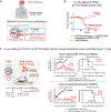
Fig. 2.
Fig. 2.. SynNotch recognition of the CNS-specific ECM molecule BCAN directs CAR antitumor activity specifically and potently to the intracerebral GBM PDX.
(A) Design of a brain-targeted CAR T cell. The α-BCAN synNotch receptor was used to drive the expression of antitumor CAR only in the brain. This tissue-specific priming circuit was designed to restrict the expression of CAR only to the CNS, preventing damage to normal, non-CNS tissues that express the CAR target antigens EphA2 and IL13Rα2. This circuit should selectively identify GBM cells, which are the only cells expressing EphA2 or IL13Rα2 in the CNS. (B) Killing of GBM6 PDX tumors in vitro. Primary CD8+ T cells transduced with the α-BCAN synNotch→α-EphA2/IL13Rα2 CAR circuit (or with the constitutively expressed α-EphA2/IL13Rα2 CAR) were cocultured with GBM6 target cells and K562 priming cells either expressing or not expressing BCAN. Relative cell survival of target GBM6 cells is shown at 72 hours (relative to untransduced T cell controls, n = 3, error bars indicate SEM). See fig. S3, A and B, for further details and controls. (C) In vivo clearance of GBM6 tumors. GBM6 tumors expressing mCherry and luciferase were orthotopically inoculated in the brains of NCG mice. Ten days after tumor inoculation, mice were infused intravenously with 2 million each of CD4+ and CD8+ T cells expressing the α-BCAN synNotch→α-EphA2/IL13Rα2 CAR circuit (n = 5) or no construct (negative control) (n = 5). Tumor size and survival were monitored over time by bioluminescence imaging. Thick line shows mean tumor size; thin lines show individual mice. Dotted line shows background bioluminescence. P = 0.018, mixed-effects analysis. Survival was analyzed over 80 days by log-rank (Mantel-Cox) test (P = 0.003). See additional repeats in fig. S3C. BCAN KO GBM6 tumors (fig. S7C) were also cleared with similar efficiency. (D) GBM6 tumor–bearing mice were euthanized 10 days after α-BCAN SynNotch→CAR T cell infusion (3 million each of CD4+ and CD8+). Representative confocal fluorescent microscopy of brain sections shows synNotch activation (green) and reveals that T cell–mediated killing (cleaved caspase 3 staining, red stain) is restricted to the tumor (adjacent neurons are not apoptotic, gray NeuN stain). Scale bars, 1 mm (left) and 200 μm (right, enlargement of outlined region). See fig. S4 for further analysis of brain-localized T cells in nontumor regions. (E) Flow cytometry of α-BCAN synNotch→α-EphA2/IL13Rα2 CAR T cells isolated from blood, spleen, and brain of a GBM6-bearing mouse at day 6 after T cell injection demonstrating the higher presence of GFP+ primed T cells in the brain compared with the blood or the spleen. See fig. S5 for further analysis of brain-localized T cells. (F) Brain-flank dual GBM6 tumor model. GBM6 tumor cells were inoculated in the brains of NCG mice, whereas BCAN KO GBM6 cells were inoculated in the flanks of the identical mice (fig. S7, A and B). Both tumors expressed the CAR-target antigens EphA2 and IL13Rα2, but BCAN was only expressed in the brain. Ten days after tumor inoculation, mice were infused intravenously with 2 million each of CD4+ and CD8+ T cells expressing no construct (control) (n = 4) or α-BCAN synNotch→α-EphA2/IL13Rα2 CAR circuit (n = 5). Tumor size in the brain and in the flank were monitored over time by bioluminescence imaging. Only the tumor inoculated in the brain was reduced over time; the flank tumor grew at the same rate as in the mice treated with nontransduced T cells. Data are representative of two independent experiments. Thick line indicates the mean and shaded area the SEM (see fig. S8 for studies showing efficient in vivo killing of brain-inoculated GBM39 tumors in the PDX line lacking BCAN expression).
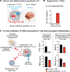
Fig. 3.
Fig. 3.. CNS-specific priming of synNotch-CAR T cells is a generalizable tool for effective killing of brain metastases such as HER2+ and TNBC brain metastases.
(A) Treatment for CNS metastases is a major unmet need, particularly for breast cancer. A CNS-specific priming circuit could be used as a general platform to target such metastases. Restricting CAR expression only to the brain could prevent damage to normal, nonbrain tissues that express target-killing antigens. Tumor-specific antigens are particularly hard to find for TNBC. Our strategy could be applied to target brain metastases for HER2+ breast cancer or TROP2+ TNBC (fig. S9). (B) Real-time in vitro killing of BT-474 breast cancer cells, a model of HER2+ breast cancer. BT-474 cells express HER2 but are negative for BCAN. Therefore, to mimic brain priming, we cocultured BT-474 cells with K562 cells expressing the priming antigen BCAN. Killing of BT-474 cells was only observed in the presence of BCAN+ K562 cells (n = 3, error bars indicate SEM). See fig. S9C for in vitro killing studies of BT-20 breast cancer cells, a model of TNBC. (C) In vivo tumor experiments with BT-474 (HER2+ breast cancer) and BT-20 (TNBC) tumors. BT-474 or BT-20 tumors expressing GFP and luciferase were orthotopically inoculated in the brains of NSG mice. Seven days after tumor inoculation, mice were infused intravenously with 3 million each of CD4+ and CD8+ T cells expressing no construct (control) (n = 5) or α-BCAN synNotch→CAR T cells (for BT-474: α-HER2 CAR; for BT-20: α-TROP2 CAR) (n = 5). Tumor size and survival were monitored over time by bioluminescence imaging. Thick line shows mean ± SEM (shaded area).
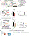
Fig. 4.
Fig. 4.. CNS-specific synNotch circuits can be programmed to produce the anti-inflammatory cytokine IL-10.
(A) CNS-specific synNotch cells could in principle be used to modulate neuroinflammation. For example, CNS priming could be used to trigger the expression of IL-10, a potent anti-inflammatory cytokine. For more analysis of optimal CNS antigens to target in neuroinflammation, see fig. S10. (B) Primary human T cells were engineered with the α-BCAN synNotch→IL-10 circuit and cocultured with K562 cells engineered to express BCAN. Supernatants were collected after 48 hours, and IL-10 was quantified by enzyme-linked immunosorbent assay (ELISA). Quantification shows the specific secretion of IL-10 only when the engineered T cells are cultured with BCAN+ K562 cells (n = 3). (C) In vitro inhibition assays of microglia and T cell activation. BV2 mouse microglia were cultured with control or therapeutic T cells (α-BCAN synNotch→IL-10) in the presence of BCAN+ K562 cells to induce payload expression. Two hours later, IFN-γ and LPS were added to induce activation of the microglia. Cells were cultured for 24 hours, and the supernatant was collected to assess inflammation by assaying for secretion of IL-6 and TNF-α by ELISA (n = 3, mean ± SD). Conventional CD25 TCR+ CD4+ T cells were sorted from MOG-specific TCR (2D2)–transgenic mice and cocultured with antigen-presenting cells presenting MOG peptide to induce their activation. To assay the inhibition of activation, MOG-specific TCR 2D2 CD4 T cells were cocultured at a 1:1 ratio with control transduced or engineered with α-BCAN synNotch→IL-10 T cells for 4 days in the presence of BCAN+ K562 cells to stimulate IL-10 induction. Activation of the MOG-specific TCR 2D2 CD4 T cells was analyzed by flow cytometry using the activation marker CD25 or by ELISA to measure IFN-γ secretion (n = 3, mean ± SD).
Fig. 5.
Fig. 5.. CNS-targeted anti-inflammatory circuit ameliorates EAE model.
(A) Schematic of the adoptive transfer EAE model. RAG-1–/– mice received an adoptive transfer of TH17–polarized CD4 T cells [20 × 106 in (B) and 25 × 106 in (C)] from P35–55 MOG–immunized C57BL/6J mice. At the indicated days after adoptive transfer (arrows), mice received primary human CD4 T cells transduced with either control (no circuit, n = 5) or α-BCAN synNotch→IL-10 circuit (n = 5) at the indicated times (10 × 106). The EAE neurological disease scoring scale is shown. (B) Treatment with α-BCAN synNotch→IL-10 T cells yields improved EAE scores and increased survival. Ten million T cells were injected on each day, as indicated by a black arrow. P < 0.05, two-way ANOVA. EAE severity was assessed by the area under the curve of each animal starting from the day of first treatment, day 7 until day 25 (n = 5, mean± SE). P < 0.05, unpaired t test. Survival curves show improved protection by the α-BCAN synNotch→IL-10 T cell treatment as analyzed by log-rank (Mantel-Cox) test. The effects of the two treatments on mouse mobility are shown in fig. S11. Additional repeats of this experiment are shown in figs. S12 and S13. (C) Treatment with α-CDH10 synNotch→IL-10 T cells yielded improved EAE scores and increased survival. Ten million T cells were injected on each day, indicated by a black arrow. EAE scores showed significant improvements with CNS-targeted α-CDH10 synNotch→IL-10 T cells. P < 0.05, two-way ANOVA. EAE severity was assessed by the area under the curve of each animal starting from the day of first treatment, day 7, until day 25 (n = 7, mean ± SE). P < 0.05, unpaired t test. Survival curves show improved protection by the α-CDH10 synNotch→IL-10 T cell treatment as analyzed by log-rank (Mantel-Cox) test. (D) Brain-sensing cells could be used as a general platform to treat a broad set of CNS diseases such as primary and secondary brain tumors, neuroinflammation, or even neurodegeneration. This customizable platform can be used to locally deliver any genetically encodable molecular therapy that is appropriate for a specific CNS disease, thereby improving on-target action and alleviating off-target toxicity.

Comment in

References

    1. Feigin VL et al., Global, regional, and national burden of neurological disorders during 1990–2015: A systematic analysis for the Global Burden of Disease Study 2015. Lancet Neurol. 16, 877–897 (2017). doi: 10.1016/S1474-4422(17)30299-5; pmid: 28931491 - DOI - PMC - PubMed
    1. Terstappen GC, Meyer AH, Bell RD, Zhang W, Strategies for delivering therapeutics across the blood-brain barrier. Nat. Rev. Drug Discov. 20, 362–383 (2021). doi: 10.1038/s41573-021-00139-y; pmid: 33649582 - DOI - PubMed
    1. Smolders J et al., Tissue-resident memory T cells populate the human brain. Nat. Commun. 9, 4593 (2018). doi: 10.1038/s41467-018-07053-9; pmid: 30389931 - DOI - PMC - PubMed
    1. Morsut L et al., Engineering customized cell sensing and response behaviors using synthetic Notch receptors. Cell 164, 780–791 (2016). doi: 10.1016/j.cell.2016.01.012; pmid: 26830878 - DOI - PMC - PubMed
    1. Choe JH et al., SynNotch-CAR T cells overcome challenges of specificity, heterogeneity, and persistence in treating glioblastoma. Sci. Transl. Med. 13, eabe7378 (2021).doi: 10.1126/scitranslmed.abe7378; pmid: 33910979 - DOI - PMC - PubMed

Publication types

MeSH terms