. 2024 Dec 5;20(12):e1012713.
doi: 10.1371/journal.ppat.1012713. eCollection 2024 Dec.

Receptor binding and structural basis of raccoon dog ACE2 binding to SARS-CoV-2 prototype and its variants

Affiliations

Receptor binding and structural basis of raccoon dog ACE2 binding to SARS-CoV-2 prototype and its variants

Chunliang Luo et al. PLoS Pathog. .

Abstract

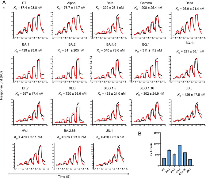
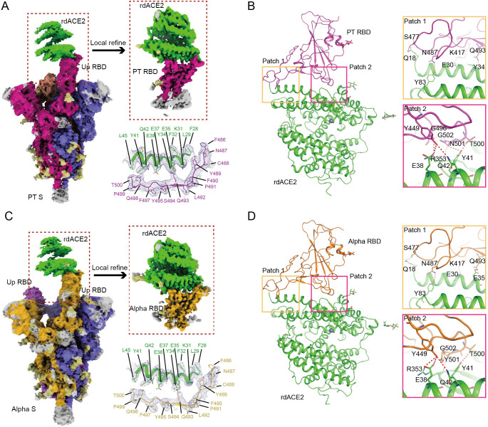
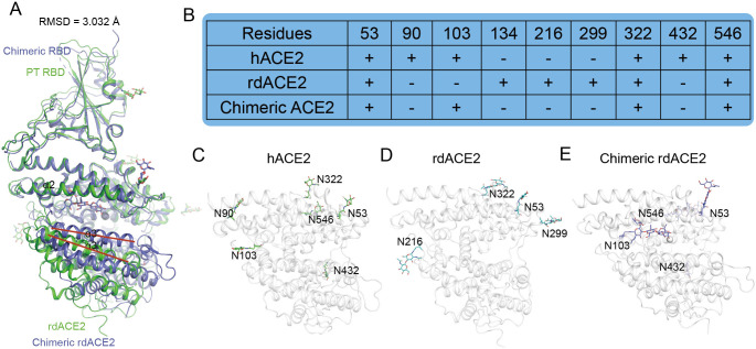
Raccoon dog was proposed as a potential host of SARS-CoV-2, but no evidence support such a notion. In our study, we investigated the binding affinities of raccoon dog ACE2 (rdACE2) to the spike (S) protein receptor binding domain (RBD) of SARS-CoV-2 prototype (PT) and its variants. It revealed that the binding affinities of RBD from SARS-CoV-2 variants were generally lower than that of the PT RBD. Through structural and functional analyses, we found amino acids H34 and M82 play pivotal roles in maintaining the binding affinity of ACE2 to different SARS-CoV-2 sub-variants. These results suggest that raccoon dogs exhibit lower susceptibility to SARS-CoV-2 compared to those animal species with a high prevalence of SARS-CoV-2 transmission.


Conflict of interest statement

The authors have declared that no competing interests exist.

Figures

Fig 1
Fig 1. SPR assays for the binding of rdACE2 to RBDs from different SARS-CoV-2 variants and the infectivity of pseudotyped SARS-CoV-2 in BHK-21 cells transient expression of rdACE2.
(A) Raw curves are represented by black lines, and fitted curves are represented by red lines. The dissociation constants (KD) are presented as mean ± SD calculated from three independent experiments. (B) Transduction of the pseudotyped SARS-CoV-2 PT and VOCs in BHK-21 cells transient expression of rdACE2. The data represent the results of three replicates.
Fig 2
Fig 2. The structures of SARS-CoV-2 PT and Alpha S protein in complex with rdACE2.
Cryo-EM density maps of the PT (A) and Alpha (C) S proteins bound to rdACE2 are depicted, with local refinement conducted on the binding interface of the S protein and rdACE2. The density maps illustrating the binding interface are presented as mesh, while the overall structures of rdACE2/PT RBD (B) and rdACE2/Alpha RBD (D) are represented as cartoons. Residues involved in the hydrogen bond networks of patch 1 and patch 2 are highlighted as sticks.
Fig 3
Fig 3. Differential N-glycosylation between hACE2 and rdACE2.
(A) Superimposition of PT RBD/rdACE2 structure and chimeric RBD/chimeric rdACE2 strcture (PDB:8VQR). (B) Statistical analysis of N-glycosylations in hACE2 and rdACE2. (C-E) N-glycosylation modifications observed in the structures of hACE2, rdACE2, and chimeric rdACE2, respectively.
Fig 4
Fig 4. Comparison of the binding interfaces between PT RBDs with hACE2 or rdACE2.
(A and B) The binding interfaces of PT RBD (A) and rdACE2 (B) in the rdACE2/PT RBD complex structure. (C and D) The binding interfaces of PT RBD (C) and hACE2 (D) in the hACE2/PT RBD complex structure. (E and F) Venn diagrams highlighting key residues at the binding interface. The red residues indicate those that interact with hACE2 but not with rdACE2. The cyan residues represent the distinct residues involved in PT RBD binding in both hACE2 and rdACE2.
Fig 5
Fig 5. The critical residues involved in the binding of rdACE2 to SARS-CoV-2 RBDs.
(A) Binding affinities of PT, Alpha, BA.2 and PT K417N RBDs to the different mutants of rdACE2. Raw curves are represented by black lines, and fitted curves are represented by red lines. (B-C) Structural comparison highlighting the designated key ACE2 residues (labeled above) that influence RBD binding, with these residues depicted as sticks. (D) Varying distances of K417/N417/T417 in the RBD to D30 (hACE2) or E30 (rdACE2).

Similar articles

References

    1. Garbino J, Crespo S, Aubert JD, Rochat T, Ninet B, Deffernez C, et al.. A prospective hospital-based study of the clinical impact of non-severe acute respiratory syndrome (Non-SARS)-related human coronavirus infection. Clin Infect Dis. 2006;43(8):1009–15. Epub 2006/09/20. doi: 10.1086/507898 ; PubMed Central PMCID: PMC7107919. - DOI - PMC - PubMed
    1. Perlman S, Netland J. Coronaviruses post-SARS: update on replication and pathogenesis. Nat Rev Microbiol. 2009;7(6):439–50. doi: 10.1038/nrmicro2147 ; PubMed Central PMCID: PMC2830095. - DOI - PMC - PubMed
    1. Fouchier RA, Hartwig NG, Bestebroer TM, Niemeyer B, de Jong JC, Simon JH, et al.. A previously undescribed coronavirus associated with respiratory disease in humans. Proc Natl Acad Sci U S A. 2004;101(16):6212–6. doi: 10.1073/pnas.0400762101 ; PubMed Central PMCID: PMC395948. - DOI - PMC - PubMed
    1. van der Hoek L, Pyrc K, Jebbink MF, Vermeulen-Oost W, Berkhout RJ, Wolthers KC, et al.. Identification of a new human coronavirus. Nat Med. 2004;10(4):368–73. doi: 10.1038/nm1024 ; PubMed Central PMCID: PMC7095789. - DOI - PMC - PubMed
    1. Woo PC, Lau SK, Chu CM, Chan KH, Tsoi HW, Huang Y, et al.. Characterization and complete genome sequence of a novel coronavirus, coronavirus HKU1, from patients with pneumonia. J Virol. 2005;79(2):884–95. Epub 2004/12/23. doi: 10.1128/JVI.79.2.884-895.2005 ; PubMed Central PMCID: PMC538593. - DOI - PMC - PubMed

MeSH terms

Substances

Supplementary concepts