. 2024 Dec 5;15(12):881.
doi: 10.1038/s41419-024-07271-8.

SENP3-FIS1 axis promotes mitophagy and cell survival under hypoxia

Affiliations

SENP3-FIS1 axis promotes mitophagy and cell survival under hypoxia

Alice Zhao et al. Cell Death Dis. .

Abstract

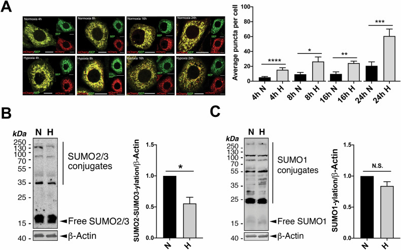
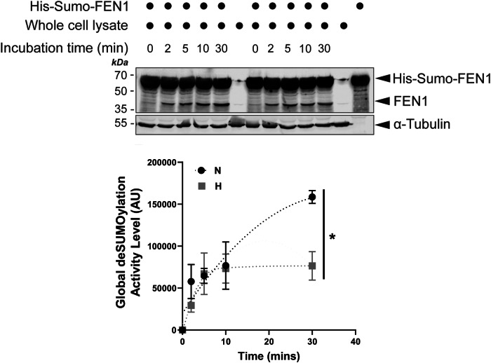
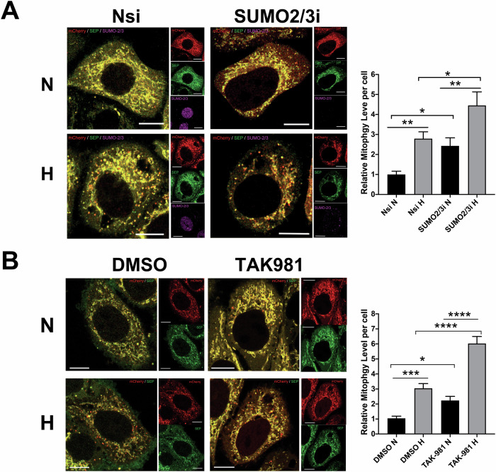
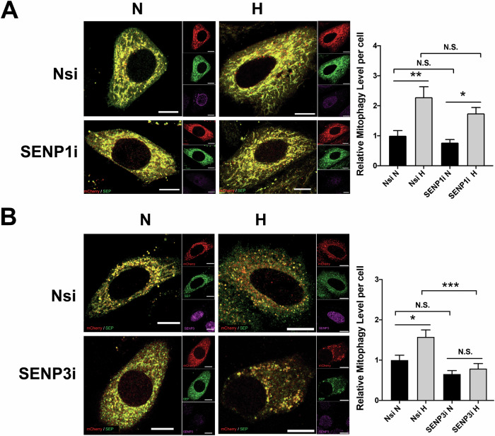
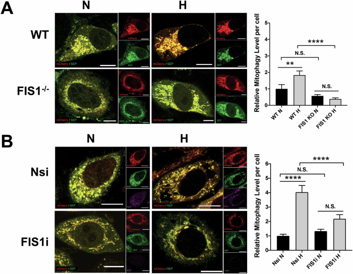
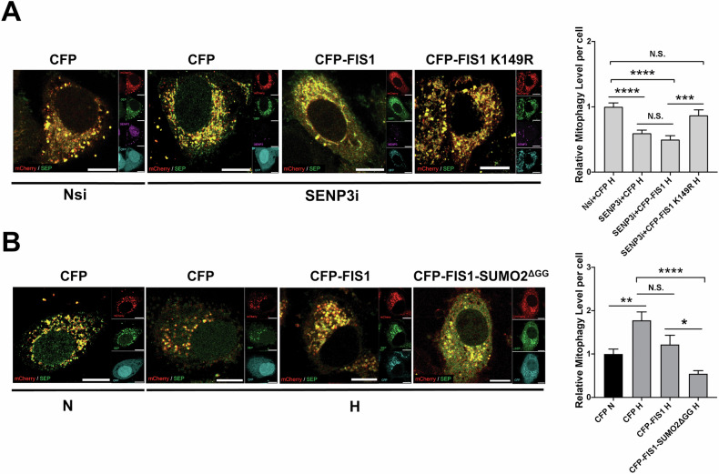
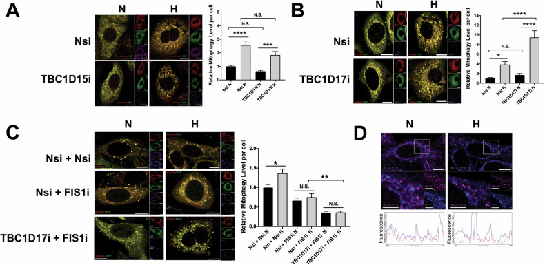
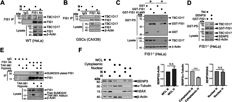
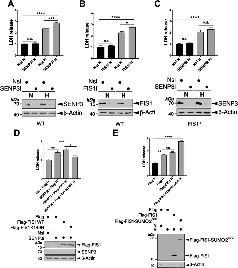
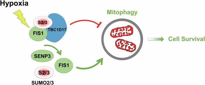
SUMOylation, the covalent attachment of the small ubiquitin-like modifier (SUMO) to target proteins, and its reversal, deSUMOylation by SUMO proteases like Sentrin-specific proteases (SENPs), are crucial for initiating cellular responses to hypoxia. However, their roles in subsequent adaptation processes to hypoxia such as mitochondrial autophagy (mitophagy) remain unexplored. Here, we show that general SUMOylation, particularly SUMO2/3 modification, suppresses mitophagy under both normoxia and hypoxia. Furthermore, we identify deSUMO2/3-ylation enzyme SENP3 and mitochondrial Fission protein 1 (FIS1) as key players in hypoxia-induced mitophagy (HIM), with SUMOylatable FIS1 acting as a crucial regulator for SENP3-mediated HIM regulation. Interestingly, we find that hypoxia promotes FIS1 SUMO2/3-ylation and triggers an interaction between SUMOylatable FIS1 and Rab GTPase-activating protein Tre-2/Bub2/Cdc16 domain 1 family member 17 (TBC1D17), which in turn suppresses HIM. Therefore, we propose a novel SUMOylation-dependent pathway where the SENP3-FIS1 axis promotes HIM, with TBC1D17 acting as a fine-tuning regulator. Importantly, the SENP3-FIS1 axis plays a protective role against hypoxia-induced cell death, highlighting its physiological significance, and hypoxia-inducible FIS1-TBC1D17 interaction is detectable in primary glioma stem cell-like (GSC) cultures derived from glioblastoma patients, suggesting its disease relevance. Our findings not only provide new insights into SUMOylation/deSUMOylation regulation of HIM but also suggest the potential of targeting this pathway to enhance cellular resilience under hypoxic stress.


Conflict of interest statement

Competing interests: The authors declare no competing interests. Ethics approval and consent to participate: All methods were conducted in accordance with relevant guidelines and regulations, including the principles outlined in the Declaration of Helsinki. Informed consent was obtained from all human subjects. Ethics approval for deriving primary GSC cultures was granted by the Yorkshire & The Humber - Leeds East Research Ethics Committee (IRB protocol 11-YH-0319/STH15598). Ethics approval for deriving primary renal cell cultures was obtained from the Research Ethical Committee (REC: 20SW0193) as part of the Ex Vivo DEtermiNed Cancer Therapy (EVIDENT) trial (NCT05231655).

Figures

Fig. 1
Fig. 1. deSUMO2/3-ylation induces mitophagy and promotes HIM.
A Hypoxia induces mitophagy. HeLa cells were transfected with Mito-pHfluorin per 35mm-dish and exposed to normoxia (N) or hypoxia (H;1% O2 for 4, 8, 16, or 24 h) (Scale bar, 10 μm). Upper panel shows that hypoxia-induced mitophagy is detectable as early as 4 h after cells exposed to 1% O2. Histogram in the right panel shows the average number of puncta per cell under indicated time points (n = 19 ~ 51 cells; *p < 0.05; **p < 0.01; ***p < 0.001; ****; unpaired t-test). B, C Hypoxia causes decreased SUMO2/3-ylation (B, n = 5, biological replicates; *p < 0.05; paired t-test) but not SUMO1-ylation (C, n = 6, biological replicates; N.S., non-significant; paired t-test) in HeLa cells. HeLa cells were exposed to 1% O2 for 24 h. Whole cell lysate samples were prepared and blotted as indicated.
Fig. 2
Fig. 2. Hypoxia affects the global deSUMOylation activity levels.
HeLa cells were cultured under either normoxic or hypoxic (1% O2) conditions for 24 h. Lysate samples from these cells were incubated with His-Sumo-FEN1 at 30 °C for varying durations. Lysate levels of FEN1, generated by SUMO protease cleavage of His-Sumo-FEN1, were detected by immunoblotting and normalised to α-Tubulin (loading control). The resulting FEN1 levels were expressed as an index of global deSUMOylation activity in arbitrary units (AU; n = 3, biological replicates; *p < 0.05; unpaired t-test).
Fig. 3
Fig. 3. Global deSUMOylation induces mitophagy and promotes HIM.
A RNAi-mediated SUMO-2/3 depletion induces mitophagy and promotes HIM. HeLa cells expressing Mito-pHfluorin were transfected with Nsi or SUMO-2/3-specific siRNA (SUMO-2/3i). 48 h post-transfection the cells were exposed to normoxia or hypoxia (1% O2) for 24 h, and the cells were analysed 72 h post-transfection (Scale bar 10 µm). Histogram in the right panel shows average number of Mito-pHfluorin red puncta per cell for cells exposed to N or H for 24 h (n = 42–71, *p < 0.05; **p < 0.01; Ordinary One-way ANOVA followed by Sidak’s multiple comparisons test). B Global SUMOylation inhibition induces mitophagy and promotes HIM. HeLa cells were transfected with Mito-pHfluorin. 48 h post-transfection the cells were treated with DMSO or TAK-981 (100 nM) and exposed to normoxia or hypoxia (1% O2) for 24 h, and the cells were analysed 72 h post-transfection (Scale bar 10 µm). Histogram in the right panel shows relative mitophagy level per cell for cells treated with DMSO or TAK-981 under and exposed to N or H for 24 h (n = 62–81, *p < 0.05; ***p < 0.001; ****p < 0.0001; Ordinary One-way ANOVA by Sidak’s multiple comparisons test).
Fig. 4
Fig. 4. SENP3 plays an essential role in mitophagy induced by hypoxia.
A RNAi-mediated SENP1 depletion does not appear to affect hypoxia-induced mitophagy in HeLa cells. HeLa cells expressing Mito-pHfluorin were transfected with Nsi or SENP1-specific siRNA (SENP1i). 48 h post-transfection the cells were exposed to normoxia or hypoxia (1% O2) for 24 h, and the cells were analysed 72 h post-transfection (Scale bar 10 µm). Histogram in the right panel shows relative mitophagy level per cell for cells exposed to N or H for 24 h (n = 32–55; N.S., non-significant; *p < 0.05; **p < 0.01; Ordinary One-way ANOVA by Sidak’s multiple comparisons test). B RNAi-mediated SENP3 depletion abolishes hypoxia-induced mitophagy in HeLa cells. HeLa cells expressing Mito-pHfluorin were transfected with Nsi or SENP3-specific siRNA (SENP3i). 48 h post-transfection the cells were exposed to normoxia or hypoxia (1% O2) for 24 h, and the cells were analysed 72 h post-transfection (Scale bar 10 µm). Histogram in the right panel shows average number of Mito-pHfluorin red puncta per cell for cells exposed to N or H for 24 h (n = 42–63, N.S., non-significant; *p < 0.05; ***p < 0.001; Ordinary One-way ANOVA by Sidak’s multiple comparisons test).
Fig. 5
Fig. 5. FIS1 is essential for mitophagy induced by hypoxia in HeLa cells.
A Genetic depletion of FIS1 abolishes hypoxia-induced mitophagy. Wild-type (WT; FIS1+/+) or FIS1 knockout (KO; FIS1−/−) HeLa cells were transfected with Mito-pHfluorin. 48 h post-transfection the cells were exposed to normoxia or hypoxia (1% O2) for 24 h, and the cells were analysed 72 h post-transfection (Scale bar 10 µm). Histogram in the right panel shows relative mitophagy level per cell for cells exposed to N or H for 24 h (n = 33–64, N.S., non-significant; **p < 0.01; ****p < 0.0001; Ordinary one-way ANOVA followed by Sidak’s multiple comparisons test). B RNAi-mediated FIS1 depletion prevents hypoxia-induced mitophagy. HeLa cells were transfected with Mito-pHfluorin and Nsi or FIS1-specific siRNA, and the cells were analysed 72 h post-transfection (Scale bar 10 µm). Histogram in the right panel shows relative mitophagy level per cell for cells exposed to N or H for 24 h (n = 54–61, N.S., non-significant; ****p < 0.0001; Ordinary one-way ANOVA followed by Sidak’s multiple comparisons test).
Fig. 6
Fig. 6. SUMOylatable FIS1 is required for SENP3 regulation of mitophagy induced by hypoxia.
A Expressing SUMOylation-deficient CFP-FIS1 K149R rescues hypoxia-induced mitophagy in SENP3-KD HeLa cells. HeLa cells expressing Mito-pHfluorin were transfected with Nsi or SENP3i (50 nM), together with CFP, CFP-FIS1 or CFP-FIS1 K149R. 48 h post-transfection the cells were exposed to hypoxia (1% O2) for 24 h, and the cells were analysed 72 h post-transfection (Scale bar 10 µm). Histogram in the right panel shows relative mitophagy level per cell for cells exposed to N or H for 24 h (n = 48-74; N.S., non-significant; ***p < 0.001 ****p < 0.0001; Ordinary one-way ANOVA followed by Sidak’s multiple comparisons test). B Expressing or CFP-FIS1-SUMO2ΔGG abolishes hypoxia-induced mitophagy. HeLa cells expressing Mito-pHfluorin were transfected CFP, CFP-FIS1 or CFP-FIS1-SUMO2ΔGG. 48 h post-transfection the cells were exposed to normoxia or hypoxia (1% O2) for 24 h. and the cells were analysed 72 h post-transfection (Scale bar 10 µm). Histogram in the right panel shows relative mitophagy level per cell for cells exposed to N or H for 24 h (n = 46–58; N.S., non-significant; *p < 0.05 **p < 0.01 ****p < 0.0001; Ordinary one-way ANOVA followed by Sidak’s multiple comparisons test).
Fig. 7
Fig. 7. FIS1 is required for TBC1D17 regulation of HIM.
A RNAi-mediated TBC1D15 depletion does not affect hypoxia-induced mitophagy in HeLa cells. HeLa cells were transfected with Mito-pHfluorin and Nsi or TBC1D15-specific siRNA (TBC1D15i; 20 nM). 48 h post-transfection the cells were exposed to normoxia or hypoxia (1% O2) for 24 h, and the cells were analysed 72 h post-transfection (Scale bar 10 µm). Histogram in the right panel shows relative mitophagy level per cell for cells exposed to N or H for 24 h (n = 39–59, N.S., non-significant; ***p < 0.001; ****p < 0.0001; Ordinary one-way ANOVA followed by Sidak’s multiple comparisons test). B RNAi-mediated TBC1D17 depletion promotes hypoxia-induced mitophagy in HeLa cells. HeLa cells were transfected with Mito-pHfluorin and Nsi or TBC1D17-specific siRNA (TBC1D17i; 20 nM). 48 h post-transfection the cells were exposed to normoxia or hypoxia (1% O2) for 24 h, and the cells were analysed 72 h post-transfection (Scale bar 10 µm). Histogram in the right panel shows relative mitophagy level per cell for cells exposed to N or H for 24 h (n = 42–56, N.S. non-significant; *p < 0.05; ****p < 0.0001; Ordinary one-way ANOVA followed by Sidak’s multiple comparisons test). C HeLa cells expressing Mito-pHfluorin were transfected with Nsi, TBC1D17i (20 nM), and/or FIS1-specific siRNA (50 nM). 48 h post-transfection the cells were exposed to normoxia or hypoxia (1% O2) for 24 h, and the cells were analysed 72 h post-transfection (Scale bar 10 µm). Histogram in the right panel shows relative mitophagy level per cell for cells exposed to N or H for 24 h (n = 52–62; N.S., non-significant; *p < 0.05; **p < 0.01; Ordinary one-way ANOVA followed by Sidak’s multiple comparisons test). D Hypoxia does not appear to affect the colocalisation of TBC1D17 with FIS1 in the Nsi/Nsi cells shown in (C). Relative fluorescence intensity of each channel as points along the white lines shown in the lower graphs for normoxia and hypoxia, respectively (Scale bar 10 µm).
Fig. 8
Fig. 8. SUMOylatable FIS1 is required for FIS1-TBC1D17 interaction.
A, B Hypoxia induces FIS1-TBC1D17 interaction. HeLa cells (A) or GSCs (B) were exposed to normoxia or hypoxia (1% O2) for 24 h. FIS1 was enriched through immunoprecipitation (IP). Lysate (input) and IP samples were immunoblotted as indicated. C TBC1D17 interacts with GST-FIS1 but not GST-FIS1 K149R mutant in FIS1 KO HeLa cells under hypoxia. FIS1 KO HeLa cells were transfected with GST, GST-FIS1 or GST-FIS1 K149R mutant. 48 h post-transfection the cells were exposed to normoxia or hypoxia (1% O2) for 24 h. GST-tagged proteins were enriched through GST-Pulldown (PD), Lysate (input) and GST-PD samples were immunoblotted as indicated. D SENP3 knockdown increases TBC1D17 interaction with GST-FIS1 in FIS1 KO HeLa cells exposed to hypoxia (1% O2) for 24 h. FIS1 KO HeLa cells expressing GST-FIS1 were transfected with Nsi or SENP3i. 48 h post-transfection the cells were lysed and GST-tagged proteins were enriched through GST-PD, Lysate (input) and GST-PD samples were immunoblotted as indicated. E Hypoxia induces FIS1 SUMO2/3-ylation in HeLa cells. HeLa cells were exposed to normoxia or hypoxia (1% O2) in the absence or presence of TAK981 (100 nM) for 24 h. FIS1 was enriched through IP. Lysate (input) and IP samples were immunoblotted as indicated. F Hypoxia reduces the levels of cytoplasmic SENP3 in HeLa cells. HeLa cells were exposed to normoxia or hypoxia (1% O2) for 24 h. Samples of whole cell lysate (WCL), cytoplasmic or nuclear fraction was prepared and blotted as indicated, SENP3 levels were normalised to β-Actin in WCL, α-Tubulin in the cytoplasmic fraction, and H2AX in the nuclear fraction (n = 5 biological replicates; N.S., not statistically significant; ***p < 0.001; Paired t-test).
Fig. 9
Fig. 9. SENP3-FIS1 axis promotes cell survival under hypoxia.
A RNAi-mediated depletion of SENP3 increases LDH release from HeLa cells exposed to hypoxia for 24 h. HeLa cells were transfected with Nsi or SENP3i. 48 h post-transfection the cells were exposed to normoxia or hypoxia (1% O2) for 24 h. Histogram in the upper panel shows relative levels of LDH release under indicated conditions (n = 3; biological replicates; N.S., non-significant; ***p < 0.001; ****p < 0.0001; Ordinary one-way ANOVA followed by Tukey’s multiple comparisons test). B RNAi-mediated depletion of FIS1 increases LDH release from HeLa cells exposed to hypoxia for 24 h. HeLa cells were transfected with Nsi or FIS1i. 48 h post-transfection the cells were exposed to normoxia or hypoxia (1% O2) for 24 h. Histogram in the upper panel shows relative levels of LDH release under indicated conditions (n = 3 biological replicates; N.S., non-significant; *p < 0.05; ****p < 0.0001; Ordinary one-way ANOVA followed by Tukey’s multiple comparisons test). C RNAi-mediated depletion of SENP3 does not increase LDH release from FIS1 KO HeLa cells exposed to hypoxia for 24 h. FIS1 KO HeLa cells were transfected with Nsi or SENP3i. 48 h post-transfection the cells were exposed to normoxia or hypoxia (1% O2) for 24 h. Histogram in the upper panel shows relative levels of LDH release under indicated conditions (n = 6 biological replicates; N.S., non-significant; ****p < 0.0001; Ordinary one-way ANOVA followed by Tukey’s multiple comparisons test). D Expressing SUMOylation-deficient FIS1 K149R mutant reverses the effect of SENP3 knockdown on hypoxia-induced LDH release. pcDNA3-Flag, Flag-FIS1 WT or Flag-FIS1 K149R mutant was transfected into HeLa cells in which SENP3 was depleted using siRNA for 48 h, and the cells were exposed to hypoxia (1% O2) for a further 24 h. Histogram in the upper panel shows relative levels of LDH release under indicated conditions (n = 5 ~ 6 biological replicates; *p < 0.05; **p < 0.01; ***p < 0.001; Ordinary one-way ANOVA followed by Tukey’s multiple comparisons test). E Expression of Flag-FIS1-SUMO2ΔGG, but not Flag-FIS1, in HeLa cells increases hypoxia-induced LDH release. pcDNA3-Flag, Flag-FIS1 WT or Flag-FIS1-SUMO2ΔGG mutant was transfected into HeLa cells for 48 h, and the cells were exposed to normoxia or hypoxia (1% O2) for 24 h. Histogram in the upper panel shows relative levels of LDH release under indicated conditions (n = 3 biological replicates; N.S., non-significant; **p < 0.01; ****p < 0.0001; Ordinary one-way ANOVA followed by Tukey’s multiple comparisons test). In (AE), whole cell lysate samples were immunoblotted as indicated in the lower panels.
Fig. 10
Fig. 10. Schematic representation of a proposed SUMO2/3-dependent mitophagy/cell survival pathway under hypoxia.
In response to hypoxia, FIS1 SUMO2/3-ylation increases due to reduced cytoplasmic SENP3. SUMO2/3-ylated FIS1 interacts with TBC1D17. With the associated FIS1, TBC1D17 inhibits the levels of hypoxia-induced mitophagy. Moreover, FIS1 deSUMO2/3-ylation mediated by residual cytoplasmic SENP3 is essential for maintaining hypoxia-induced mitophagy for cell survival.

References

    1. Russell OM, Lightowlers RN, Turnbull DM. Applying the Airbrakes: Treating Mitochondrial Disease with Hypoxia. Mol Cell. 2016;62:5–6. - PubMed
    1. Solaini G, Baracca A, Lenaz G, Sgarbi G. Hypoxia and mitochondrial oxidative metabolism. Biochimica et biophysica acta. 2010;1797:1171–7. - PubMed
    1. Zhang W, Ren H, Xu C, Zhu C, Wu H, Liu D, et al. Hypoxic mitophagy regulates mitochondrial quality and platelet activation and determines severity of I/R heart injury. eLife. 2016;5:e21407. - PMC - PubMed
    1. Fuhrmann DC, Brüne B. Mitochondrial composition and function under the control of hypoxia. Redox Biol. 2017;12:208–15. - PMC - PubMed
    1. Wang CY, She JX. SUMO4 and its role in type 1 diabetes pathogenesis. Diabetes/Metab Res Rev. 2008;24:93–102. - PubMed

LinkOut - more resources