. 2024 Dec 5;15(1):10392.
doi: 10.1038/s41467-024-54694-0.

Synaptic connectome of the Drosophila circadian clock

Affiliations

Synaptic connectome of the Drosophila circadian clock

Nils Reinhard et al. Nat Commun. .

Abstract

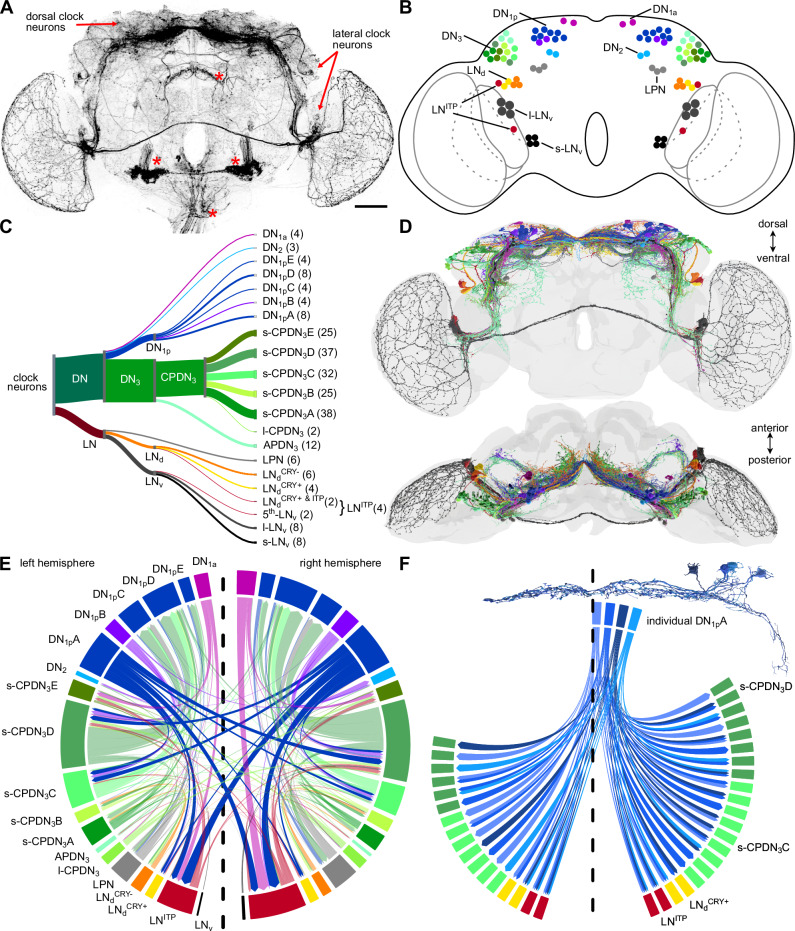
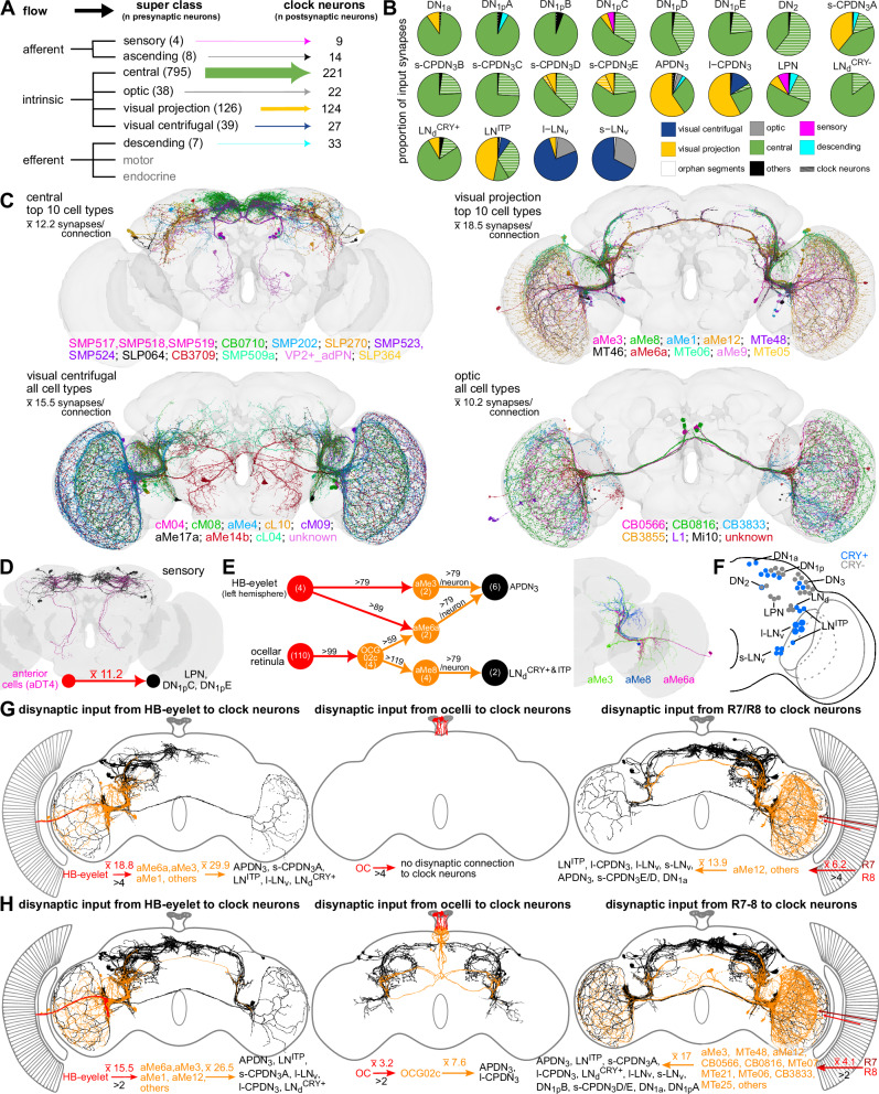
The circadian clock and its output pathways play a pivotal role in optimizing daily processes. To obtain insights into how diverse rhythmic physiology and behaviors are orchestrated, we have generated a comprehensive connectivity map of an animal circadian clock using the Drosophila FlyWire brain connectome. Intriguingly, we identified additional dorsal clock neurons, thus showing that the Drosophila circadian network contains ~240 instead of 150 neurons. We revealed extensive contralateral synaptic connectivity within the network and discovered novel indirect light input pathways to the clock neurons. We also elucidated pathways via which the clock modulates descending neurons that are known to regulate feeding and reproductive behaviors. Interestingly, we observed sparse monosynaptic connectivity between clock neurons and downstream higher-order brain centers and neurosecretory cells known to regulate behavior and physiology. Therefore, we integrated single-cell transcriptomics and receptor mapping to decipher putative paracrine peptidergic signaling by clock neurons. Our analyses identified additional novel neuropeptides expressed in clock neurons and suggest that peptidergic signaling significantly enriches interconnectivity within the clock network.


Conflict of interest statement

Competing interests: The authors declare no competing interests.

Figures

Fig. 1
Fig. 1. Drosophila melanogaster circadian clock network.
A Clk856-Gal4 drives GFP expression in most of the circadian clock neurons and some non-clock neurons (asterisk). Only a few DN3 are included in this Gal4-line. Scale bar = 50μm. B Drosophila clock neurons can be divided into four classes each of Dorsal neurons (DN) and Lateral neurons (LN) based on their cell body location. These can be further subdivided into different cell types based on their morphology and gene expression. Subtypes of clock neurons are color-coded. This color code is used throughout the manuscript. C Classification and numbers of all clock neurons identified in the FlyWire connectome. Refer to Table 1 for details. D The morphology of identified clock neurons in the connectome largely resembles the morphology of clock neurons marked by Clk856-Gal4. E Broad synaptic interconnectivity between different cell types within the clock network. The direction of the arrow indicates the flow of information. Strong connectivity is observed from DN1a to LNITP, from s-CPDN3C and s-CPDN3D to DN1pC-E, as well as from DN1pA to LNITP, LNdCRY+, s-CPDN3C and s-CPDN3D. Note extensive contralateral connectivity between different cell types, most of which can be accounted for by the DN1pA. The dashed line indicates the brain midline. F Individual DN1pA in the right hemisphere forms both ipsilateral and contralateral connections with s-CPDN3C, s-CPDN3D, LNdCRY+, and LNITP. Source data for panels E and F are provided in the Source Data file. Brain mesh is from Dorkenwald et al., 2024.
Fig. 2
Fig. 2. Morphology of newly identified s-CPDN3 and DN1p subtypes.
A Schematic cycling of Period (PER) and Vrille (VRI) protein abundance in clock neurons. B tim-(UAS)-Gal4 drives GFP expression in all DN3, many of which coexpress PER and about 12-19 coexpress VRI. When VRI staining is strong, PER staining is weak or not detectable. Image based on a female brain fixed at ZT1. Representative images are based on expression analyzed in 5 male and 5 female brains. Scale bar = 10 μm (C) DN3 are closely associated with the lateral horn. D The number of DN3 per hemisphere accounts for s-CPDN3, l-CPDN3, and APDN3 identified in the connectome. On average, females have slightly more DN3 (83 ± 5 standard deviation) compared to males (76 ± 6 standard deviation), which could be attributed to DN3 being more densely packed (and thus difficult to quantify) in males. n = 10 hemispheres from 5 brains. Error bars depict standard deviation. EI s-CPDN3 cell types identified in the FlyWire dataset. Numbers in brackets represent the number of neurons in the total brain. s-CPDN3 could be subdivided into five subtypes. Each subtype comprises two to eight different cell types that have unique morphological characteristics. J Clk4.1M-Gal4 reliably drives GFP expression in 14-15 DN1p per hemisphere. K Multi-color flip-out (MCFO) analysis reveals previously characterized DN1pA (4-5 neurons/hemisphere) and DN1pB (2 neurons) subtypes. MCFO analysis additionally reveals the morphology of uncharacterized DN1p subtypes. DN1pC and DN1pE comprise two cells each per hemisphere L, N. DN1pD comprises four cells per hemisphere two of which project over the midline while the other two do not, suggesting that the DN1pD is comprised of two subtypes M. Scale bar = 50μm. Source data for panel D are provided in the Source Data file. Brain mesh is from Dorkenwald et al., 2024.
Fig. 3
Fig. 3. Light and other inputs to the clock network.
A Input to the clock neurons grouped by the nine neuronal super classes annotated in the FlyWire connectome. Intrinsic neurons, specifically the central and visual projection neurons, are the largest groups providing inputs to the clock. B Breakdown of inputs to different types of clock neurons. C Neurons from the four different super classes (central, visual projection, visual centrifugal, and optic) which provide major inputs (based on the number of neurons) to the clock. D Anterior cells (AC, magenta) providing temperature inputs to LPN, DN1pC, and DN1pE. (black). E aMe neurons provide the strongest inputs to clock neurons. Upstream partners of aMe neurons with strong connectivity include HB-eyelet and OCG02c. Numbers above the arrows indicate the number of synapses and numbers in circles indicate the number of neurons. Note that the connection from the OC to OCG02c is below the threshold of 5 synapses/connection. Neuronal reconstructions of aMe3, aMe6a, and aMe8 are shown in one hemisphere. F Cryptochrome (CRY)-positive clock neurons are shown in blue. G Disynaptic inputs to the clock from the three types of extrinsic photoreceptors (HB-eyelet, ocelli, photoreceptor cells of the compound eye) using a threshold of >4 synapses. H Disynaptic inputs to the clock from the three types of extrinsic photoreceptors using a threshold of >2 synapses. For C, D, G, and H, numbers represent the average number of synapses. All cell types are listed according to their input strength (from high to low) to clock neurons. Note: HB eyelet was only identified in the left hemisphere. HB Hofbauer-Buchner, OC ocellar retinula cells, OCG ocellar ganglion cell. Source data for panels BD, G, and H are provided in the Source Data file. Brain mesh is from Dorkenwald et al., 2024.
Fig. 4
Fig. 4. Direct and indirect clock output pathways.
A Clock output grouped by the nine neuronal super classes annotated in the FlyWire connectome. Intrinsic neurons, specifically the central neurons, are the largest group of neurons downstream of clock neurons. B Breakdown of outputs from different types of clock neurons. All clock neurons, except for l-LNv, provide a majority of their output to central neurons. l-LNv mostly provides inputs to optic neurons. C Neurons from the four different super classes (central, optic, visual projection, and visual centrifugal) which receive inputs from the clock. D Individual postsynaptic partners of DN1pA and s-CPDN3C-D are sorted based on the number of synapses. Postsynaptic clock neurons are colored based on their identity. E CRY-positive DN1pA provide major inputs to CRY-negative DN1pC-E via s-CPDN3C-D. Contralateral connections from DN1pA to s-CPDN3 are stronger than ipsilateral connections. F APDN3 forms the most output synapses in the clock network and is highly connected to several types of Clamp neurons (CL). G DN1pB-E provides strong outputs to Tubercle-innervating neurons. H Several clock neurons provide strong inputs to neurons from diverse neuropils. Mono- and di-synaptic clock inputs to neurons associated with the I central complex, J mushroom bodies, and K neurosecretory cells. Note that there are only a few direct connections from the clock to downstream higher brain centers. Numbers above the arrows indicate the number of synapses and numbers in circles indicate the number of neurons. Note: all numbers refer to neurons across both hemispheres. For CI, cell types are listed according to the strength of the clock input (from high to low). EB ellipsoid body, FB fan-shaped body, NO noduli, EPG ellipsoid body-protocerebral bridge-gall neuron, PFGs protocerebral bridge glomerulus-fan-shaped body-ventral gall surround, KC Kenyon cell, DAN dopaminergic neuron, MBON mushroom body output neuron, SEZ-NSC suboesophageal neurosecretory cells, l-NSC lateral neurosecretory cells, m-NSC medial neurosecretory cells. Source data for panels BK are provided in the Source Data file. Brain mesh is from Dorkenwald et al., 2024.
Fig. 5
Fig. 5. Clock output to descending neurons.
A Mono- and di-synaptic clock output to descending neurons. B Novel and C peptidergic descending neurons receiving direct clock input. All cell types are listed according to the strength of the clock input (from high to low). DF Neurotransmitters expressed in clock neurons and their pre- and post-synaptic partners. D Neurotransmitter prediction from Eckstein et al. 2024 based on electron microscopic data for presynaptic neurons of the clock network (left), the clock neurons (middle), and their postsynaptic neurons (right). E Most of the lateral clock neurons are cholinergic and most of the dorsal clock neurons are glutamatergic, while no clock neurons are GABAergic. Electron microscopy-based neurotransmitter predictions (Em) of clock neurons agree with F anatomical (An) expression mapping with T2A-Gal4 lines for neurotransmitter markers. G Weighted direct connections of clock neurons to descending neurons colored according to their neurotransmitter identity. s-CPDN3 and LPN are strongly connected to three types of descending neurons. H Proportion of input synapses of the three descending neuron types that receive the strongest input from clock neurons. Each cell type gets about 25% input from clock neurons. I Schematic showing pathways via which CCAP-positive DNpe048 integrate time cues from the clock network with hunger and thirst signals to regulate feeding behavior. J Reconstructions of neurons that transmit time cues (black) and hunger and thirst-related signals (grey) to DNpe048 (magenta). K Pathways from clock neurons to descending neurons which could modulate the timing of reproductive behaviors. ACh acetylcholine, Glut glutamate, GABA γ-Aminobutyric acid, ChAT choline acetyltransferase, VGlut glutamate vesicle transporter, CCAP crustacean cardioactive peptide; Scale bar represents 5 µm. Source data for panels A, D, E, G, H, and K are provided in the Source Data file. Brain mesh is from Dorkenwald et al., 2024.
Fig. 6
Fig. 6. Circadian clock neuropeptidome.
A Single-cell RNA sequencing of clock neurons reveals 16 distinct clock neuron clusters (shown in a t-SNE plot) that can be reliably identified based on known markers. B, C Clock neurons express at least 12 different neuropeptides. Based on average scaled expression (red = high and grey = low). D DH44 is a new clock-related neuropeptide which is expressed in APDN3, DN1a, LNd, and LPN (arrowheads). E Proctolin is a new clock-related neuropeptide which is expressed in one DN1p (open arrowhead) and two DN2 (filled arrowheads). F AstC is expressed in DN2 in addition to other clock neurons. Double arrowheads indicate DN3 labeled by anti-AstC, filled arrowheads indicate DN2 and open arrowheads indicate DN1p. Scale bars = 50 μm for overview and 10 μm for higher magnification images. PER Period, TIM Timeless, VRI Vrille, PDF Pigment dispersing factor, DH44 Diuretic hormone 44, AstC Allatostatin-C. G Single-cell transcriptomes of NSC express receptors for clock-related neuropeptides. Based on average scaled expression (red = high and grey = low). Source data for panels A, C, and G are provided in the Source Data file.
Fig. 7
Fig. 7. Paracrine signaling within the clock network.
A Expression of clock-related neuropeptide receptors in different clock clusters was identified using single-cell transcriptome analysis. B t-SNE plots showing the expression of receptors in different clock clusters. Clustering is based on Fig. 6A. C Pdfr[RA]-T2A-Gal4 drives GFP expression in all clock cell types. Arrowheads indicate different types of clock neurons that express PDFR[RA]. Scale bars = 20 μm. PDF, Pigment dispersing factor, TIM Timeless. D Schematics depicting expression of receptors in different clock neurons. The schematics are based on GFP expression using T2A-Gal4 lines for different receptors (see Supplementary Data 4 for confocal images). The numbers refer to neurons expressing that receptor in one hemisphere. E Chord diagrams showing synaptic (filled boxes) and putative peptidergic paracrine connections (arrows) between clock neurons. This figure is based on synaptic connectivity in Fig. 1E, and peptide and receptor expression (following thresholding) mapping reported in Figs. 6B–F, 7A–D, Supplementary Fig. 15, and Supplementary Data 3, 4. See Supplementary Fig.16 for a detailed explanation of the filtering of putative paracrine connections. Source data for panels A, B, and E are provided in the Source Data file.
Fig. 8
Fig. 8. Parallels in Drosophila and vertebrate clock input and output pathways.
Direct and indirect light input pathways to the Drosophila clock (comprised of LN and DN) and vertebrate suprachiasmatic nucleus (SCN). Note that the pineal gland receives light input only in non-mammalian vertebrates. Drosophila and vertebrate clocks utilize different neuropeptides (grey box). Output from the clock to downstream targets is either synaptic (direct in red and indirect in orange) or paracrine (grey arrow). CRY Cryptochrome, MPS Melanopsin, RHT Retinohypothalamic tract, ipRGC Intrinsically-photosensitive retinal ganglion cells, aMe Accessory medulla neurons, NSC neurosecretory cell, AVP arginine vasopressin, CCK cholecystokinin, ENK met-enkephalin, GRP gastrin-releasing peptide, NMS neuromedin S, PROK2 prokineticin 2, SST somatostatin, VIP vasoactive intestinal peptide.

References

    1. Dunlap, J. C. Molecular bases for circadian clocks. Cell96, 271–290 (1999). - PubMed
    1. Takahashi, J. S. Transcriptional architecture of the mammalian circadian clock. Nat. Rev. Genet.18, 164–179 (2017). - PMC - PubMed
    1. Mohawk, J. A., Green, C. B. & Takahashi, J. S. Central and peripheral circadian clocks in mammals. Annu Rev. Neurosci.35, 445–462 (2012). - PMC - PubMed
    1. Liu, A. C. et al. Intercellular coupling confers robustness against mutations in the SCN circadian clock network. Cell129, 605–616 (2007). - PMC - PubMed
    1. Maywood, E. S. et al. Synchronization and maintenance of timekeeping in suprachiasmatic circadian clock cells by neuropeptidergic signaling. Curr. Biol.16, 599–605 (2006). - PubMed

Publication types

LinkOut - more resources