. 2024 Dec 5;15(1):10624.
doi: 10.1038/s41467-024-54997-2.

Cell competition drives bronchiolization and pulmonary fibrosis

Affiliations

Cell competition drives bronchiolization and pulmonary fibrosis

Rachel Warren et al. Nat Commun. .

Abstract

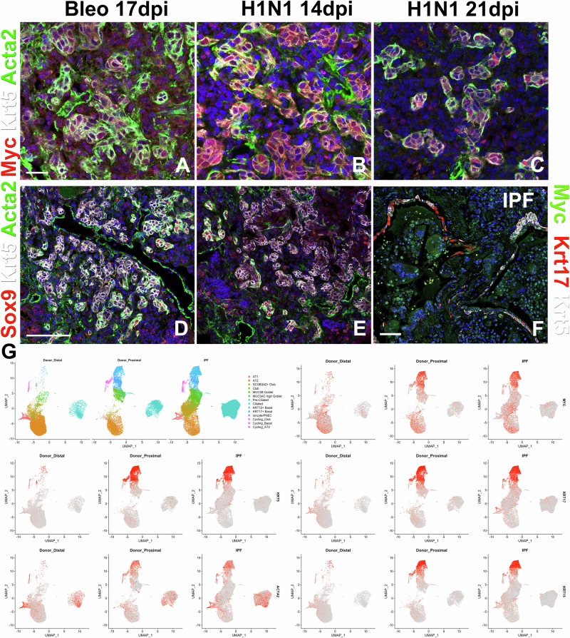
Idiopathic pulmonary fibrosis (IPF) is a progressive respiratory scarring disease arising from the maladaptive differentiation of lung stem cells into bronchial epithelial cells rather than into alveolar type 1 (AT1) cells, which are responsible for gas exchange. Here, we report that healthy lungs maintain their stem cells through tonic Hippo and β-catenin signaling, which promote Yap/Taz degradation and allow for low-level expression of the Wnt target gene Myc. Inactivation of upstream activators of the Hippo pathway in lung stem cells inhibits this tonic β-catenin signaling and Myc expression and promotes their Taz-mediated differentiation into AT1 cells. Vice versa, increased Myc in collaboration with Yap promotes the differentiation of lung stem cells along the basal and myoepithelial-like lineages allowing them to invade and bronchiolize the lung parenchyma in a process reminiscent of submucosal gland development. Our findings indicate that stem cells exhibiting the highest Myc levels become supercompetitors that drive remodeling, whereas loser cells with lower Myc levels terminally differentiate into AT1 cells.


Conflict of interest statement

Competing interests: The authors declare no competing interests.

Figures

Fig. 1
Fig. 1. Club cells compete with BLCs to regenerate vs bronchiolize the lung parenchyma upon catastrophic injury to the lung parenchyma.
Scgb1a1CreERT;mTmG and Scgb1a1CreERT;Mycf/f;mTmG were placed on tamoxifen containing chow at 8 weeks of age for 3 weeks to inactivate Myc and permanently label all Club cells/BASCs and their offspring with GFP. After a 3 week wash-out period, mice were infected with H1N1 influenza virus, and lungs were harvested at 6 weeks post injury. Coimmunostaining for GFP (lineage label), Keratin 5 (Krt5; basal and BLCs), and Keratin 8 (Krt8; BLCs and transitional cells) on Scgb1a1CreERT;mTmG (A) and Scgb1a1CreERT;Mycf/f;mTmG (E) lung sections. Coimmunostaining for GFP (lineage label), surfactant protein C (Sftpc; AT2 cells), Rage (AT1 cells) on Scgb1a1CreERT;mTmG (B, C) and Scgb1a1CreERT;Mycf/f;mTmG (F, G) lung sections. White boxes in B and F are enlarged in (C, G), respectively. D Diagram demonstrating that normal Club cells inhibit BLCs and give rise to alveolar epithelial cells. H Diagram demonstrating that Myc deficient Club cells are outcompeted by Myc sufficient BLCs. Created in BioRender. Warren (2024) https://BioRender.com/ k46i357. I Nanostring nCounter analysis on RNA from Scgb1a1CreERT;mTmG (n = 4) and Scgb1a1CreERT;Mycf/f;mTmG (n = 7) lungs for BLC genes Keratin 17 (p = 0.03, Log2 fold change = −1.87), Sox9 (p = 0.03, Log2 fold change = −1.04), and Krt5 (p = 0.047, Log2 fold change = −1.86). Data are Log2 normalized. J Lineage tracing analysis on immunostaining in B (n = 7), E (n = 2) using Aivia machine learning software (Sftpc/gfp p = 0.003, RAGE/gfp p = 0.6, Sum/gfp p = 0.03). K Hydroxyproline analysis on Cre- controls (n = 17) and Scgb1a1CreERT;Mycf/f;mTmG (n = 20) lungs normalized to control (p = 0.04). L Image analysis of total area of basal cells in control (n = 11) and Scgb1a1CreERT;Mycf/f;mTmG (n = 9) lung sections (p = 0.03). Data are presented as mean values +/− SEM. Scale bar: 250 μm. Two two-tailed unpaired T-test was used to determine significance. F test was used to determine equal variances and unreported F values indicate equal variance. *p < 0.05, **p < 0.01.
Fig. 2
Fig. 2. High expression of Myc in BLCs in IPF.
AE Coimmunostaining for myoepithelial cell markers Myc (AC) or Sox9 (D, E), Krt5, and Acta2 (smooth muscle actin) on honeycomb regions in mouse lungs 17 days after bleomycin (AD) and 14 or 21 days after H1N1 injury (B, C, E). F Coimmunostaining for Myc, Krt17, and Krt5 on honeycomb regions in human IPF tissue. (G) scRNAseq analysis of myoepithelial cell genes Krt5, Krt17, Myc, and Acta2 expression in human IPF vs donor lungs. Uniform Manifold Approximation and Projection (UMAP) of 10x scRNAseq data on human control (Donor Distal) and control including microdissected proximal airways (donor proximal) and IPF distal lungs demonstrating high Myc, Krt5, Krt17, Krt15, Acta2 expression in bronchiolized epithelium in IPF and proximal airway basal cells. Scale bar: 50 μm (AC), 100 μm (DF).
Fig. 3
Fig. 3. Basal cell pod development resembles submucosal gland development.
A, B Nkx2.1Flpo;Acta2-Frt-STOP-FrtCreERT2;mTmG mice were place on tamoxifen containing chow for 3 weeks. Following a 3 week washout period, mice were infected with H1N1. At 6 weeks after injury, left lung lobes and trachea were inflation fixed, embedded in paraffin, and sectioned. Coimmunostaining for myoepithelial cell markers Acta2, Krt5, and lineage label GFP on Nkx2.1Flpo;Acta2-Frt-STOP-FrtCreERT2;mTmG trachea (E) and lung (F). C, D Nkx2.1Flpo;Acta2-Frt-STOP-FrtCreERT2;mTmG mice were intranasally administered H1N1. At 2 weeks after injury mice were placed on tamoxifen containing chow. At 6 weeks after injury, left lung lobes and trachea were inflation fixed, embedded in paraffin, and sectioned. Coimmunostaining for Krt5, Krt8, and GFP on Nkx2.1Flpo;Acta2-Frt-STOP-FrtCreERT2;mTmG lungs. E Trp63DreERT2;Acta2CreERT2;RLTG mice were intranasally administered H1N1 and placed on tamoxifen containing chow at 2 weeks after injury. tdTomato is induced in only Trp63DreERT2 expressing cells and GFP is induced only when both Trp63DreERT2 and Acta2CreERT2 are expressed (myoepithelial cells). Coimmunostaining for RFP (tdtomato) and GFP on lungs at 6 weeks after injury. F Sox9CreERT2;tdTomato mice were intranasally administered H1N1 and placed on tamoxifen containing chow at the injury. Coimmunostaining for RFP (tdTomato) and Krt5 on lungs at 6 weeks after injury. GL Nkx2.1Flpo; Acta2CreERT2;FLTG mice were intranasally administered H1N1 and placed on tamoxifen containing chow at 2 weeks after injury with additional tamoxifen shots at 14 and 16 days after injury. tdTomato is induced in only Nkx2.1Flpo expressing cells and GFP is induced only when both Nkx2.1Flpo and Acta2CreERT2 are expressed (myoepithelial cells). G Coimmunostaining for GFP and Krt5 on lungs at 6 weeks after injury and (H) image analysis using Aivia machine learning software (n = 4 animals). Coimmunostaining for GFP and (I) Dclk1 (tuft cells), (J) Foxj1 (ciliated cells), and (K, L) Scgb1a1 (secretory cells) and Krt5 (BLCs). Data are presented as mean values +/− SEM. Scale bar: 200 μm (A) 100 μm (D, F), 50 μm (B, G), 25 μm (C, E, IL).
Fig. 4
Fig. 4. Airway epithelial Myc is required for bronchiolization.
A Mice were placed on tamoxifen chow for 3 weeks to inactivate Myc and permanently lineage label bronchial epithelial cells and their offspring. Following a 3-week washout period, mice were injured with intratracheal administration of bleomycin (BK) or intranasal administration of H1N1 (LU) and lungs were harvested at 6 weeks post injury. (B, E, L, O) Immunostaining for Krt5 (basal and BLCs) and Acta2 (smooth muscle actin; myofibroblasts) on bleomycin (B, E) and influenza (L, O) injured Sox2CreERT2;mTmG and Sox2CreERT2;Mycf/f;mTmG. (C, F, M, P) Immunostaining for Rage (AT1 cells), GFP (lineage label), and Sftpc (AT2 cells) on Sox2CreERT2;mTmG and Sox2CreERT2;Mycf/f;mTmG. (D, G, N, Q) Models demonstrating that Myc sufficient Club cells inhibit BLCs and give rise to alveolar epithelial cells after bleomycin (D) and influenza (N) injury but Myc insufficient BLCs fail to give rise to basal cell pods (G, Q). Created in BioRender. Warren (2024) https://BioRender.com/j04r129. H Hydroxyproline analysis on bleomycin injured Cre- controls (n = 26) and Sox2CreERT2;Mycf/f (n = 19) (p = 0.04). I Nanostring nCounter analysis on RNA from bleomycin injured Cre- controls (n = 17) and Sox2CreERT2;Mycf/f;mTmG (n = 6) lungs BLC genes (Muc5ac p = 0.01, Muc5b p = 0.003, Krt5 p = 0.046). Data are Log2 normalized. J Lineage tracing analysis on bleomycin injured lungs from immunostaining in (C) (n = 2), (F) (n = 5) using Aivia machine learning software (p = 0.008). K Image analysis of the total area of basal cells in bleomycin injured control (n = 3) and Sox2CreERT2;Mycf/f;mTmG (n = 3) lung sections (p = 0.04). R Hydroxyproline analysis on influenza injured Cre- controls (n = 8) and Sox2CreERT2;Mycf/f (n = 5) (p = 0.004). S qPCR analysis on RNA from influenza injured Cre- controls (n = 12) and Sox2CreERT2;Mycf/f;mTmG (n = 7) lungs for BLC genes (Krt5 (p = 0.04, F = 3.32×10⁻⁹) and Tp63 (p = 0.01, F = 6.29×10⁻⁸)). T Lineage tracing analysis on influenza-injured lungs from immunostaining in M (n = 4), (P) (n = 5) using Aivia machine learning software (p = 0.04). U Image analysis of total area of basal cells in influenza injured control (n = 5) and Sox2CreERT2;Mycf/f;mTmG (n = 13) lung sections (p = 0.007). Data are presented as mean values +/− SEM. Scale bars: 100 µm. Magnification insets are 400x larger. Two tailed unpaired T-test was used to determine significance. F test was used to determine equal variances and unreported F values indicate equal variance. *p < 0.05, **p < 0.01.
Fig. 5
Fig. 5. Myc drives basal cell pod expansion and maintenance after H1N1 injury.
C, K Krt5CreERT2;mTmG and Krt5CreERT2;Mycf/f;mTmG were infected with H1N1 at 8 weeks of age. At 2 weeks after injury, mice were placed on tamoxifen chow to inactivate Myc and permanently label all BLCs and their offspring with GFP and lungs were harvested at 6 (A-H) or 12 (I-Q) weeks post injury. A, B Coimmunostaining for Keratin 8 (Krt8; BLCs and transitional cells), GFP (lineage label), and Keratin5 (Krt5; basal and BLCs), (D, E) and coimmunostaining for Muc5b (mucus-producing secretory cells) and GFP (lineage label) on Krt5CreERT2;mTmG (A, D) and Krt5CreERT2;Mycf/f;mTmG (B, E). F qPCR analysis for Gfp, Krt5, and Muc5b Cre- control (n = 5) and Krt5CreERT2;Mycf/f (n = 5). Values are graphed as ratios (GFP/Krt5 p = 0.046, GFP/Muc5b p = 0.79, Krt5/Muc5b p = 0.006). G Hydroxyproline analysis on Cre- control (n = 10) and Krt5CreERT2;Mycf/f (n = 16) (p = 0.01). H Image analysis of total area of basal cells in influenza injured control (n = 6) and Krt5CreERT2;Mycf/f;mTmG (n = 8) lung sections (p = 0.02). I, J Coimmunostaining for Rage (AT1 cells) and GFP (lineage label) on Krt5CreERT2;mTmG and Krt5CreERT2;Mycf/f;mTmG lungs at 12 weeks post injury with magnification in OQ. L Lineage tracing analysis on influenza injured lungs from immunostaining in I and (J) (n = 3). Area of GFP (p = 0.02), Sftpc (p = 0.03), and Rage (p = 0.02) were determined using Aivia machine learning software. M, N Diagram depicting that Myc deficient basal cells pods are smaller than Myc sufficient basal cell pods and can differentiating into Rage+ AT1 cells at 12 weeks after influenza injury. Created in BioRender. Warren. (2024) https://BioRender.com/ w28w946. Data are presented as mean values +/− SEM. Scale bars: 250 µm (A, B, D, E), 125 µm (I, J), 25 µm (OQ). The two-tailed unpaired T-test was used to determine significance. F test was used to determine equal variances and unreported F values indicate equal variance. *p < 0.05, **p < 0.01.
Fig. 6
Fig. 6. Myc overexpression in Club cells promotes SCMC status and bronchiolization.
A Mice were placed on tamoxifen chow for 3 weeks and following a 3-week washout period, mice were injured with intratracheal administration of bleomycin and placed on doxycycline containing chow to induce Myc overexpression and harvested at 6 weeks post injury. BD, HJ Coimmunostaining for markers associated with fibrosis and bronchiolization (Col1a1, Krt5, and Acta2) on control (BD) and Scgb1a1CreERT;LSL-rtTA;Tet-Myc (HJ). Coimmunostaining for myoepithelial cell-like markers Sox9, Krt5, and Myc on control (E, F) and Scgb1a1CreERT;LSL-rtTA;Tet-Myc (K, L). G, M Diagram demonstrating that Myc sufficient Club cells inhibit basal cells and give rise to alveolar epithelial cells after bleomycin injury while Myc overexpressing Club cells dedifferentiate into basal cells and promote their invasion into the alveolus. Created in BioRender. Warren, R. (2024) https://BioRender.com/ l07p577. N Immunostaining for Muc5b (mucus producing secretory cells), Krt5 (BLCs), and Scgb1a1 (Club cells/BASCs) on Scgb1a1CreERT;LSL-rtTA;Tet-Myc. O Hydroxyproline analysis on Cre- control (n = 31) and Scgb1a1CreERT;LSL-rtTA;Tet-Myc (n = 30) normalized to control (p = 0.003). P Log2 normalized values for RNA expression for BLC (Krt5, Krt17, Scgb3a2, Muc5b) and AT2 cell (Sftpc) genes from NanoString analysis on control (n = 9 Krt5 p = 0.0000008, Krt17 p = 0.000001, Scgb3a2 p = 0.02, Sftpc p = 0.02 F = 0.02, Muc5b p = 0.002), Scgb1a1CreERT;LSL-rtTA;Tet-Myc (n = 5), and Scgb1a1CreERT;Yapf/f;LSL-rtTA;Tet-Myc (n = 8 Krt5 p = 0.0001, Krt17 p = 0.00006, Scgb3a2 p = 0.0007, Sftpc p = 0.04,Muc5b p = 0.02). Values are normalized to control and p values are compared to Scgb1a1CreERT;LSL-rtTA;Tet-Myc. Data are presented as mean values +/− SEM. Scale bars: 500 µm (B, G, L) and 100 µm (CF, HK). Two tailed unpaired T-test was used to determine significance. F test was used to determine equal variances and unreported F values indicate equal variance. *p < 0.05, **p < 0.01, ***p < 0.001, ****p < 0.0001.
Fig. 7
Fig. 7. Cytoplasmic Yap/Taz in airway epithelial cells inhibits bronchiolization and pulmonary fibrosis.
A Mice were placed on tamoxifen chow for 3 weeks to inactivate Yap1 and/or Wwtr1 or Stk3/4 and permanently label all Sox2+ cells and their offspring with GFP. Following a 3 week washout period, mice were injured with intratracheal administration of bleomycin and harvested at 6 weeks post injury. Coimmunostaining for Rage (AT1 cells), GFP (lineage label), and Sftpc (AT2 cells) on Sox2CreERT2;mTmG (B), Sox2CreERT2;Stk3f/f;Stk4f/f;mTmG (D), Sox2CreERT2;Yap1f/f;mTmG (F), Sox2CreERT2;Wwtr1f/f;mTmG (H), and Sox2CreERT2;Yap1f/f;Wwtr1f/f;mTmG (J) lungs. C, E, G, I, K diagrams illustrating the direction of airway epithelial cell differentiation in figures (B, D, F, H, J). Created in BioRender. Warren (2024) https://BioRender.com/ a02e401. L Lineage tracing analysis on bleomycin injured lungs from immunostaining in B (n = 3) and D (n = 4) using Zeiss Zen Intellesis machine learning software to trace (p = 0.57). M Lineage tracing analysis on bleomycin-injured lungs from immunostaining in B (n = 3), F (n = 5, p = 0.92), H (n = 4, p = 0.002), and J (n = 4, p = 0.004) using Aivia machine learning software. N Hydroxyproline analysis on Cre- control, Sox2CreERT2;Yap1f/f;Wwtr1f/f (n = 19, p = 0.04), Sox2CreERT2;Yap1f/f (n = 25, p = 0.01), Sox2CreERT2;Wwtr1f/f (n = 9, p = 0.04), and Sox2CreERT2;Stk3f/f;Stk4f/f (n = 18, p = 0.03). O qPCR analysis for bronchiolization and fibrosis genes (Krt5, p63, Col1a1, Col3a1 and Muc5b) on Cre- control (n = 22), Sox2CreERT2;Yap1f/f;Wwtr1f/f (n = 8, Krt5 p = 0.03, F = 0.00001, p63 p = 0.03, F = 0.01, Col1a1 p = 0.2, Col3a1 p = 0.008, and Muc5b p = 0.05), Sox2CreERT2;Yap1f/f (n = 5, Krt5 p = 0.01, F = 0.00007, p63 p = 0.002, F = 0.005, Col1a1 p = 0.002, Col3a1 p = 0.0005 and Muc5b p = 0.8), Sox2CreERT2;Wwtr1f/f (n = 10, Krt5 p = 0.27, p63 p = 0.05, Col1a1 p = 0.03 F = 0.02, Col3a1 p = 0.004 and Muc5b p = 0.05), and Sox2CreERT2;Stk3f/f;Stk4f/f (n = 10, Krt5 p = 0.009, F = 5.7 × 10⁻⁸, p63 p = 0.01 F = 0.05, Col1a1 p = 0.95, Col3a1 p = 0.16 and Muc5b p = 0.00009 F = 0.003). qPCR for Myc on control (n = 23), Sox2CreERT2;Yap1f/f;Wwtr1f/f (n = 14, p = 0.05) and Sox2CreERT2;Stk3f/f;Stk4f/f (n = 10, p = 0.04). Values are represented as 2-ΔΔCt normalized to Control. Values are normalized to control. Data are presented as mean values +/− SEM. Scale bar: 200 µm. Two tailed unpaired T-test was used to determine significance. F test was used to determine equal variances and unreported F values indicate equal variance. *p < 0.05, **p < 0.01, ***p < 0.001.
Fig. 8
Fig. 8. Taz is required for AT1 cell differentiation.
AP SftpcCreERT2;mTmG, SftpcCreERT2;Nf2f/f;mTmG, SftpcCreERT2;Nf2f/f;Yap1f/f;mTmG, and SftpcCreERT2;Nf2f/f;Wwtr1f/f;mTmG were placed on tamoxifen containing chow at 8 weeks of age for 3 weeks to inactivate Nf2 and/or Yap1 and/or Wwtr1 and permanently lineage label AT2 stem cells and their offspring. At 9 weeks after being placed on normal chow left lung lobes were inflation fixed, embedded in paraffin, and sectioned. Coimmunostaining for GFP (lineage label), Rage (AT1 cells), and Sftpc (AT2 cells). Q Image analysis for lineage labeled AT1 and AT2 cells in A-P using Aivia machine learning software (SftpcCreERT2;mTmG (n = 6), SftpcCreERT2;Nf2f/f;mTmG (n = 8, p = 0.00003), SftpcCreERT2;Nf2f/f;Yap1f/f;mTmG (n = 5, p = 0.0009), and SftpcCreERT2;Nf2f/f;Wwtr1f/f;mTmG (n = 6, p = 0.13)). Data are presented as mean values +/− SEM. Two tailed unpaired T-test was used to determine significance. F test was used to determine equal variances. Scale bar: 200μm. *p < 0.05.
Fig. 9
Fig. 9. Myc and Yap1 increase stem cell competitiveness to drive bronchiolization.
Stem cells exhibiting the highest Myc and Yap1 levels become supercompetitors that drive remodeling, whereas Taz promotes terminal differentiation into AT1 cells by inhibiting Myc. Created in BioRender. Warren, R. (2024) https://BioRender.com/d35d050.

Update of

References

    1. Barkauskas, C. E. & Noble, P. W. Cellular mechanisms of tissue fibrosis. 7. New insights into the cellular mechanisms of pulmonary fibrosis. Am. J. Physiol. Cell Physiol.306, C987–C996 (2014). - PMC - PubMed
    1. King, T. E. Jr., Pardo, A. & Selman, M. Idiopathic pulmonary fibrosis. Lancet378, 1949–1961 (2011). - PubMed
    1. Steele, M. P. & Schwartz, D. A. Molecular mechanisms in progressive idiopathic pulmonary fibrosis. Annu. Rev. Med.64, 265–276 (2013). - PubMed
    1. Parimon, T., Yao, C., Stripp, B. R., Noble, P. W. & Chen, P. Alveolar Epithelial Type II cells as drivers of lung fibrosis in idiopathic pulmonary fibrosis. Int J. Mol. Sci.21, 2269 (2020). - PMC - PubMed
    1. Dressen, A. et al. Analysis of protein-altering variants in telomerase genes and their association with MUC5B common variant status in patients with idiopathic pulmonary fibrosis: a candidate gene sequencing study. Lancet Respir. Med.6, 603–614 (2018). - PMC - PubMed

Publication types

MeSH terms

Substances

Associated data

LinkOut - more resources