. 2024 Dec 5;14(1):30306.
doi: 10.1038/s41598-024-81020-x.

Bacterial products initiation of alpha-synuclein pathology: an in vitro study

Affiliations

Bacterial products initiation of alpha-synuclein pathology: an in vitro study

Octavian Costin Ioghen et al. Sci Rep. .

Abstract

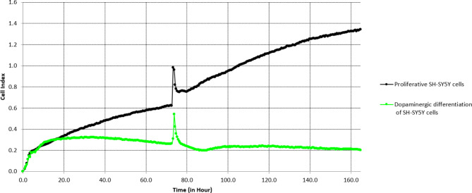
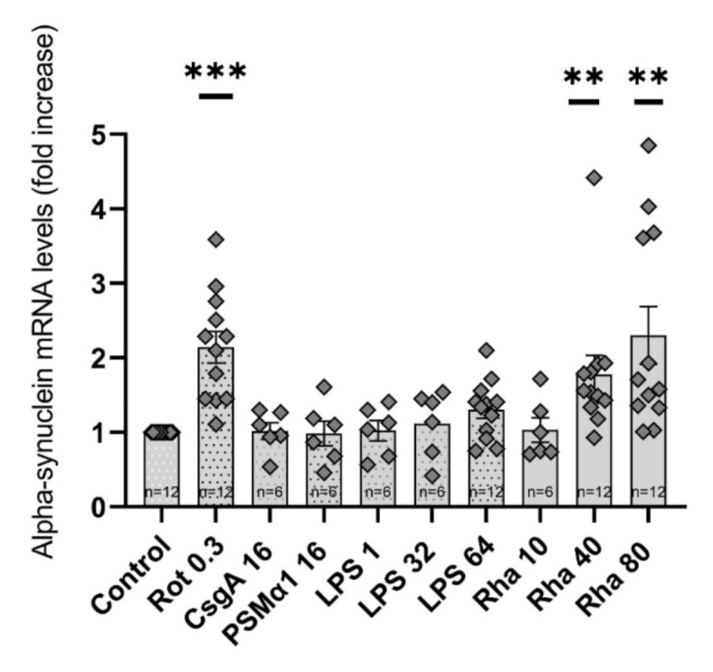
Parkinson's Disease (PD) is a prevalent and escalating neurodegenerative disorder with significant societal implications. Despite being considered a proteinopathy, in which the aggregation of α-synuclein is the main pathological change, the intricacies of PD initiation remain elusive. Recent evidence suggests a potential link between gut microbiota and PD initiation, emphasizing the need to explore the effects of microbiota-derived molecules on neuronal cells. In this study, we exposed dopaminergic-differentiated SH-SY5Y cells to microbial molecules such as lipopolysaccharide (LPS), rhamnolipid, curli CsgA and phenol soluble modulin α-1 (PSMα1). We assessed cellular viability, cytotoxicity, growth curves and α-synuclein levels by performing MTS, LDH, real-time impedance readings, qRT-PCR and Western Blot assays respectively. Statistical analysis revealed that rhamnolipid exhibited concentration-dependent effects, reducing viability and inducing cytotoxicity at higher concentrations, increasing α-synuclein mRNA and protein levels with negative effects on cell morphology and adhesion. Furthermore, LPS exposure also increased α-synuclein levels. Curli CsgA and PSMα-1 showed minimal or no changes. Our findings suggest that microbiota-derived molecules, particularly rhamnolipid and LPS, impact dopaminergic neurons by increasing α-synuclein levels. This study highlights the potential involvement of gut microbiota in initiating the upregulation of α-synuclein that may further initiate PD, indicating the complex interplay between microbiota and neuronal cells.

Keywords: Alpha-synuclein overexpression; Curli CsgA; Lipopolysaccharide; Microbiota; Microbiota-derived molecules; Parkinson’s disease; Phenol soluble modulin α-1; Rhamnolipid.


Conflict of interest statement

Declarations. Competing interests: The authors declare no competing interests.

Figures

Fig. 1
Fig. 1
Stepwise graphical representation of SH-SY5Y dopaminergic differentiation protocol with corresponding phase contrast microscopy images (40× objective). In undifferentiated cells (A), clusters of cells, various shapes of neuronal bodies, especially round and ovoid, short neurites, and few synapses are noticed. At the end of the differentiation protocol (C), significant changes such as uniform distribution of the cells, pyramidal and stellate neuronal bodies, numerous and long neurites, and an increase in the number of synapses are observed.
Fig. 2
Fig. 2
MTS and LDH assays were performed on dopaminergic-differentiated SH-SY5Y cells 48 h after treatment with rhamnolipid (A), lipopolysaccharide (B), curli CsgA (C), PSMα1 (D) and rotenone (E), used as a positive control for the upregulation of α-synuclein expression. The data were expressed as the mean ± SEM of two independent experiments in triplicate (n = 6). Statistical analyses used fold-change calculation as fold increase to control followed by statistical significance testing (One-Way ANOVA followed by Dunnett’s Test), *p < 0.05, **p < 0.01, ***p < 0.001.
Fig. 3
Fig. 3
Real-time cell monitoring of SH-SY5Y cells in the proliferative state using growth medium (supplemented with 10% fetal bovine serum) vs. SH-SY5Y cells that follow the dopaminergic differentiation protocol. The continuous increase in cell index in the proliferative SH-SY5Y cells reflects cell proliferation. In contrast the horizontal curve without significant cell index changes in the cells that follow the dopaminergic differentiation protocol reflects the stop of proliferation. The spikes after 72 h represent the pause of readings while changing the medium. In the cells that follow the dopaminergic differentiation protocol, a differentiation medium was added from the beginning to prevent the detachment of cells from the E-16 plates when changing the medium after 24 h of incubation (excluding day 0 presented in the differentiation protocol). To more accurately depict variations in the cell index throughout the course of the cell monitoring period, normalisation was not carried out. Representative image of triplicate wells.
Fig. 4
Fig. 4
Real-time cellular impedance monitoring of dopaminergic-differentiated SH-SY5Y cells treated with microbiota-derived molecules. Based on the MTS and LDH end-point assay results, several concentrations were chosen to evaluate their effects for 120 h, such as 10 µg/ml, 40 µg/ml, and 80 µg/ml of rhamnolipid (A), 1 µg/ml, 32 µg/ml and 64 µg/ml of LPS (B), 16 µg/ml of curli CsgA (C) and 16 µg/ml of PSMα1 (D). As in the previous experiments, rotenone was chosen as a positive control in concentrations of 0.1 µM, 0.3 µM, and 10 µM. The cell index was normalized to 1 immediately after treatment addition. The impedance measurements presented in the images are from the moment of treatment addition, while the dopaminergic differentiation was not shown here. The images are representative of two independent experiments, each performed in duplicate wells (n = 4).
Fig. 5
Fig. 5
QRT-PCR for α-synuclein mRNA performed 48 h after treatment with bacterial compounds (curli CsgA 16 µg/ml, PSMα1 16 µg/ml, LPS 1 µg/ml, 32 µg/ml and 64 µg/ml, rhamnolipid 10 µg/ml, 40 µg/ml and 80 ug/ml) on dopaminergic-differentiated SH-SY5Y cells. Rotenone was used as a positive control for α-synuclein overexpression. The data are shown as a fold increase to control. The data were normalized to GAPDH. Data are expressed as the mean ± SEM of three independent experiments carried out at least in duplicate. Statistical analyses used fold-change calculation as fold increase to control followed by statistical significance testing (for Rha 40 µg/ml and Rha 80 µg/ml which were non-parametric data we used Kruskal–Wallis test and Dunn’s multiple comparison test, while for the other parametric data we used One-Way ANOVA followed by Dunnett’s Test) *p < 0.05, **p < 0.01, ***p < 0.001.
Fig. 6
Fig. 6
Western Blot for α-synuclein performed 48 h after treatment with bacterial compounds (curli CsgA 16 µg/ml, PSMα1 16 µg/ml, LPS 1 µg/ml, 32 µg/ml and 64 µg/ml, rhamnolipid 10 µg/ml, 40 µg/ml and 80 µg/ml) on dopaminergic-differentiated SH-SY5Y cells. The blots were cut prior to hybridisation with antibodies due to technical reasons to ensure they fit the incubation boxes. Rotenone 0.3 µM was used as a positive control for α-synuclein overexpression. Controls were established as treatment with the vehicle of the stock solution of the bacterial compounds (PBS, DMSO, PBS + 33% glycerol). Quantification of the α-synuclein levels is represented in (A) and immunoblots in (B) (three different blots are inserted in the same picture, each of the blots with the vehicle control of the treatments). The data of intracellular α-synuclein protein were normalized to GAPDH. The data are shown as fold increase to control, each of the controls is considered = 1.0, which explains why in (A) there is only one control bar and in (B) there are three control samples (CTRL Rha LPS, CTRL CsgA PSM, CTRL Rot). The data are shown as a fold increase to control. Data are expressed as the mean ± SEM of four independent experiments carried out in duplicate (for curli CsgA 16 µg/ml and PSMα1 16 µg/ml – three independent experiments carried out in duplicate). Statistical analyses used fold-change calculation as fold increase to control followed by statistical significance testing (One-Way ANOVA followed by Dunnett’s Test), *p < 0.05, **p < 0.01, ***p < 0.001.

References

    1. Dorsey, E. R., Sherer, T., Okun, M. S. & Bloemd, B. R. The emerging evidence of the Parkinson pandemic. J. Parkinsons. Dis.8, S3. 10.3233/JPD-181474 (2018). - PMC - PubMed
    1. Salamon, A., Zádori, D., Szpisjak, L., Klivényi, P. & Vécsei, L. Neuroprotection in Parkinson’s disease: Facts and hopes. J. Neural Transm.127, 821–829. 10.1007/S00702-019-02115-8 (2020). - PMC - PubMed
    1. Marsh, A. P. Molecular mechanisms of proteinopathies across neurodegenerative disease: a review. Neurol. Res. Pract.10.1186/S42466-019-0039-8 (2019). - PMC - PubMed
    1. Tofaris, G. K. Initiation and progression of α-synuclein pathology in Parkinson’s disease. Cell. Mol. Life Sci.10.1007/S00018-022-04240-2 (2022). - PMC - PubMed
    1. Stefanis, L. et al. How is alpha-synuclein cleared from the cell?. J. Neurochem.150, 577–590. 10.1111/JNC.14704 (2019). - PubMed

MeSH terms

LinkOut - more resources