. 2024 Dec 5;14(1):30345.
doi: 10.1038/s41598-024-81509-5.

New insights into the structural role of EMILINs within the human skin microenvironment

Affiliations

New insights into the structural role of EMILINs within the human skin microenvironment

Alvise Schiavinato et al. Sci Rep. .

Abstract

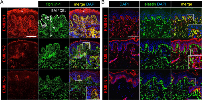
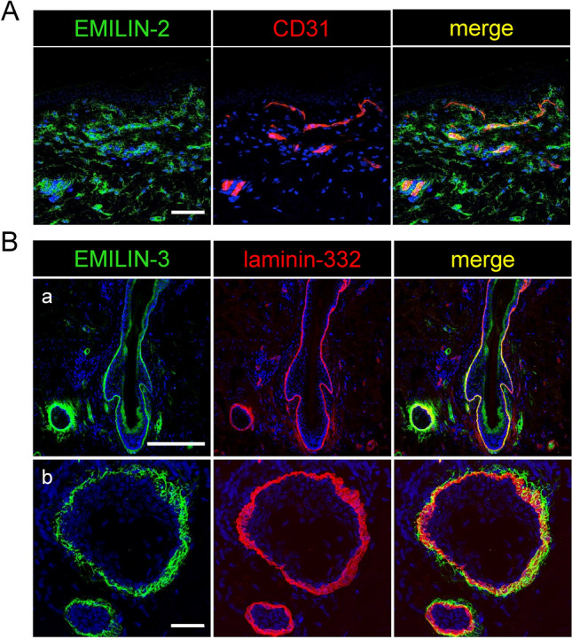
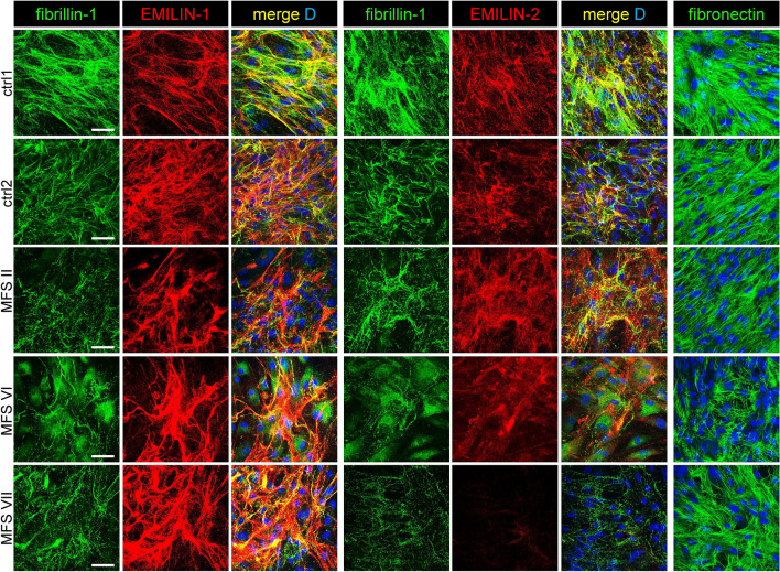
Supramolecular extracellular matrix (ECM) networks play an essential role in skin architecture and function. Elastin microfibril interface-located proteins (EMILINs) comprise a family of three extracellular glycoproteins that serve as essential structural components of the elastin/fibrillin microfibril network, and exert crucial functions in cellular signaling. Little is known about the structural nature of EMILIN networks in skin. We therefore investigated the spatiotemporal localization of EMILIN-1, -2, -3 in human skin induced by aging, UV-exposure, fibrosis, and connective tissue disorder. Confocal immunofluorescence and immunogold electron microscopy analysis identified all EMILINs as components of elastic fibers and elastin-free oxytalan fibers inserted into the basement membrane (BM). Further, our ultrastructural analysis demonstrates cellular contacts of dermally localized EMILIN-1 positive fibers across the BM with the surface of basal keratinocytes. Analysis of skin biopsies and fibroblast cultures from fibrillin-1 deficient Marfan patients revealed that EMILINs require intact fibrillin-1 as deposition scaffold. In patients with scleroderma and the bleomycin-induced murine fibrosis model EMILIN-2 was upregulated. EMILIN-3 localizes to the tips of candelabra-like oxytalan fibers, and to specialized BMs engulfing hair follicles and sebaceous glands. Our data identify EMILINs as important markers to monitor rearrangements of the dermal ECM architecture induced by aging and pathological conditions.

Keywords: Basement membrane; Connective tissue disease; Dermal matrix; Elastic fibers; Extracellular matrix proteins; Human skin; Oxytalan fibers; Skin fibrosis; UV exposure.


Conflict of interest statement

Declarations. Competing interests: The authors declare no competing interests.

Figures

Fig. 1
Fig. 1
Single-cell RNA sequencing analysis reveals expression of EMILIN transcripts in distinct skin cell clusters. Mean expression levels of EMILIN-1, -2, and -3 transcripts (shown in rose, green, and light blue, respectively) across 12 identified skin cell clusters in the analyzed dataset. Cell clusters were characterized based on the expression of specific marker genes (Supplementary Figs. S3 and S4, Supplementary Table S1). Mean expression levels were calculated as the average number of reads per cell for each EMILIN gene across clusters.
Fig. 2
Fig. 2
EMILINs have a specific localization and distribution in human skin as shown by confocal immunofluorescence and electron microscopy. EMILIN-1 and EMILIN-3, but not EMILIN-2, co-localize with fibrillin-1 and elastin. EMILIN-1 is distributed throughout the entire dermis, whereas EMILIN-3 is predominantly located at the tips of oxytalan fibers in close proximity to the basement membrane. (A) Co-localization of EMILINs with fibrillin-1 in dermis (parieto-occipital region, male donor, 15 years). Scale bar: 250 μm. Asterisks indicate nonspecific epidermal staining observed with the anti-EMILIN-1 antibody. (B) Co-localization of EMILINs with elastin (gluteal region, male donor, 29 years). Scale bar: 75 μm. Dotted white line indicates location of BM and DEJ. BM basement membrane, DEJ dermal epidermal junction, HF hair follicle, PD papillary dermis, RD reticular dermis.
Fig. 3
Fig. 3
Localization of EMILIN-2 and -3 to specialized microenvironments of the skin. EMILIN-2 shows a perivascular localization in human dermis while EMILIN-3 was detected in skin appendages. (A) EMILIN-2 was co-stained with the endothelial cell marker CD31 and detected by confocal immunofluorescence microscopy on skin sections from the anterior axillary line of a 78-year old male donor. Scale bar: 75 μm. (B) EMILIN-3 and laminin-332 were co-stained on human skin sections and signals for both proteins were visualized by confocal immunofluorescence microscopy. Detected signals for EMILIN-3 localize to laminin-332 positive basement membranes surrounding (a) hair follicles and (b) sebaceous glands. Scale bars: 250 μm and 35 μm.
Fig. 4
Fig. 4
Specific localization of EMILIN-1 and -3 at the DEJ. EMILIN-1 and EMILIN-3 appear to localize within the BM. (A) Confocal immunofluorescence microscopy showing co-localization of EMILIN-1 and EMILIN-3 with laminin-332 and collagen VII at the DEJ (anterior axillary line, male donor, 78 years). White arrows point to EMILIN-1 signals beyond the BM. Scale bar: 75 μm. (B) Immunogold localization of EMILIN-1 and -3 in 33 year- (cheek), 19 month- (digit), and 23 year-old neurofibroma skin showing BM duplication. Scale bar: 500 nm. AF: anchoring fibrils, BM: basement membrane.
Fig. 5
Fig. 5
Altered localization of EMILINs in aged and sun-exposed skin. Ageing and photoageing lead to fragmentation of EMILIN-1 and EMILIN-3 fibers. (A) Localization of EMILINs in skin from donors of increasing age. (B) Localization of EMILINs in sun-exposed skin. Donor age and localization of skin biopsy are indicated. (C) (left) Localization of EMILIN-1 at the DEJ. Elongated EMILIN-1 fibers inserting from the dermis into the BM are not yet prominent at 4 month of age. (middle) In aged but sun-protected skin EMILIN-1 positive fibers appear to be often fragmented and show especially at the position of oxytalan fibers a punctate pattern. (right) In aged and sun-exposed skin elongated EMILIN-1 positive fibers are absent from the DEJ. Overall EMILIN-1 distribution appears to be more diffuse without a clear indication of linear deposition into discrete fibers. Images shown in left and right represent three-fold magnifications of the areas marked by white dashed boxes shown in (A) and (B). DAPI (D) marks nuclei. Ages of donors and anatomical localizations of taken biopsies are indicated. Scale bars: 250 μm.
Fig. 6
Fig. 6
The presence of mutant fibrillin-1 affects the integrity of EMILIN networks in skin of patients with MFS. Fibrillin-1 deficiency negatively impacts the structural integrity of EMILIN-1, -2, and -3 fiber networks. EMILINs were detected together with fibrillin-1 in the dermis of three MFS patients (I-III) by confocal immunofluorescence microscopy. The appearance of non-colocalizing dotted signals suggests that degradation of fibrillin fibers due to fbrillin-1 deficiency leads to dissociation of EMILINs from this network. (A) Colocalization studies of EMILIN-1, -2, and -3 with fibrillin-1 showed impaired EMILIN network integrity when fibrillin-1 is deficient. The age of each patient is indicated: (III) 48 years, (VI) 20 years (V) 20 years. Merged images on the right represent a 5.7-fold magnification of the marked area. Scale bar: 250 μm. (B) Co-localization of EMILIN-1 and EMILIN-3 with fibrillin-1 in dermis of MFS patient (58 years) at the DEJ. Right: 3.6-fold magnification of marked area. Scale bar: 25 μm. Arrows point to punctate signals of EMILINs colocalizing (closed arrows) and non-colocalizing (open arrows) with fibrillin-1.
Fig. 7
Fig. 7
Fibrillin-1 deficiency affects EMILIN-1 and -2 fiber assembly in cultures of primary fibroblasts of MFS. ECM analysis of MFS dermal fibroblasts showed an abnormal ECM deposition of EMILINs when fibrillin-1 assembly is deficient. EMILIN-1 formed abnormally thick fibers that co-localized with the only sparsely formed fibrillin-1 fiber network. However, EMILIN-2 was only deposited as amorphous material with no evidence of fibril formation, while fibronectin assembly was not majorly affected in all analyzed cultures. Dermal fibroblasts were plated at confluence and cultured for one week followed by analysis of the ECM by confocal immunofluorescence microscopy using antibodies specific for fibrillin-1, EMILIN-1, -2, and fibronectin. Scale bar: 50 μm.
Fig. 8
Fig. 8
Altered localization of EMILINs within skin lesions from scleroderma patients. Confocal immunofluorescence analysis shows increased EMILIN-2 deposition in fibrotic lesions from scleroderma patients. In addition, a minor increase in EMILIN-1 signals was observed alongside a partial disruption of its typical fibrillar arrangement, whereas EMILIN-3 fibrils at the DEJ were predominantly absent in the affected areas. Skin biopsies from scleroderma patients were taken from the following anatomical locations: ctrl: anterior axillary line, SCL I: left lower arm, SCL II: right lower arm/dorsal, SCL III: left lower arm, SCL IV: left lower arm. Clinical information regarding the biopsies is summarized in supplementary Table S1. Donor age is indicated. D: DAPI. Scale bars: 250 μm.
Fig. 9
Fig. 9
EMILIN-2 is upregulated in experimental mouse model of bleomycin-induced skin fibrosis. Increased EMILIN-2 protein deposition and synthesis upon bleomycin application in murine skin. (A) Deposition of EMILINs in mouse skin after 4 weeks of intradermal bleomycin (bleo) or 0.9% NaCl injection. (B) EMILIN-2 immunoblot analysis of skin proteins isolated during the course of bleomycin or NaCl injection. Donor age is indicated. DAPI (D) marks nuclei. Scale bar: 50 μm.

References

    1. Danussi, C. et al. EMILIN1-alpha4/alpha9 integrin interaction inhibits dermal fibroblast and keratinocyte proliferation. J. Cell Biol.195, 131–145. 10.1083/jcb.201008013 (2011). - PMC - PubMed
    1. Spessotto, P. et al. beta 1 Integrin-dependent cell adhesion to EMILIN-1 is mediated by the gC1q domain. J. Biol. Chem.278, 6160–6167. 10.1074/jbc.M208322200 (2003). - PubMed
    1. Spessotto, P. et al. EMILIN1 represents a major stromal element determining human trophoblast invasion of the uterine wall. J. Cell Sci.119, 4574–4584. 10.1242/jcs.03232 (2006). - PubMed
    1. Colombatti, A. et al. The EMILIN/Multimerin family. Front. Immunol.2, 93. 10.3389/fimmu.2011.00093 (2011). - PMC - PubMed
    1. Schiavinato, A. et al. EMILIN-3, peculiar member of elastin microfibril interface-located protein (EMILIN) family, has distinct expression pattern, forms oligomeric assemblies, and serves as transforming growth factor beta (TGF-beta) antagonist. J. Biol. Chem.287, 11498–11515. 10.1074/jbc.M111.303578 (2012). - PMC - PubMed

LinkOut - more resources