. 2024 Dec 5;22(1):588.
doi: 10.1186/s12964-024-01964-5.

Extrajunctional CLDN10 cooperates with LAT1 and accelerates clear cell renal cell carcinoma progression

Affiliations

Extrajunctional CLDN10 cooperates with LAT1 and accelerates clear cell renal cell carcinoma progression

Akifumi Onagi et al. Cell Commun Signal. .

Abstract

Background & aims: In addition to their adhesive properties, cell adhesion molecules such as claudins (CLDNs) exhibit signaling ability to organize diverse cellular events. Although the CLDN-adhesion signaling stimulates or inhibits cancer progression, the underlying mechanism remains poorly established. Here, we verified whether and how CLDN10 promotes intracellular signals and malignant phenotypes in clear cell renal cell carcinoma (ccRCC).

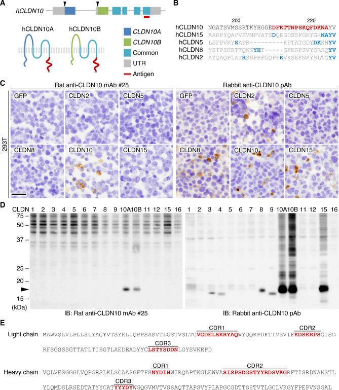
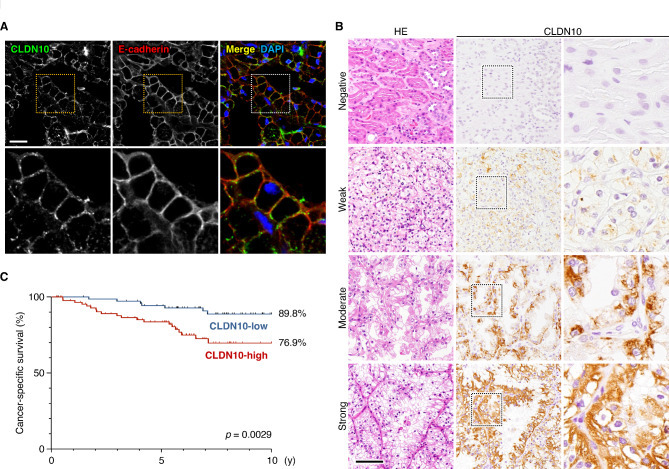
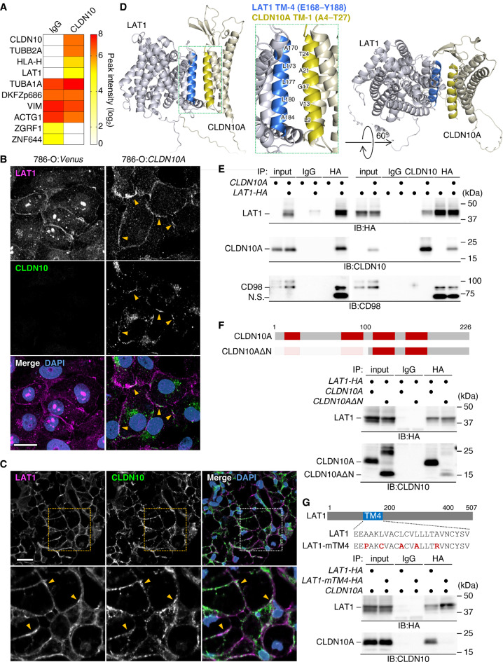
Methods: We developed a novel monoclonal antibody that specifically recognizes CLDN10. By immunohistochemistry using this antibody, the clinicopathological significance of aberrant CLDN10 expression in 165 ccRCC patients was determined. We next generated the ccRCC cells (786-O, ACHN, and OS-RC-2) expressing CLDN10, and compared their phenotypes with those of control cells. Immunoprecipitation-mass spectrometry was used to identify a CLDN10-interacting protein, followed by evaluation of its association with CLDN10 and loss-of-functions in ccRCC cells.

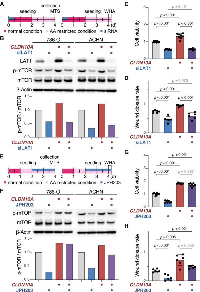
Results: High CLDN10 expression predicted poor outcome in ccRCC patients and represented an independent prognostic marker for cancer-specific survival. Cell surface CLDN10 promoted cell viability, proliferation, and migration of ccRCC cells, as well as their tumor growth. CLDN10 also activated mTOR signaling and expression of downstream targets, including MYC target genes. Notably, we found that CLDN10 forms a complex with an amino acid transporter, LAT1, and that CLDN10-LAT1 signaling facilitates malignant phenotypes in ccRCC cells. Structural prediction and immunoprecipitation analysis results strongly suggest an interaction between CLDN10-TM1 (transmembrane domain 1) and LAT1-TM4.

Conclusions: We conclude that CLDN10-LAT1 signaling drives ccRCC progression. Taken together with our previous findings on CLDN-Src-family kinases signaling, CLDNs propagate distinct intracellular signals depending on their association with different binding partners.

Keywords: CD98; Cell adhesion signal; Claudin; JPH203; Renal cancer; Tight junction; mTOR.


Conflict of interest statement

Declarations. Ethics approval and consent to participate: All animal experiments conformed to the National Health Guide for the Care and Use of Laboratory Animals and were approved by the Animal Experiments Committee of Fukushima Medical University (approval code, 067 and 2022097; approval date, Jul 1, 2017 and Oct 27, 2022). The human studies were approved by the Research Ethics Committee of Fukushima Medical University (approval code, 2021-098; approval date, 29 June 2021) and were conducted in accordance with the 1964 Helsinki Declaration or comparable standards. Consent for publication: Not applicable. Competing interests: The authors declare no competing interests.

Figures

Fig. 1
Fig. 1
Establishment and characterization of rat anti-human CLDN10 mAbs. (A) Gene structure and protein topology of hCLDN10. Arrowheads reveal transcription start sites, and the antigen region is indicated in red. UTR, untranslated region. (B) Amino acid sequences of the C-terminal cytoplasmic domains of hCLDN10 and the corresponding regions of the closely related hCLDN members. Conserved amino acids and the antigen region are shown in light blue and red, respectively. (C, D) Immunohistochemical and Western blot analyses showing the specificity of the anti-hCLDN10 mAb (clone #25). 293T cells were transfected with the indicated expression vectors and subjected to analyses using the indicated anti-CLDN10 Abs. Scale bar, 50 μm. (E) The complementarity-determining regions (CDRs) of the anti-hCLDN10 mAb (clone #25)
Fig. 2
Fig. 2
High CLDN10 expression is associated with poor prognosis in ccRCC patients. (A) Confocal images of the indicated proteins in ccRCC tissues. Scale bar, 20 μm. (B) Representative immunohistological images showing negative/weak/moderate/strong signal intensity for CLDN10 expression in ccRCC tissues. HE, hematoxylin-eosin. Scale bar, 200 μm. (C) Cancer-specific survival in the CLDN10-low and CLDN10-high expression groups of ccRCC patients
Fig. 3
Fig. 3
Cell surface CLDN10 promotes malignant behavior of the ccRCC cell line 786-O. (A) The construct of the hCLDN10A expression vector. EF-1α, elongation factor-1α; IRES, internal ribosome entry site. (B) Western blot for the indicated proteins in the revealed 786-O cells. (C) Confocal images of CLDN10 in the indicated cells. Scale bar, 20 μm. (D-F) Quantitative cell viability, proliferation, and wound healing assays in the indicated cells. The levels are plotted and shown in the histograms (mean ± SD). n = 8, 4, and 18 for D, E, and F, respectively. (G) GSEA showing the enrichment of mTOR and PI3K/AKT/mTOR signalings, as well as gene sets of MYC targets in 786-O:CLDN10A cells
Fig. 4
Fig. 4
CLDN10 forms a complex with LAT1 in 786-O:CLDN10A and 293T + CLDN10A cells. (A) Immunoprecipitation-mass spectrometry (IP-MS) analysis revealing that CLDN10, TUBB2A, HLA-H, and LAT1 are potentially associated with CLDN10 in 786-O:CLDN10A cells. (B, C) Confocal images of the indicated proteins in the revealed 786-O cells (B) and ccRCC tissues (C). Yellow arrowheads show colocalization of CLDN10A and LAT1 on cell membranes. Scale bars, 20 μm. (D) Three-dimensional structural analysis using PyMOL indicating that the first transmembrane domain of CLDN10A (CLDN10A -TM1) binds to LAT1-TM4. (E) IP-IB analysis showing the CLDN10A/LAT1 and CD98/LAT1 complexes in 293T cells. (F) IP-IB analysis revealing that the association between CLDN10A∆N and LAT1 is markedly decreased in 293T cells. Transmembrane domains are shown in red. (G) IP-IB analysis indicating that LAT1-mTM4 hardly form a complex with CLDN10A in 293T cells. The substituted amino acids are shown in red. IP, immunoprecipitation; IB, immunoblot; HA, hemagglutinin
Fig. 5
Fig. 5
CLDN10–LAT1 signaling promotes ccRCC progression. (A, E) Culture conditions for 786-O and ACHN cells (A for B–D, and E for F–H). Cells were grown in the presence or absence of LAT1 siRNA (both #1 and #2) or JPH203. (B, F) Western blot for the indicated proteins in the revealed 786-O and ACHN cells. The protein levels are normalized to the rehybridized β-actin levels, and the relative levels are shown in the histograms. (C, D, G, H) Quantitative cell viability and wound healing assays of the indicated 786-O cells. The levels are plotted and shown in the histograms (mean ± SD). n = 8 for C and G, and n = 6 for D and H
Fig. 6
Fig. 6
A schema showing the expression and biological functions of CLDN10 in normal kidney and ccRCC tissues. CLDN10 is exclusively localized at tight junctions of TAL and proximal tubule epithelial cells in normal kidney tissues, and contributes to paracellular transport or barrier. On the other hand, CLDN10 is distributed on the whole cell membranes of CLDN-high ccRCC cells, and cooperates with LAT1, resulting in tumor progression. ccRCC, clear cell renal cell carcinoma; PCT, proximal convoluted tubule; PST, proximal straight tubule; TAL, thick ascending limb; T, tumor

References

    1. Bray F, Laversanne M, Sung H, Ferlay J, Siegel RL, Soerjomataram I, et al. Global cancer statistics 2022: GLOBOCAN estimates of incidence and mortality worldwide for 36 cancers in 185 countries. CA Cancer J Clin. 2024;74:229–63. - PubMed
    1. Woods P. Kidney cancer statistics [Internet]. WCRF International. 2022 [cited 2024 Apr 10]. http://www.wcrf.org/cancer-trends/kidney-cancer-statistics/
    1. Capitanio U, Montorsi F. Renal cancer. Lancet. 2016;387:894–906. - PubMed
    1. Stewart GD, Klatte T, Cosmai L, Bex A, Lamb BW, Moch H, et al. The multispeciality approach to the management of localised kidney cancer. Lancet. 2022;400:523–34. - PubMed
    1. Hsieh JJ, Purdue MP, Signoretti S, Swanton C, Albiges L, Schmidinger M, et al. Renal cell carcinoma. Nat Rev Dis Primers. 2017;3:17009. - PMC - PubMed

MeSH terms