. 2024 Dec 5;23(1):368.
doi: 10.1186/s12936-024-05202-8.

Human liver organoids are susceptible to Plasmodium vivax infection

Affiliations

Human liver organoids are susceptible to Plasmodium vivax infection

Norapat Nitaramorn et al. Malar J. .

Erratum in

Abstract

Background: The eradication of Plasmodium vivax malaria is complicated due to the presence of hypnozoites, the hidden dormant form of the parasite that is present in the liver. Currently available drug regimens are effective at killing hypnozoites but cause side effects and are difficult to administer. Studies testing drugs for liver-stage malaria remain rare and mainly rely on the use of cancerous or immortalized hepatic cells and primary hepatocytes.

Methods: Organoids were used as platform to model liver-stage vivax malaria. Hepatic endoderm cells, endothelial progenitor cells and mesenchymal cells were generated from human induced pluripotent stem cells and self-assembled into liver organoids on top of Matrigel layer. Liver characteristic and maturity were examined through genes and proteins expression of liver markers, and liver functional tests before infected with Plasmodium vivax sporozoites. The infection was then verified by the detection of parasitophorous vacuole membrane proteins, Upregulated in Infectious Sporozoite 4 (UIS4), and blood-stage infection following co-culture with human reticulocytes.

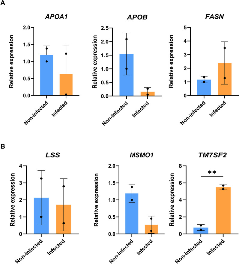
Results: Generated liver organoids showed upregulation of liver specific transcripts including hepatic nuclear factor 4A (HNF4A), alpha-fetoprotein (AFP), and albumin (ALB) which also confirmed by the protein expression. Furthermore, those organoids resembled mature hepatocytes in terms of albumin secretion, fat and glycogen storage and cytochrome activity. Following invasion of P. vivax sporozoites, PvUIS4 was detected and the hepatic merozoites could develop into ring-stage and early trophozoites in human reticulocytes. Moreover, differential expression patterns of genes involved in lipid and cholesterol synthesis were also detected.

Conclusions: Stem cell-derived liver organoids resemble mature liver cells in terms of liver functions and are susceptible to infection with P. vivax sporozoites, paving the way for studies on the mechanism of hypnozoite formation and testing of possible hypnozoitocidal drugs.

Keywords: Plasmodium vivax; Disease model; Hepatocyte; Induced pluripotent stem cells; Liver organoid; Malaria.


Conflict of interest statement

Declarations. Ethics approval and and consent to participate: To generate liver organoids in this study, a human iPSC line, MUSIi001-A, established in a previous study [26], was used. Human erythrocytes were obtained from the peripheral venous blood of the subjects after obtaining informed consent. Protocols related to iPSCs, blood collection and cell preparation were approved by the Human Research Protection Unit, Faculty of Medicine Siriraj Hospital, Mahidol University (COA no. Si 953/2023, 924/2566(IRB3), and 400/2567(Exempt)). P. vivax sporozoites were used in accordance with biosafety guidelines, and the corresponding protocols were approved by the Faculty of Medicine Siriraj Hospital, Mahidol University (approval no. SI 2024–003). Consent for publications: Not applicable. Competing interests: The authors declare that they have no competing interests.

Figures

Fig. 1
Fig. 1
iPSC differentiation and liver organoid formation. A Schematic diagram showing liver organoid formation in a 3D culture system. The maturity of the liver organoids was assessed from day 33 onward. Created in BioRender. https://BioRender.com/e26k372B Microscopy images of iPSC-derived hepatic endoderm cells (HEs), endothelial cells (ECs), and septum transversum mesenchyme (27) cells on day 8 post-differentiation. Scale bars = 100 µm. C Levels of transcripts specific to each cell type, including α-fetoprotein (AFP) and hepatic nuclear factor 4 α (HNF4α) for hepatic endoderm cells, vascular endothelial cadherin (VE-cadherin) and platelet endothelial cell adhesion molecule (PECAM1) for endothelial cells, and endoglin (ENG) and Thy-1 cell surface antigen (THY1) for STM cells. The data are presented as the means ± standard deviations (SDs) (n = 2, technical replicates) and were statistically analysed using Student’s t test. Significant differences are indicated by *p < 0.05, ***p < 0.001, and ****p < 0.0001. D Microscopy images of liver organoids at 24 h, 48 h, 72 h, 33 days, and 69 days post-seeding. Scale bars = 100 µm
Fig. 2
Fig. 2
Liver organoid characterization. A Gene expression profile of liver organoids (LOs) on day 33 and day 69 showing the mature hepatocyte markers α-fetoprotein (AFP), albumin (ALB), hepatic nuclear factor 4 α (HNF4α), and cytochrome P3A4 (CYP3A4), as well as host cell receptors for Plasmodium sporozoites (CD81, SR-BI). Relative expression levels were calculated using the 2−ΔΔCT method. The data are presented as the means ± SDs (n = 2, technical replicates of the iPSCs and HepG2 cells; n = 3, biological replicates of the LOs) and were statistically analysed using Student’s t test. Significant differences are indicated by *p < 0.05, **p < 0.01, and ***p < 0.001. B Confocal microscopic observation of AFP, ALB, CYP3A4, and SR-BI. Scale bars = 100 µm. C Total amount of albumin (µg/mL) secreted into the culture medium of day 33 liver organoids compared with iPSCs and HepGe2 cells (***p < 0.001, Student’s t-test). D Gene expression of the cytochrome P2D6 transcript (CYP2D6). The data are presented as the means ± SDs (n = 2, technical replicates of the iPSCs and HepG2 cells; n = 3, biological replicates of the 33-day LOs and 69-day LOs). E Drug metabolism of Tafenoquine in liver organoids. The amount of tafenoquine (ng/mL) in the culture medium was measured at different time points, including 4, 24, 48, 96, and 144 h after addition of the drug. The control group included drugs without liver organoids. F, G Lipid and glycogen staining of day 33 liver organoids. Representative images are shown. Scale bars = 10 µm for lipid staining and 50 µm for glycogen staining
Fig. 3
Fig. 3
P. vivax infection in liver organoids. A Schematic diagram showing P. vivax infection in liver organoids. Liver organoids collected on day 33 were used for infection. Created in BioRender. https://BioRender.com/r42z460B Sporozoite gliding assay showed the time lapse images of sporozoite captured by holotomography techniques. Red pointers indicated same sporozoite in different positions at different time points. Scale bar = 2 µm
Fig. 4
Fig. 4
Liver-stage and blood-stage infection of P. vivax in liver organoids. A Confocal microscopic observation of P. vivax UIS4 in the liver organoids. Sporozoites were inoculated with the LOs. At day 6 post-inoculations, the LOs were collected and subjected to prepare a tissue section for immunofluorescence. Under the confocal images, nuclei were stained with DAPI (blue), while P. vivax UIS4 was detected using anti-PvUIS4 (green). Bright field (BF) images show the edge of the cell layer. Scale bar = 10 µm. B Representative microscopic images of Plasmodium-parasitized reticulocytes. Human reticulocytes were added into the LOs that were inoculated with P. vivax sporozoite for 6 days. At 24 h post reticulocyte adding, thin blood smears were prepared and stained with Giemsa. Scale bar = 10 µm
Fig. 5
Fig. 5
Gene expression of lipogenesis-related genes in infected and noninfected liver organoids. A Gene expression of transcripts related to lipid metabolism, including apolipoprotein A1 (APOA1), apolipoprotein B (APOB), and fatty acid synthase (FASN). B Gene expression of transcripts related to cholesterol metabolism, including lanosterol synthase (LSS), methyl sterol monooxygenase 1 (MSMO1) and transmembrane 7 superfamily 2 (TM7SF2). Human GAPDH was used as an endogenous control. Relative expression levels were calculated using the 2−ΔΔCT method. The data are presented as the means ± SDs (n = 2, biological replicate) and were statistically analysed using Student’s t test. Significant differences are indicated by **p < 0.01

References

    1. Venugopal K, Hentzschel F, Valkiūnas G, Marti M. Plasmodium asexual growth and sexual development in the haematopoietic niche of the host. Nat Rev Microbiol. 2020;18:177–89. - PMC - PubMed
    1. Hovlid ML, Winzeler EA. Phenotypic screens in antimalarial drug discovery. Trends Parasitol. 2016;32:697–707. - PMC - PubMed
    1. Siqueira-Neto JL, Wicht KJ, Chibale K, Burrows JN, Fidock DA, Winzeler EA. Antimalarial drug discovery: progress and approaches. Nat Rev Drug Discov. 2023;22:807–26. - PMC - PubMed
    1. Tripura R, Peto TJ, Chalk J, Lee SJ, Sirithiranont P, Nguon C, et al. Persistent Plasmodium falciparum and Plasmodium vivax infections in a western Cambodian population: implications for prevention, treatment and elimination strategies. Malar J. 2016;15(1):181. - PMC - PubMed
    1. McKenzie FE, Jeffery GM, Collins WE. Plasmodium vivax blood-stage dynamics. J Parasitol. 2002;88:521–35. - PMC - PubMed

LinkOut - more resources