. 2024 Dec 5;22(1):589.
doi: 10.1186/s12964-024-01969-0.

A fully human IgG1 antibody targeting connexin 32 extracellular domain blocks CMTX1 hemichannel dysfunction in an in vitro model

Affiliations

A fully human IgG1 antibody targeting connexin 32 extracellular domain blocks CMTX1 hemichannel dysfunction in an in vitro model

Abraham Tettey-Matey et al. Cell Commun Signal. .

Erratum in

Abstract

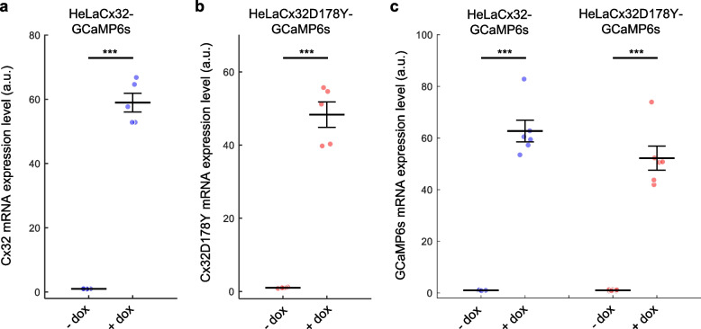
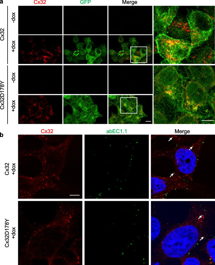
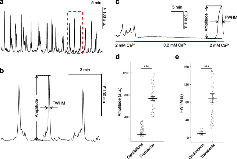
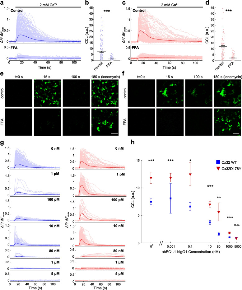
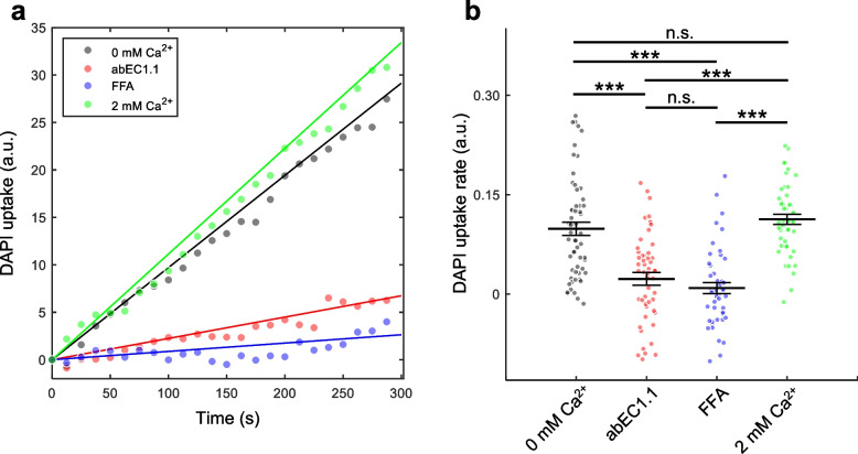
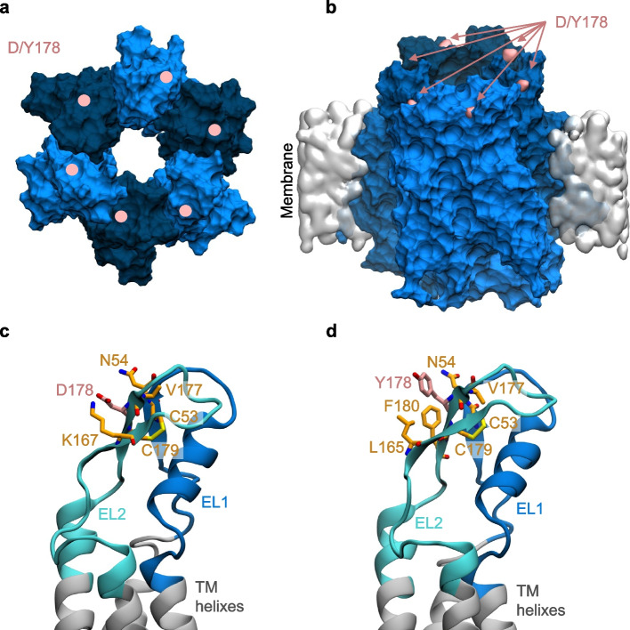
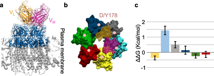
Connexins (Cxs) are fundamental in cell-cell communication, functioning as gap junction channels (GJCs) that facilitate solute exchange between adjacent cells and as hemichannels (HCs) that mediate solute exchange between the cytoplasm and the extracellular environment. Mutations in the GJB1 gene, which encodes Cx32, lead to X-linked Charcot-Marie-Tooth type 1 (CMTX1), a rare hereditary demyelinating disorder of the peripheral nervous system (PNS) without an effective cure or treatment. In Schwann cells, Cx32 HCs are thought to play a role in myelination by enhancing intracellular and intercellular Ca2+ signaling, which is crucial for proper PNS myelination. Single-point mutations (p.S85C, p.D178Y, p.F235C) generate pathological Cx32 HCs characterized by increased permeability ("leaky") or excessive activity ("hyperactive").We investigated the effects of abEC1.1-hIgG1, a fully human immunoglobulin G1 (hIgG1) monoclonal antibody, on wild-type (WT) and mutant Cx32D178Y HCs. Using HeLa DH cells conditionally co-expressing Cx and a genetically encoded Ca2+ biosensor (GCaMP6s), we demonstrated that mutant HCs facilitated 58% greater Ca2+ uptake in response to elevated extracellular Ca2+ concentrations ([Ca2+]ex) compared to WT HCs. abEC1.1-hIgG1 dose-dependently inhibited Ca2+ uptake, achieving a 50% inhibitory concentration (EC50) of ~ 10 nM for WT HCs and ~ 80 nM for mutant HCs. Additionally, the antibody suppressed DAPI uptake and ATP release. An atomistic computational model revealed that serine 56 (S56) of the antibody interacts with aspartate 178 (D178) of WT Cx32 HCs, contributing to binding affinity. Despite the p.D178Y mutation weakening this interaction, the antibody maintained binding to the mutant HC epitope at sub-micromolar concentrations.In conclusion, our study shows that abEC1.1-hIgG1 effectively inhibits both WT and mutant Cx32 HCs, highlighting its potential as a therapeutic approach for CMTX1. These findings expand the antibody's applicability for treating diseases associated with Cx HCs and inform the rational design of next-generation antibodies with enhanced affinity and efficacy against mutant HCs.

Keywords: ATP release; Ca2+ uptake; Charcot–Marie–Tooth diseases; Connexons; Cx32; Dye uptake; Molecular dynamics; Monoclonal antibodies.

Plain language summary

Connexins (Cxs) are proteins essential for communication between cells, forming gap junction channels (GJCs) that connect neighboring cells and hemichannels (HCs) that link the inside of cells to their surroundings. Mutations in the GJB1 gene, which makes Cx32, cause X-linked Charcot-Marie-Tooth type 1 (CMTX1), a rare nerve disorder that currently has no cure. Our research studied the effects of a specially designed antibody, abEC1.1-hIgG1, on both normal and mutated Cx32 HCs using a well-established cell model for studying these channels. We discovered that the antibody significantly reduced the movement of calcium ions (Ca2+), which are vital for nerve health, in both normal and mutated channels. Further analysis showed that the antibody’s success might be due to how it binds to certain areas of the channels. Our findings suggest that abEC1.1-hIgG1 could be developed into a treatment for CMTX1 and similar conditions. Moreover, this research could help in creating even more effective antibodies for targeting faulty hemichannels.


Conflict of interest statement

Declarations. Ethics approval and consent to participate: Not applicable. Consent for publication: This study does not contain any individual participant data. No consent to publish is required. Competing interests: G. Yang, F. Zonta and F. Mammano report a patent family: “WO2017128880 – Fully human antibody specifically inhibiting connexin 26”, Inventors: Qu Z, Yang G, Mammano F, Zonta F, International application number: PCT/CN2016/109847, granted to ShanghaiTech University; and a patent family “WO2020237491 – Composition and Methods to treat Ectodermal Dysplasia 2, Clouston Type”, Inventors: Mammano F, Yang G, Zonta F, International Application No.: PCT/CN2019/088689, pending to ShanghaiTech University. The other authors declare no competing interests.

Figures

Fig. 1
Fig. 1
Validation of Tet-On bicistronic lentiviral vectors. Dot plots showing mean ± s.e.m. of mRNA expression levels measured by qPCR in cells exposed to doxycycline for 48 h (+ dox) or not (− dox): a Cx32, b Cx32D178Y, and c GCaMP6s. Asterisks indicate statistical significance (p-value, Kruskal–Wallis (KW) test); n > 3 independent cell cultures per condition
Fig. 2
Fig. 2
Immunofluorescence characterization of Cx expression. a Representative confocal images of fixed and permeabilized cells expressing human WT Cx32 (upper panels) or mutant Cx32D178Y (lower panels), either exposed to doxycycline for 48 h (+ dox) or not exposed (− dox). Cells were labeled with anti-Cx32 (red) and anti-GFP (green) antibodies (the GFP epitope recognized by the antibody is also present in GCaMP6s). Scale bar: 10 µm. Right panels: higher magnification of boxed areas, showing fluorescent puncta at cell–cell contacts; scale bar: 10 µm. b Representative confocal images showing immunofluorescence labeling with abEC1.1 and anti-Cx32. Live cells expressing human WT Cx32 (upper panels) or mutant Cx32D178Y (lower panels), exposed to doxycycline for 48 h (+ dox), were treated with 10 nM abEC1.1 antibody. Cells were then fixed (but not permeabilized) and counterstained with a secondary antibody selective for the human Fc domain of IgG (green). After permeabilization, cells were stained with anti-Cx32 (red) and counterstained with DAPI (blue) to visualize nuclei. Scale bar: 5 µm
Fig. 3
Fig. 3
Functional characterization of GCaMP6s expression. a Representative trace of GCaMP6s fluorescence (F) emission from HeLa-Cx32-GCaMP6s cells during spontaneous [Ca2+]cyt oscillations in 2 mM extracellular Ca2+ ([Ca2+]ex). b Detail of the oscillations highlighted by the dashed red box in (a) with indication of the parameters used for quantitative data analysis. c Effect of reducing [Ca2+]ex from 2 mM to 0.2 mM, followed by a return to 2 mM. d, e Dot plots with mean ± s.e.m. of amplitude (d) and full width at half maximum (FWHM) (e) of spontaneous [Ca2+]cyt oscillations in 2 mM [Ca2+]ex (left) and [Ca2+]cyt transients evoked by increasing [Ca2+]ex from 0.2 mM to 2 mM (right). Asterisks indicate statistical significance (p-value, Kruskal–Wallis test); n = 40 to 60 cells per condition, 5 independent experiments
Fig. 4
Fig. 4
Ca2+ uptake through Cx32 and Cx32D178Y HCs. a, c Mean GCaMP6s ΔFFmax traces (thick lines) in response to a Ca2+ stimulus, overlaid with individual cell responses (light lines) from doxycycline-induced HeLa-Cx32-GCaMP6s (a) or HeLa-Cx32D178Y-GCaMP6s (c) cell cultures. Cells were maintained in ECM (Control, upper panels; n > 140 cells) or ECM containing 100 µM FFA (lower panels; n > 260 cells). b, d Dot plots showing cytosolic Ca2+ load (CCL), measured as the area under ΔFFmax traces from t = 0 to t = 100 s, with mean ± s.e.m. superimposed. e, f Representative sequences of fluorescence images taken at the indicated time points (corresponding to the x-axis in panels a and c, from Video S2-S5); scale bar: 100 µm. g Mean GCaMP6s ΔFFmax traces (thick lines) overlaid with individual cell responses (light lines) from doxycycline-induced HeLa-Cx32-GCaMP6s (left panels, blue) and HeLa-Cx32D178Y-GCaMP6s (right panels, red) cell cultures in ECM supplemented with increasing concentrations of abEC1.1-hIgG1. h Dose-dependent effect of abEC1.1-hIgG1 on the CCL induced by Ca2+ uptake through Cx32 HCs (blue squares) and Cx32D178Y HCs (red triangles), based on traces in (g). Data are presented as mean ± s.e.m. Asterisks indicate statistical significance (p-value, Kruskal–Wallis test); n > 15 cells per condition
Fig. 5
Fig. 5
DAPI uptake through Cx32D178Y HCs. a Kinetics of DAPI uptake 48 h post-dox induction. The plots represent the time course of the mean fluorescence intensity from ROIs encompassing the nuclei of n = 40 to 60 cells per condition, with data fitted using a least-squares linear regression through the origin. Experiments were performed in 0 Ca2+ ZCM, with 1 μM abEC1.1-hIgG1 or 50 μM FFA added as indicated. b Dot plots showing DAPI uptake rates, with mean ± s.e.m. superimposed. Asterisks indicate statistical significance (p-value, Kruskal–Wallis test); n = 40 to 60 cells per condition
Fig. 6
Fig. 6
ATP release through Cx32D178Y HCs. a Kinetics of ATP release, shown as mean luminescence signals (solid lines) ± s.e.m. (dashed lines) over time. Gaps in the data following the addition of 2 μM 4-Br-A23187 are due to instrumentation limitations. b Dot plots representing the quantification of luminescence data from panel (a), with mean ± s.e.m. superimposed. Asterisks indicate statistical significance (p-value, ANOVA test); n = 5 cell cultures per condition
Fig. 7
Fig. 7
Atomistic models of WT and D178Y mutant Cx32 HCs. a Top view of a Cx32 HC from the extracellular perspective, rendered in surface mode. The six connexin subunits are shown in alternating dark and light blue. The positions of the D/Y178 residues in each connexin are indicated by pink dots. b Side view of a Cx32 HC embedded in the lipid membrane, with D/Y178 residues highlighted in pink. c, d Close-up views of the residues interacting with D178 (c) and Y178 (d) within a connexin. Both panels depict the extracellular loops (EL1, EL2) and parts of the transmembrane (TM) helices, showing residues within 3 Å of D/Y178. D/Y178 residues are colored in pink, while the surrounding interacting residues are shown in orange
Fig. 8
Fig. 8
Model of abEC1.1-hIgG1 Fab bound to WT or mutant Cx32 HC. a Final configuration of the HC-Fab complex after 100 ns of MDS, shown from the extracellular view (top) and side view (bottom). Proteins are rendered in surface mode, with connexins depicted in alternating dark and light blue, and the Fab heavy chain and light chain colored in pink and orange, respectively. For clarity, only the extracellular loops and portions of the transmembrane (TM) helices are shown for the HCs. b Close-up of the interaction between the D/Y178 residue and the Fab. Only one of the six D/Y178 residues directly interacts with the S56 residue of the Fab. Proteins are displayed in cartoon representation. c Comparison of the RMSD during the MDS for the WT and D178Y complexes. Results are presented as the mean (solid line) with standard deviation (shaded area) from three independent replicas
Fig. 9
Fig. 9
Binding energy difference (∆∆G) induced by the p.D178Y mutation. a Side view of the Cx32 HC bound to the Fab region of abEC1.1-hIgG1. Only the colored portion of the HC was used for FEP calculations. The Fab’s heavy chain and light chain are shown in purple and orange, respectively (VL = light chain variable domain, VH = heavy chain variable domain). b Top view of the HC model. The HC is displayed in surface mode, with each Cx protomer rendered in a different color. Fab residues that interact with the HC (within a distance of < 4 Å) are depicted in wire-frame surface mode, with the heavy chain in purple and the light chain in orange. Pink dots mark the location of the D178Y mutation. c Binding energy differences (∆∆G) resulting from the p.D178Y mutation for each Cx protomer, presented as mean ± s.e.m. The color of each bar corresponds to the respective connexin protomer colors shown in panel b

References

    1. Fridman V, Saporta MA. Mechanisms and treatments in demyelinating CMT. Neurotherapeutics. 2021;18:2236–68. - PMC - PubMed
    1. Hertzog N, Jacob C. Mechanisms and treatment strategies of demyelinating and dysmyelinating Charcot-Marie-Tooth disease. Neural Regen Res. 2023;18:1931–9. - PMC - PubMed
    1. Panosyan FB, Laura M, Rossor AM, Pisciotta C, Piscosquito G, Burns J, Li J, Yum SW, Lewis RA, Day J, et al. Cross-sectional analysis of a large cohort with X-linked Charcot-Marie-Tooth disease (CMTX1). Neurology. 2017;89:927–35. - PMC - PubMed
    1. Vance JM. Hereditary motor and sensory neuropathies. J Med Genet. 1991;28:1–5. - PMC - PubMed
    1. Suter U, Snipes GJ. Biology and genetics of hereditary motor and sensory neuropathies. Annu Rev Neurosci. 1995;18:45–75. - PubMed

MeSH terms

LinkOut - more resources