. 2024 Dec 5;43(1):318.
doi: 10.1186/s13046-024-03240-3.

Inhibition of the chemokine receptors CXCR1 and CXCR2 synergizes with docetaxel for effective tumor control and remodeling of the immune microenvironment of HPV-negative head and neck cancer models

Affiliations

Inhibition of the chemokine receptors CXCR1 and CXCR2 synergizes with docetaxel for effective tumor control and remodeling of the immune microenvironment of HPV-negative head and neck cancer models

Lucas A Horn et al. J Exp Clin Cancer Res. .

Abstract

Background: Relapsed head and neck squamous cell carcinoma (HNSCC) unrelated to HPV infection carries a poor prognosis. Novel approaches are needed to improve the clinical outcome and prolong survival in this patient population which has poor long-term responses to immune checkpoint blockade. This study evaluated the chemokine receptors CXCR1 and CXCR2 as potential novel targets for the treatment of HPV-negative HNSCC.

Methods: Expression of IL-8, CXCR1, and CXCR2 was investigated in HNSCC tissues and human cell line models. Inhibition of CXCR1/2 with the clinical stage, small molecule inhibitor, SX-682, was evaluated in vitro and in vivo using human xenografts and murine models of HNSCC, both as a monotherapy and in combination with the taxane chemotherapy, docetaxel.

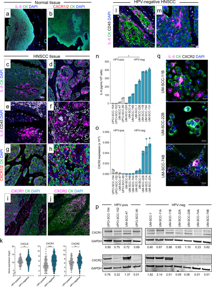
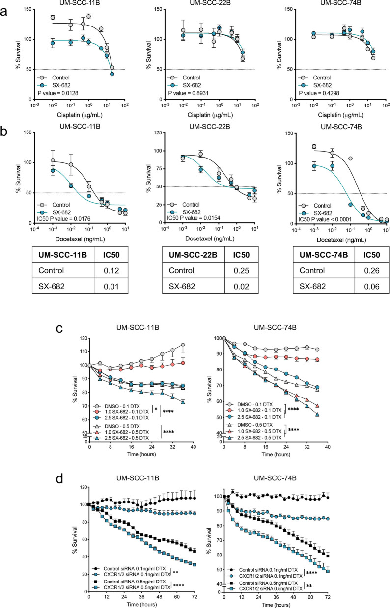
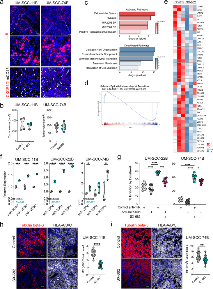
Results: High levels of IL-8, CXCR1, and CXCR2 expression were observed in HPV-negative compared to HPV-positive HNSCC tumors or cell lines. Treatment of HPV-negative HNSCC cell lines in vitro with SX-682 sensitized the tumor cells to the cytotoxic activity of docetaxel. In vivo, treatment of HNSCC xenograft models with the combination of SX-682 plus docetaxel led to strong anti-tumor control resulting in tumor cures. This phenomenon was associated with an increase of microRNA-200c and a decreased expression of its target, tubulin beta-3, a protein involved in resistance to microtubule-targeting chemotherapies. In vivo treatment of a murine syngeneic model of HNSCC with SX-682 plus docetaxel led to potent anti-tumor efficacy through a simultaneous decrease in suppressive CXCR2+ polymorphonuclear, myeloid-derived suppressor cells and an increase in cytotoxic CD8+ T cells in the combination therapy treated tumors compared to controls.

Conclusions: This study reports, for the first time, mechanistic findings through which the combination of CXCR1/2 inhibition and docetaxel chemotherapy exhibits synergy in models of HPV-negative HNSCC. These findings provide rationale for the use of this novel combination approach to treat HPV-negative HNSCC patients and for future combination studies of CXCR1/2 inhibition, docetaxel, and immune-based therapies.

Keywords: CXCR1; CXCR2; Docetaxel; HNSCC; IL-8; Immunotherapy.


Conflict of interest statement

Declarations. Ethics approval and consent to participate: All animal procedures reported in this study were approved by the NCI Animal Care and Use Committee (ACUC) and in accordance with federal regulatory requirements and standards. All components of the intramural NIH ACU program are accredited by Association for Assessment and Accreditation of Laboratory Animal Care (AAALAC) International. Consent for publication: Not applicable. Competing interests: The authors from Syntrix Pharmaceuticals are employees or officers of the company. CP discloses spouse’s employment and holdings in MacroGenics, Inc. All other authors from the National Cancer Institute, NIH, do not have any competing interests to disclose. The NCI, NIH has an ongoing Cooperative Research and Development Agreement (CRADA) with Syntrix Pharmaceuticals.

Figures

Fig. 1
Fig. 1
Expression of IL-8 and CXCR1/2 in HNSCC. a-h Representative images of tissues from a head and neck cancer tumor tissue array stained for IL-8 mRNA (pink), pan-cytokeratin (CK, green), CXCR1 and CXCR2 mRNA (CXCR1/2, red), or CD45 (white). a, b Representative cases corresponding to normal pharyngeal mucosa (a) and normal tongue (b). c-h Representative cases of squamous cell carcinoma of larynx stage IVA (c, e), epiglottis stage III (d, f), two cases of squamous cell carcinoma of larynx stage III (g, h). i A case of squamous cell carcinoma of the laryngopharynx stage III stained with antibodies against CXCR1 (pink) and CK (green). j A case of nasopharynx stage I stained with antibodies against CXCR2 (pink) and CK (green). k RNAseq data from the TCGA head and neck cancer database analyzed for expression of CXCL8, CXCR1, and CXCR2 in HPV-negative (n = 410) and HPV-positive (n = 90) tumors. l, m Representative images of HPV-negative tumor tissues from a head and neck cancer tumor tissue array stained for IL-8 mRNA (pink), pan-cytokeratin (CK, green), and CD45 (white) corresponding to squamous cell carcinoma of pharynx stage I (l) and pharynx stage II (m). n IL-8 secreted by HPV-negative and HPV-positive HNSCC cell lines in 48-h culture supernatants determined by ELISA and normalized to cell counts at time of collection. o RT-PCR analysis of CXCR2 gene expression in HNSCC cell lines, separated by HPV status. p Expression of CXCR1 and CXCR2 proteins in HPV-positive and HPV-negative HNSCC cell lines via immunoblot analysis. GAPDH was detected on the same membranes as a loading control. Numbers below blots indicate expression of the protein of interest as a ratio compared to the corresponding GAPDH. q Representative images of HPV-negative HNSCC cell lines stained for IL-8 mRNA (pink), pan-cytokeratin (CK, green), and CXCR2 mRNA (white). DAPI (blue) was used to stain nuclei in all representative images. Values graphed in (n, o) represent the mean ± SEM of technical replicates (n = 2 for ELISA assay; n = 3 for RT-PCR). Results are representative of 2 independent experiments. * p < 0.05, ** p < 0.01, **** p < 0.0001 for Student’s t-test in (k, n)
Fig. 2
Fig. 2
Inhibition of the CXCR1/2 pathway improves susceptibility of HPV-negative HNSCC to docetaxel in vitro. a, b Indicated cell lines were pre-treated with DMSO (Control) or SX-682 (10 µM) for 72 h, followed by treatment with DMSO or SX-682 and a range of cisplatin (a) or docetaxel (b) concentrations for 72 h. A CellTiterGlo assay was used to determine % survival of cells and GraphPad Prism was used to calculate an IC50 value. Values represent the mean ± SEM of technical replicates and the solid line is a calculated nonlinear regression. Dotted horizontal line plotted at 50% survival. c Proliferation of indicated cell lines pre-treated with DMSO, 1 µM SX-682 (orange), or 2.5 µM SX-682 (teal) and treated with either 0.1 ng/mL docetaxel (circles) or 0.5 ng/mL docetaxel (triangles). d Proliferation of indicated cell lines transfected with control siRNA (black) or a pool of CXCR1/2 siRNA (teal) and treated with either 0.1 ng/mL docetaxel (circles) or 0.5 ng/mL docetaxel (triangles). Values represent the mean ± SEM of technical replicates. Results in a, b, c, and d are representative of 2 independent experiments. IC50 values were compared using an extra sum-of-squares F test in (a, b). * p < 0.05, ** p < 0.01, **** p < 0.0001 for 2-way ANOVA in (c, d)
Fig. 3
Fig. 3
SX-682 modulates the phenotype of HPV-negative HNSCC xenografts and upregulates miR-200. a UM-SCC-11B and UM-SCC-74B xenografts grown in NSG-MHC I/II knockout mice were evaluated via RNA in situ hybridization for expression of IL-8 or CXCR1/2 mRNA (red) and staining for CD45 (white). b Final tumor volumes of UM-SCC-11B and UM-SCC-74B xenografts on day 15 post-tumor implantation in mice receiving a control or a SX-682-containing feed starting on day 7 or day 6, respectively. c RNAseq analysis on control and SX-682 treated UM-SCC-74B xenografts. Shown are selected activated and deactivated GO, HALLMARK, and C3 pathways when comparing SX-682 to control. d GSEA Enrichment plot showing normalized enrichment score by ranked genes for HALLMARK Epithelial-Mesenchymal Transition pathway. e Heatmap of leading-edge genes identified in (d). Data represents n = 3 mice per group. f Cell lines treated with SX-682 (teal) or DMSO control (gray) for 24–48 h, followed by analysis of miR-200a, miR-200b, and miR-200c by RT-PCR. Expression was normalized to expression of snoRNA135 and graphed as relative to DMSO cells. g UM-SCC-22B and UM-SCC-74B cells were pre-treated with SX-682 and transfected with anti-miR200c or a control anti-miR. Cells were subsequently treated with docetaxel (0.75 ng/mL and 0.5 ng/mL respectively) with or without SX-682. Growth inhibition was determined after 72 or 48 h respectively. h-i Tumors from control and SX-682 treated UM-SCC-11B (h) and UM-SCC-74B xenografts (i) from (b) were collected at endpoint and stained for expression of tubulin beta-3 (red), HLA-A/B/C (white), and DAPI (blue) (n = 4 tumors per group). Quantification of the MFI of tubulin beta-3 expression within the regions of HLA-A/B/C expression is also shown; each dot represents an individual region of interest (ROI). Results in f and g are representative of 2 independent experiments. * p < 0.05, ** p < 0.01, *** p < 0.001, **** p < 0.0001 for Student’s t-test in (f, h, i) and for 1-way ANOVA followed by Tukey’s post hoc test in (g)
Fig. 4
Fig. 4
CXCR1/2 inhibition increases susceptibility of HNSCC xenografts to docetaxel in vivo. a, d, g Schematics depicting the timeline of therapy with the UM-SCC-11B, UM-SCC-74B, and UPCI-SCC-90 models, respectively. Control diet or SX-682 feed (200 mg/kg) was administered to mice starting on day 6 or 7, as indicated, for the duration of the experiment. Docetaxel injections (5 mg/kg) were delivered by intraperitoneal (i.p.) injection on indicated days. Average tumor growth and tumor volumes at the end of study, respectively, for the UM-SCC-11B (b, c), UM-SCC-74B (e, f), and UPCI-SCC-90 (h, i) models (n = 6–7 mice/group in all studies). Gray shaded area marks the administration of SX-682 while vertical dotted lines denote docetaxel injections. UM-SCC-11B and UM-SCC-74B data are representative of 1 of 2 independent experiments. Values graphed in (b, e, h) represent the mean ± SEM; * p < 0.05, ** p < 0.01, *** p < 0.001, **** p < 0.0001 for 2-way ANOVA in (b, e, h), and for 1-way ANOVA followed by Tukey’s post hoc test in (c, f, i)
Fig. 5
Fig. 5
CXCR1/2 inhibition increases susceptibility of murine HNSCC models to docetaxel leading to enhanced anti-tumor immunity. a MOC1 tumors were evaluated via RNA in situ hybridization for the expression of murine IL-8 homologs CXCL1 (red) and CXCL2 (white) or the receptor CXCR2 (red) and immunofluorescence for pan-cytokeratin (CK, green) and CD45 (white). Nuclear staining was performed by DAPI (blue). b Schematic depicting the timeline of therapy. Control or SX-682 feed (200 mg/kg) was administered to mice starting on day 6 and remained for the duration of the experiment. Docetaxel (5 mg/kg) was delivered by i.p. injection on indicated days. c Average tumor growth (n = 7–8 mice/group). Data are representative of 1 of 2 independent experiments. d Flow cytometry analysis of indicated immune infiltrating cells in the tumors of MOC1 treated mice. e Representative scatter plots depicting the expression of CXCR2 on Ly6G+ tumor infiltrating cells in control and SX-682 plus docetaxel groups. f Orthotopic MOC1 tumors were evaluated via RNA in situ hybridization for the expression of CXCL1 (red, left) and CXCR2 (red, right) and immunofluorescence for pan-cytokeratin (CK, green) and CD45 (white). Nuclear staining was performed by DAPI (blue). g Schematic depicting the timeline of therapy for treatment of orthotopic MOC1 tumors with dosing as in (b). h Average tumor growth (n = 9–10 mice/group). i Tumor volumes recorded at the final timepoint of the experiment. j Flow cytometry analysis of indicated immune infiltrating cells in the tumors from (h, i). k Bulk RNAseq transcriptome analysis was performed on mRNA isolated from control, SX-682, docetaxel, and SX-682 plus docetaxel treated MOC1 tumors from (c); n = 5 mice per group. Bubble plots depicting the top activated and deactivated GO and HALLMARK pathways when comparing SX-682, docetaxel, and SX-682 plus docetaxel treated tumors to control tumors are shown. l Average tumor growth of MOC1 tumors untreated or treated with SX-682 plus docetaxel with or without administration of depleting antibodies for CD8+ or Ly6G.+ cells; n = 8 mice in control, 9 mice in SX-682 plus docetaxel, and 6 mice in both CD8 and Ly6G depletion groups. Values graphed in (c, h, l) represent the mean ± SEM; * p < 0.05, ** p < 0.01, *** p < 0.001, **** p < 0.0001 for 2-way ANOVA in (c, h, l), and for 1-way ANOVA followed by Tukey’s post hoc test in (d, i, j)

References

    1. Johnson DE, Burtness B, Leemans CR, Lui VWY, Bauman JE, Grandis JR. Head and neck squamous cell carcinoma. Nat Rev Dis Primers. 2020;6(1):92. - PMC - PubMed
    1. Bhatia A, Burtness B. Treating head and neck cancer in the age of immunotherapy: a 2023 update. Drugs. 2023;2023:217–48. - PubMed
    1. Bauml J, Seiwert TY, Pfister DG, Worden F, Liu SV, Gilbert J, et al. Pembrolizumab for Platinum- and Cetuximab-Refractory Head and Neck Cancer: Results From a Single-Arm. Phase II Study J Clin Oncol. 2017;35(14):1542–9. - PMC - PubMed
    1. Burtness B, Harrington KJ, Greil R, Soulieres D, Tahara M, de Castro G, Jr, et al. Pembrolizumab alone or with chemotherapy versus cetuximab with chemotherapy for recurrent or metastatic squamous cell carcinoma of the head and neck (KEYNOTE-048): a randomised, open-label, phase 3 study. Lancet. 2019;394(10212):1915–28. - PubMed
    1. Cohen EEW, Bell RB, Bifulco CB, Burtness B, Gillison ML, Harrington KJ, et al. The Society for Immunotherapy of Cancer consensus statement on immunotherapy for the treatment of squamous cell carcinoma of the head and neck (HNSCC). J Immunother Cancer. 2019;7(1):184. - PMC - PubMed

MeSH terms