. 2024 Dec 6;19(1):167.
doi: 10.1186/s13020-024-01036-3.

Siwu decoction mitigates radiation-induced immune senescence by attenuating hematopoietic damage

Affiliations

Siwu decoction mitigates radiation-induced immune senescence by attenuating hematopoietic damage

Mingyue Huang et al. Chin Med. .

Abstract

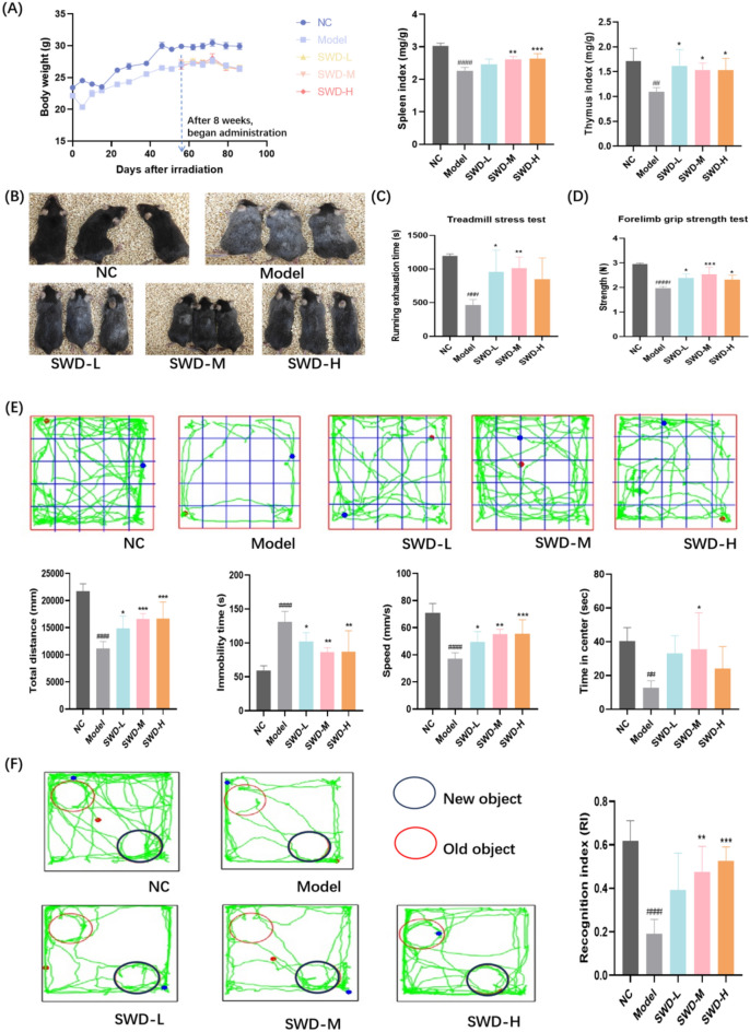
Background: To investigate the long term effects of ionizing radiation (IR) on hematopoietic stem/progenitor cells (HSPCs), immune tissues and cells, and the effects of Siwu decoction (SWD) on immune senescence mice.

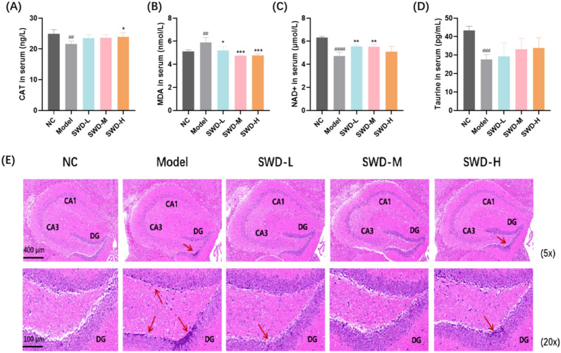
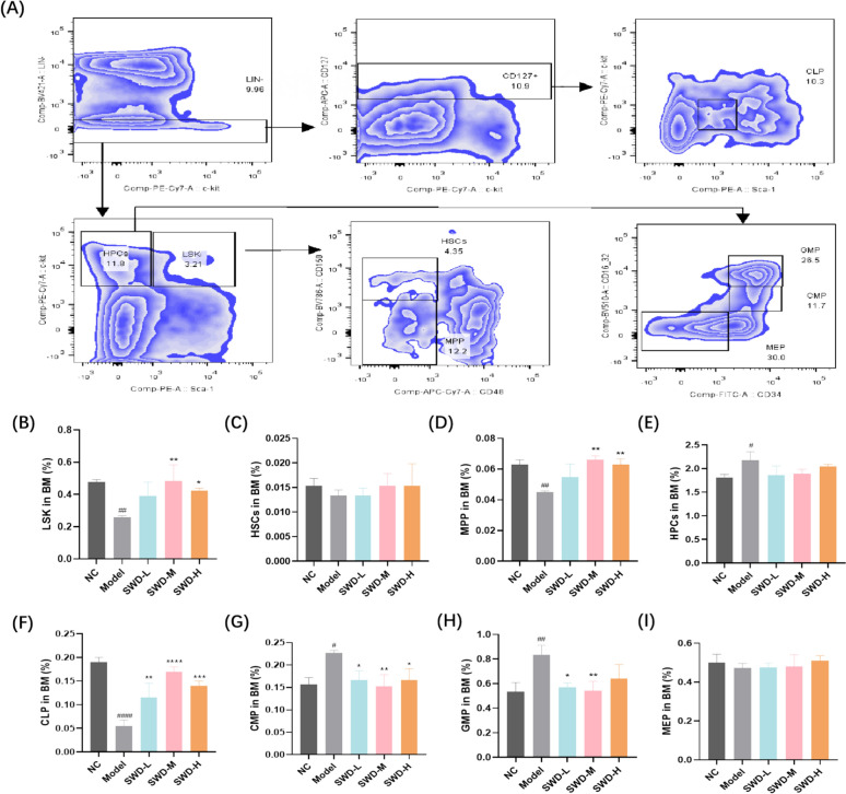
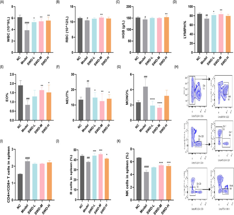
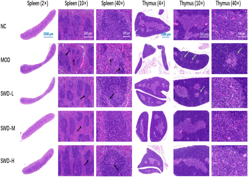
Methods: C57BL/6 J mice were exposed to 6.0 Gy 60Co γ irradiation. After 8-weeks of IR, SWD (5, 10, 20 g/kg/d) was administered for 30 days. The changes of HSPCs in bone marrow (BM) and T, B type lymphocyte and natural killer (NK) cells in spleen were detected by flow cytometry. The changes of peripheral blood cells were also examined. Hematoxylin-eosin staining were used to detect the pathological lesions of hippocampus, spleen and thymus tissues. Absolute mouse telomere length quantification qPCR assay kit was used to measure the telomere length of BM cells. The expression of factors associated with inflammation and aging such as p16, β-galactosidase in spleen, thymus and BM was determined.

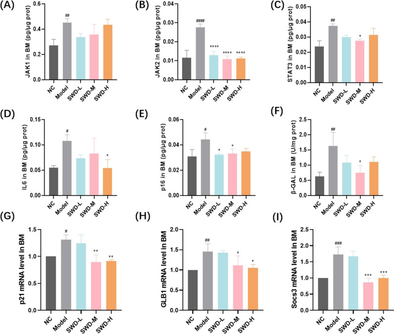
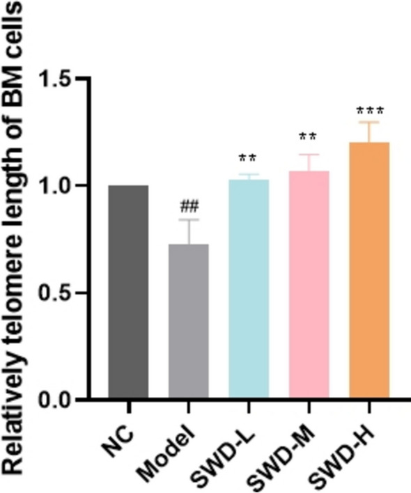
Results: Administration of SWD could increase the proportion of LSK (Lin-, Sca-1 + , c-Kit-), multipotent progenitor cells and multipotent progenitor cells and decrease the proportion of common myeloid progenitors and granulocyte-macrophage progenitors in BM. The proportion of B cells and NK cells in spleen and the content of white blood cells, red blood cells, hemoglobin, lymphocytes and eosinophils in peripheral blood were increased, at the same time, the proportion of neutrophils and monocytes was reduced by SWD. The pathological lesions of hippocampus, spleen and thymus were improved. The expression of p16 and β-galactosidase in spleen, thymus and BM was reduced and shortening of the telomere of BM cells was inhibited after administration. In addition, SWD could reduce the content of Janus activated kinase (JAK) 1, JAK2 and signal transducer and activator of transcription 3 (STAT3) in BM and spleen.

Conclusions: SWD could slow down IR-induced immune senescence by improving hematopoietic and immunologic injury. SWD might reduce the inflammation level of BM hematopoietic microenvironment by acting on JAK/STAT signaling pathway, while the immune damage of mice was improved by affecting the differentiation of HSPCs. The remission of hematopoietic and immunologic senescence was further demonstrated at the overall level.

Keywords: HSPCs; Immune; Ionizing radiation; Senescence; Siwu decoction.


Conflict of interest statement

Declarations. Ethics approval and consent to participate: The experiment was performed in accordance with the guidelines of the European Community and approved by the Institution of animal Care and Use Committee of AMMS: IACUC-DWZX-2023–547. Consent for publication: Not applicable. Competing interests: The authors declare that they have no known competing financial interests or personal relationships that could have appeared to influence the work reported in this paper.

Figures

Fig. 1
Fig. 1
Schematic of the experimental procedure for mice experiencing IR and SWD intervention
Fig. 2
Fig. 2
SWD relieved IR-induced senescence in mice. A Body weight, spleen index and thymus index (n = 6); B Snapshot comparison of mice from each group respectively (n = 3); C Running exhaustion time (n = 5); D Grip strength (n = 5); E Representative trace in OFT of each group of mice and total travel distance, immobility time, speed as well as time of entry into the central area in OFT (n = 6); F Representative trace and RI of each group of mice in NOP. RI = T2/(T1 + T2) × 100%, where T1 is the time spent exploring familiar objects and T2 is the time spent exploring new objects (n = 5). ##p < 0.01, ###p < 0.001, ####p < 0.0001 versus the control group; *p < 0.05, **p < 0.01, ***p < 0.001 versus the model group (x¯ ± s)
Fig. 3
Fig. 3
SWD reduced the level of oxidative stress in mice serum and the pathological damage of hippocampus. The content of (A) CAT; B MDA; C NAD + ; D Taurine in mice serum. ##p < 0.01, ###p < 0.001, ####p < 0.0001 versus the control group; *p < 0.05, **p < 0.01, ***p < 0.001 versus the model group (n = 5, x¯ ± s); E H&E staining results of hippocampus in each group (n = 3). Mice exposed to IR exhibited hippocampal abnormalities, including intensified cell staining in the DG region, indistinct nuclear and cytoplasmic boundaries, and markedly decreased white matter (red arrow)
Fig. 4
Fig. 4
SWD regulated the imbalance of lymphoid-myeloid differentiation. A Flow cytometry gating strategy; The proportion of (B) LSK; C HSCs; D MPP; E Hematopoietic progenitor cells (HPCs); F CLP; G CMP; H GMP; I Megakaryocytic-erythroid progenitors (MEP) in BM. #p < 0.05, ##p < 0.01, ####p < 0.0001 versus the control group; *p < 0.05, **p < 0.01, ***p < 0.001, ****p < 0.0001 versus the model group (n = 3, x¯ ± s)
Fig. 5
Fig. 5
SWD ameliorated the immune imbalance of peripheral blood and spleen. The content of (A) WBC; B RBC; C HGB; D LYMPH%; E EO%; F NEUT%; G MONO% in peripheral blood (AG, n = 5); H Flow cytometry gating strategy for splenic immune cells; I CD4 + /CD8 + T cells; J B cells; K NK cells in the spleen (IK, n = 3). #p < 0.05, ##p < 0.01, ###p < 0.001, ####p < 0.0001 versus the control group; *p < 0.05, **p < 0.01, ***p < 0.001, ****p < 0.0001 versus the model group (x¯ ± s)
Fig. 6
Fig. 6
SWD reduced the expression of inflammatory/aging related factors in BM. The protein ratio (the content of protein/total protein concentration) of (A) JAK1; B JAK2; C STAT3; D IL-6; E p16; F The activity of β-galactosidase; The expression level of (G) p21; H GLB1; I Socs3 in BM. #p < 0.05, ##p < 0.01, ###p < 0.001, ####p < 0.0001 versus the control group; *p < 0.05, **p < 0.01, ***p < 0.001, ****p < 0.0001 versus the model group (n = 3, x¯ ± s)
Fig. 7
Fig. 7
SWD inhibited the telomere shortening of BM cells. ##p < 0.01 versus the control group; **p < 0.01, ***p < 0.001 versus the model group (n = 3, x¯ ± s)
Fig. 8
Fig. 8
H&E staining results of spleen and thymus in each group. The spleen and thymus of irradiated mice were atrophied, the spleen bodies were significantly reduced, and the volume of lymph nodules became smaller (black arrow). The thymus medulla was prominently reduced, and the number of thymus corpuscles and lymphocytes was decreased (green arrow)
Fig. 9
Fig. 9
SWD reduced the expression of aging related factors in spleen and thymus. The protein ratio (the content of protein/total protein concentration) of (A) JAK1; B JAK2; C STAT3; D IL-6; E p16; F The activity of β-galactosidase in spleen; G Representative images and semi-quantitative analysis of p16 (green) and β-galactosidase (red) in spleen and thymus in each group. At least three 40-fold fields of view were randomly selected for each slice in each group. When capturing images, it was ensured that the background lighting remained consistent across all photos. The Image J 2.0 software was utilized to convert the green/red fluorescent monochrome images into black and white, with a standardized selection of black for fluorescence assessment. The integrated optical density (IOD) and pixel AREA of the tissue were measured for each image, and the mean density value IOD/AREA was calculated. #p < 0.05, ##p < 0.01, ###p < 0.001, ####p < 0.0001 versus the control group; *p < 0.05, **p < 0.01, ***p < 0.001, ****p < 0.0001 versus the model group (n = 3, x¯ ± s)

References

    1. Kritsilis M, Rizou SV, Koutsoudaki PN, Evangelou K, Gorgoulis VG, Papadopoulos D. Ageing, cellular senescence and neurodegenerative disease. Int J Mol Sci. 2018;19(10):2937. - PMC - PubMed
    1. Carante MP, Ramos RL, Ballarini F. Radiation damage in biomolecules and cells 2.0. Int J Mol Sci. 2023;24(4):3238. - PMC - PubMed
    1. Omatsu Y, Nagasawa T. Identification of microenvironmental niches for hematopoietic stem cells and lymphoid progenitors-bone marrow fibroblastic reticular cells with salient features. Int Immunol. 2021;33(12):821–6. - PubMed
    1. Yang D, de Haan G. Inflammation and aging of hematopoietic stem cells in their niche. Cells. 2021;10(8):1849. - PMC - PubMed
    1. Thambyrajah R, Bigas A. Notch signaling in HSC emergence: when, why and how. Cells. 2022;11(3):358. - PMC - PubMed

LinkOut - more resources