. 2024 Nov 19:35:e2408022.
doi: 10.4014/jmb.2408.08022.

Aromadendrin Ameliorates Airway Inflammation in Experimental Mice with Chronic Obstructive Pulmonary Disease

Affiliations

Aromadendrin Ameliorates Airway Inflammation in Experimental Mice with Chronic Obstructive Pulmonary Disease

Jinseon Choi et al. J Microbiol Biotechnol. .

Abstract

Aromadendrin (ARO) is an active plant compound that exerts anti-inflammatory effects. However, its ameliorative effects on chronic obstructive pulmonary disease (COPD) remain unclear. Therefore, we investigated the inhibitory effects of ARO on bronchial inflammation using an experimental model of COPD. In vivo analysis confirmed a notable increase in the number of neutrophils/macrophages and the formation of reactive oxygen species (ROS), myeloperoxidase (MPO), interleukin (IL)-6/IL-1β, and monocyte chemoattractant protein (MCP)-1 in the bronchoalveolar lavage (BAL) fluid of COPD mice, which was attenuated by oral gavage of ARO. In addition, hematoxylin and eosin staining showed a notable cell influx in the lungs of the COPD group, which was ameliorated by ARO. Western blotting revealed that ARO decreased the upregulation of neutrophil elastase expression in the lungs of the COPD group. Furthermore, periodic acid-Schiff staining showed that increased mucus formation in the lungs of the COPD group was downregulated by ARO. ARO also blocked CREB activation in the lungs of COPD mice. This in vivo, anti-inflammatory effect of ARO was accompanied by its modulatory effect on the activation of the MAPK/NF-κB/NLRP3 inflammasome. In summary, our study demonstrated that ARO has protective effects on bronchial inflammation by attenuating immune cell accumulation, toxic molecule/cytokine/chemokine formation, and MAPK/NF-κB/NLRP3 inflammasome activation, suggesting the potential development of ARO as an adjuvant for the prevention and treatment of COPD.

Keywords: COPD; NF-κB; NLRP3 inflammasome; aromadendrin; cytokines.


Conflict of interest statement

Conflict of Interests

The authors have no financial conflicts of interest to declare.

Figures

Fig. 1
Fig. 1. ARO inhibits neutrophils/macrophages influx in experimental COPD mice.
(A) The images of neutrophils/macrophages were obtained using Diff-Quik staining and microscopy (magnification, x400; scale bar, 25 μm). Number of (B) neutrophils and (C) macrophages in BAL fluid of mice (green arrows indicate the neutrophils and red arrows indicate the macrophages). Data are expressed as the mean ± SD (#p < 0.05 for comparison with normal control; *p < 0.05 for comparison with COPD group). NC: normal control mice; COPD: cigarette smoke exposed/LPS-administered mice; ROF: 5 mg/kg ROF-treated COPD mice; ARO 5: 5 mg/kg ARO-treated COPD mice; and ARO 10: 10 mg/kg ARO-treated COPD mice.
Fig. 2
Fig. 2. ARO reduces the concentration of inflammatory molecules in experimental COPD mice.
(A) Total cellular ROS level in BAL fluid cells was determined using 2',7'-dichlorodihydrofluorescein diacetate (DCF-DA). Levels of inflammatory molecules, such as (B) MPO (C) IL-6, (D) IL-1β, and (D) MCP-1 in BAL fluid, were detected by ELISA. Data are expressed as the mean ± SD (#p < 0.05 for comparison with normal control; *p < 0.05 for comparison with COPD group). NC: normal control mice; COPD: cigarette smoke exposed/LPS-administered mice; ROF: 5 mg/kg ROF-treated COPD mice; ARO 5: 5 mg/kg ARO-treated COPD mice; and ARO 10: 10 mg/kg ARO-treated COPD mice.
Fig. 3
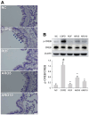
Fig. 3. Cell accumulation and elastase expression were reduced by ARO in experimental COPD mice.
(A) The histological changes in lungs of mice, which show cell accumulation, were assessed using H&E staining (left panel: magnification, 100×; scale bar, 100 μm; right panel: magnification, 400×; scale bar, 25 μm). (B) The expression of neutrophil elastase was analyzed using western blotting. Quantitative analysis of neutrophil elastase was performed using ImageJ software. Data are expressed as the mean ± SD (#p < 0.05 for comparison with normal control; *p < 0.05 for comparison with COPD group). NC: normal control mice; COPD: cigarette smoke exposed/LPS-administered mice; ROF: 5 mg/kg ROF-treated COPD mice; ARO 5: 5 mg/kg ARO-treated COPD mice; and ARO 10: 10 mg/kg ARO-treated COPD mice.
Fig. 4
Fig. 4. ARO attenuates mucus formation and CREB activation in experimental COPD mice.
(A) The histological changes in lungs of mice, which indicate mucus formation, were assessed using PAS staining (magnification, 400×; scale bar, 25 μm). (B) The activation of CREB was analyzed using western blotting. Quantitative analysis of phosphorylated (p)-CREB was performed using ImageJ software. Data are expressed as the mean ± SD (#p < 0.05 for comparison with normal control; *p < 0.05 for comparison with COPD group). NC: normal control mice; COPD: cigarette smoke exposed/LPSadministered mice; ROF: 5 mg/kg ROF-treated COPD mice; ARO 5: 5 mg/kg ARO-treated COPD mice; and ARO 10: 10 mg/kg ARO-treated COPD mice.
Fig. 5
Fig. 5. ARO inhibits MAPK activation in experimental COPD mice.
(A) The activation of JNK, p38, and ERK in lungs of mice was analyzed using western blotting. Quantitative analysis of (B) p-JNK, (C) p-p38, and (D) p-ERK was performed using ImageJ software. Data are expressed as the mean ± SD (#p < 0.05 for comparison with normal control; *p < 0.05 for comparison with COPD group). NC: normal control mice; COPD: cigarette smoke exposed/LPS-administered mice; ROF: 5 mg/kg ROF-treated COPD mice; ARO 5: 5 mg/kg ARO-treated COPD mice; and ARO 10: 10 mg/kg ARO-treated COPD mice.
Fig. 6
Fig. 6. ARO inhibits NF-κB activation in experimental COPD mice.
(A) The activation of NF-κB p65 and IκBα was analyzed using western blotting. Quantitative analysis of (B) p- NF-κB p65 and (C) p-IκBα was performed using ImageJ software. Data are expressed as the mean ± SD (#p < 0.05 for comparison with normal control; *p < 0.05 for comparison with COPD group). NC: normal control mice; COPD: cigarette smoke exposed/LPS-administered mice; ROF: 5 mg/kg ROF-treated COPD mice; ARO 5: 5 mg/kg ARO-treated COPD mice; and ARO 10: 10 mg/kg ARO-treated COPD mice.
Fig. 7
Fig. 7. ARO inhibits NLRP3 activation in experimental COPD mice.
(A) The expression of NLRP3, ASC, and caspase-1 was analyzed using western blotting. Quantitative analysis of (B) NLRP3, (C) ASC, and (D) caspase-1 was performed using ImageJ software. Data are expressed as the mean ± SD (#p < 0.05 for comparison with normal control; *p < 0.05 for comparison with COPD group). NC: normal control mice; COPD: cigarette smoke exposed/LPS-administered mice; ROF: 5 mg/kg ROF-treated COPD mice; ARO 5: 5 mg/kg ARO-treated COPD mice; and ARO 10: 10 mg/kg ARO-treated COPD mice.
Fig. 8
Fig. 8. Ameliorative effects of ARO on bronchial inflammation in experimental COPD mice.
Oral administration of ARO reduces neutrophil/macrophage inflow and ROS/MPO/elastase/IL-6/IL-1β/MCP-1 formation in COPD mice. These effects of ARO are accompanied by its modulatory effect on MAPK/NF-κB/NLRP3 activation.

Similar articles

References

    1. Kahnert K, Jorres RA, Behr J, Welte T. The diagnosis and treatment of COPD and its comorbidities. Dtsch Arztebl. Int. 2023;120:434–444. doi: 10.3238/arztebl.m2023.0027. - DOI - PMC - PubMed
    1. Christenson SA, Smith BM, Bafadhel M, Putcha N. Chronic obstructive pulmonary disease. Lancet. 2022;399:2227–2242. doi: 10.1016/S0140-6736(22)00470-6. - DOI - PubMed
    1. Butler A, Walton GM, Sapey E. Neutrophilic inflammation in the pathogenesis of chronic obstructive pulmonary disease. COPD. 2018;15:392–404. doi: 10.1080/15412555.2018.1476475. - DOI - PubMed
    1. Park HY, Man SF, Tashkin D, Wise RA, Connett JE, Anthonisen NA, Sin DD. The relation of serum myeloperoxidase to disease progression and mortality in patients with chronic obstructive pulmonary disease (COPD) PLoS One. 2013;8:e61315. doi: 10.1371/journal.pone.0061315. - DOI - PMC - PubMed
    1. Zheng S, Kummarapurugu AB, Bulut GB, Syed A, Kang L, Voynow J.A. Neutrophil elastase activates the release of extracellular traps from COPD blood monocyte-derived macrophages. Clin. Transl. Sci. 2023;16:2765–2778. doi: 10.1111/cts.13671. - DOI - PMC - PubMed

MeSH terms