. 2025 Mar;116(3):643-655.
doi: 10.1111/cas.16418. Epub 2024 Dec 5.

Cancer-associated fibroblast-derived MMP11 promotes tumor progression in pancreatic cancer

Affiliations

Cancer-associated fibroblast-derived MMP11 promotes tumor progression in pancreatic cancer

Zhuoyin Wang et al. Cancer Sci. 2025 Mar.

Abstract

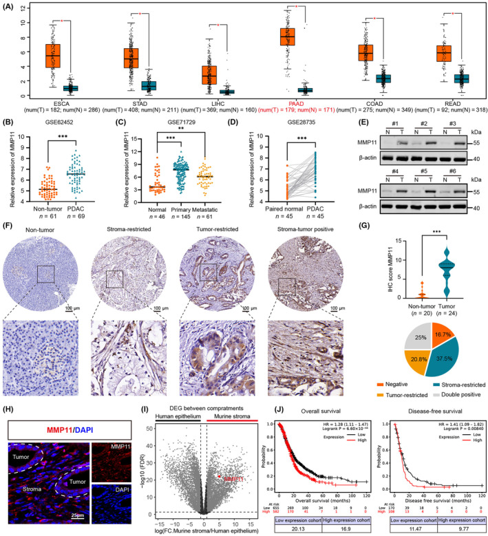
Matrix metalloproteinase 11 (MMP11), a zinc-dependent endopeptidase involved in extracellular matrix degradation and remodeling, has been identified as a tumor promoter in multiple cancer types. However, its expression pattern and role in pancreatic ductal adenocarcinoma (PDAC) remain unclear. In this study, elevated MMP11 expression was identified in PDAC tissues and was associated with diminished survival. Integrated single-cell RNA sequencing and co-immunofluorescence staining revealed that MMP11 was predominantly expressed in cancer-associated fibroblasts (CAFs). Mechanistically, cancer cell-derived TGF-β1 mediated CAF activation via the pSmad2/3 pathway and accompanied by MMP11 production. Additionally, MMP11 knockdown in CAFs impaired the proliferative and invasive abilities of AsPC-1 and BxPC-3 cells in vitro; which could be rescued by adding recombinant MMP11. Similarly, co-injection of AsPC-1 cells with MMP11-knockdown CAFs into nude mice significantly suppressed tumor growth and liver metastasis compared with tumors bearing unmodified CAFs. Furthermore, we confirmed that CAF-derived MMP11 may drive the epithelial-mesenchymal transition process of PDAC cells to promote tumor invasion via the PI3K/AKT pathway rather than extracellular matrix remodeling. Collectively, we uncovered a crosstalk between cancer cells and CAFs mediated by TGF-β1 and MMP11 that drives the progression of PDAC.

Keywords: CAFs; EMT; MMP11; PDAC; tumor progression.


Conflict of interest statement

The authors declare no conflict of interest.

Figures

FIGURE 1
FIGURE 1
MMP11 is highly expressed in the stroma of PDAC and correlates with a poor prognosis. (A) MMP11 expression profiles in various gastrointestinal malignancy tissues and corresponding normal tissues were assessed using the GEPIA2 database. (B–D) MMP11 mRNA levels in PDAC tissues and non‐tumor tissues (GSE62452 and GSE71729 ), or PDAC tissues and paired normal tissues (GSE28735). (E) The protein levels of MMP11 in six paired non‐tumor tissues (N) and PDAC tissues (T). (F) Representative IHC images of MMP11 in non‐tumor pancreas tissues and PDAC tissues. (G) Violin plot displaying the IHC scores of MMP11 expression in non‐tumor tissues and PDAC tissue (upper panel). The pie chart exhibits the proportions of tumors with MMP11‐positive staining in stromal or epithelial compartments (lower panel). (H) Representative IF images of MMP11 in PDAC tissues. The tumor and stromal compartments were divided by dashed lines. (I) Volcano plot showing differentially expressed genes between the stromal and epithelial compartments. (J) Overall survival and disease‐free survival for patients with high or low MMP11 expression were analyzed using a Kaplan–Meier plotter.
FIGURE 2
FIGURE 2
CAFs are responsible for elevated MMP11 expression in PDAC. (A, B) MMP11 expression levels in various cell types within PDAC. (C) Representative images of co‐immunofluorescence staining for the target molecule MMP11, pan‐CK, and α‐SMA. (D) Morphology of CAFs and NFs under light microscopy. (E) CAFs and NFs were validated by western blot for α‐SMA and vimentin, as well as E‐cadherin. (F, G) qRT‐PCR and western blot analysis of MMP11 expression among PDAC cell lines and CAFs. (H) The subcellular localization of MMP11 in CAFs was determined by IF.
FIGURE 3
FIGURE 3
PDAC cell‐secreted TGF‐β1 drives CAF activation and induces MMP11 expression via pSmad2/3 signaling. (A) GSEA of TGF‐β pathway enriched in PDAC patients with high MMP11 expression in TCGA‐PAAD cohort (n = 174). (B) Schematic of PSCs (HPaSteC) following indicated treatment. (C–E) CAF activation markers (α‐SMA, vimentin) and MMP11 expression levels in HPaSteC treated with CM from PDAC cells or rhTGF‐β1 were assessed by western blot (C) or IF (D, E). (F) The correlation between MMP11 and Smad2 or Smad3. (G–I) Western blot analysis of α‐SMA, MMP11, and pSmad2/3 in HPaSteC treated with CM from PDAC cells and rhTGF‐β1 alone or combined with 10 ng/mL TGF‐β1 receptor inhibitors (SB431542 or SD208) for 72 h.
FIGURE 4
FIGURE 4
MMP11 knockdown in CAFs impaired the proliferation, migration, and invasion of PDAC cells in vitro. (A–C) The proliferative abilities of AsPC‐1 and BxPC‐3 treated with CM from CAF‐shcontrol and CAF‐shMMP11 were assessed using the CCK‐8 assay (A), colony‐formation assay (B), and EdU assay (C). (D) Representative images of wound‐healing assays in AsPC‐1 and BxPC‐3 cells treated with CM from CAF‐shcontrol or CAF‐shMMP11 for 48 h. (E) Schematic diagram of transwell assays. (F) Representative images of AsPC‐1 and BxPC‐3 migration and invasion induced by CAFs with or without MMP11 knockdown.
FIGURE 5
FIGURE 5
MMP11 knockdown in CAFs suppresses tumor growth and metastasis in vivo. (A) Schematic diagram of xenograft tumor establishment in nude mice. (B) Representative images of subcutaneous xenografts (n = 6 per group). (C) Tumor burden and weight of subcutaneous xenografts were quantified. (D) Growth curve of subcutaneous xenografts. (E) Representative images of H&E and Ki‐67 staining in subcutaneous xenografts. (F) Schematic of liver metastasis models. (G) Representative bioluminescence images of liver metastasis (n = 5 per group). (H) Representative images of whole‐liver H&E‐stained sections from liver metastasis models. (I) Incidence and number of liver metastatic nodules are shown.
FIGURE 6
FIGURE 6
CAF‐derived MMP11 induces EMT to promote PDAC invasion via the PI3K/AKT pathway rather than ECM remodeling. (A) Representative images of α‐SMA, Sirius Red, and Masson's trichrome staining in subcutaneous xenografts. (B) The positive areas of α‐SMA, Sirius Red, and Masson's trichrome were quantified. (C) GSEA of the EMT between patients with high or low MMP11 expression in TCGA‐PAAD cohort (n = 174). (D) Western blot analysis of EMT‐related proteins (E‐cadherin, N‐cadherin, vimentin, and Slug) in AsPC‐1 and BxPC‐3 cells treated with rhMMP11 at 200 ng/mL for 48 h. (E) Representative IHC images of EMT‐related proteins (E‐cadherin, N‐cadherin, and vimentin) staining in subcutaneous xenografts. (F) Volcano plot of genes strongly associated with MMP11 (P adjust <0.05, |ρ| ≥ 0.3). (G) KEGG analysis of MMP11‐related genes (P adjust <0.05, |rho| ≥ 0.3, n = 2446). (H) Western blot analysis of protein levels of PI3K, AKT, and its phosphorylated forms in PDAC cells following treatment with rhMMP11.
FIGURE 7
FIGURE 7
Schematic diagram of crosstalk between PDAC cell and CAF mediated by TGF‐β1 and MMP11.

Similar articles

References

    1. Rahib L, Wehner MR, Matrisian LM, Nead KT. Estimated projection of US cancer incidence and death to 2040. JAMA Netw Open. 2021;4(4):e214708. - PMC - PubMed
    1. Siegel RL, Miller KD, Wagle NS, Jemal A. Cancer statistics, 2023. CA Cancer J Clin. 2023;73(1):17‐48. - PubMed
    1. Halbrook CJ, Lyssiotis CA, di Magliano MP, Maitra A. Pancreatic cancer: Advances and challenges. Cell. 2023;186(8):1729‐1754. - PMC - PubMed
    1. Sherman MH, Beatty GL. Tumor microenvironment in pancreatic cancer pathogenesis and therapeutic resistance. Annu Rev Pathol. 2023;18(1):123‐148. - PMC - PubMed
    1. Nan P, Dong X, Bai X, et al. Tumor‐stroma TGF‐β1‐THBS2 feedback circuit drives pancreatic ductal adenocarcinoma progression via integrin α(v)β(3)/CD36‐mediated activation of the MAPK pathway. Cancer Lett. 2022;528:59‐75. - PubMed

MeSH terms

Substances