. 2024 Nov 21:37:13796.
doi: 10.3389/ti.2024.13796. eCollection 2024.

Elevated PD-L1 and PECAM-1 as Diagnostic Biomarkers of Acute Rejection in Lung Transplantation

Affiliations

Elevated PD-L1 and PECAM-1 as Diagnostic Biomarkers of Acute Rejection in Lung Transplantation

Rene Novysedlak et al. Transpl Int. .

Abstract

Acute cellular rejection (ACR) frequently occurs following lung transplantation (LuTx) and represents a risk factor for the development of chronic lung allograft dysfunction (CLAD) as well as long-term survival. The histopathological diagnosis of ACR carries a burden of interobserver variability. The widespread utilization and cost-effectiveness of immunohistochemistry (IHC) was proven beneficial in diagnosing rejection in human kidney transplantations and LuTx rat models. However, its potential for ACR detection in patients remains unexplored. We analyzed surface markers (CD3, CD4, CD8, CD20, CD68, CD47, PD-1, PD-L1, and CD31/PECAM-1) on lung tissue cryobiopsy samples collected within 6 months post-LuTx from 60 LuTx recipients, 48 of whom were diagnosed with ACR. Additionally, serum samples from 51 patients were analyzed using a multiplex bead-based Luminex assay. The cytokines and markers included PD-L1, IL2, TNFα, IFNγ, and Granzyme B. We observed a significant increase in PD-L1 tissue expression within the rejection group, suggesting a concerted effort to suppress immune responses, especially those mediated by T-cells. Furthermore, we noted significant differences in PECAM-1 levels between ACR/non-ACR. Additionally, peripheral blood C-reactive-protein levels tended to be higher in the ACR group, while Luminex serum analyses did not reveal any significant differences between groups. In conclusion, our findings suggest the potential value of PECAM-1 and PD-L1 markers in diagnosing ACR.

Keywords: acute cellular rejection; checkpoint inhibitors; immunohistochemistry; luminex; lung transplantation.


Conflict of interest statement

LC declares being a senior Clinical Research Fellow of the Research Foundation-Flanders (FWO) (#18E2B24N) and supported by a KU Leuven University Chair funded by Medtronic, unrelated to the study. The remaining authors declare that the research was conducted in the absence of any commercial or financial relationships that could be construed as a potential conflict of interest.

Figures

FIGURE 1
FIGURE 1
Scatter plots of peripheral blood cell profile comparing rejection group to control group. Median and interquartile range is visualized.
FIGURE 2
FIGURE 2
ROC curves for IHC markers CD3, CD4, CD8, CD20 a CD68, along with corresponding AUC values and their 95% confidence interval. All 95% confidence intervals include 0.5, which shows that none of the markers are good predictors.
FIGURE 3
FIGURE 3
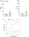
(A) Scatter plots of PD-L1+ and PD-1+ immune cells counts (positive immune cells per 1 mm2) in lung tissue biopsy. Median and interquartile range is visualized. (B) ROC curves for IHC markers PD-L1 and PD-1. Area under ROC-curves (AUC) (and associated 95% confidence interval) based on marked values for PD-L1 is 0.80 (0.65; 0.94) and for PD-1 is 0.65 (0.44; 0.86).
FIGURE 4
FIGURE 4
(A) IHC staining of CD31+ cells in control group and A1-A3 rejection groups. (B) CD31+ immune cells (orange arrow) and endothelia cells (green arrow). Only immune cells were counted. (C) Endothelium exhibiting CD31 positivity, presumably indicative of endothelial swelling associated with endothelitis, a characteristic frequently observed in A3.
FIGURE 5
FIGURE 5
(A) Scatter plot of PECAM-1/CD31 immune cells in lung tissue biopsy. Median and interquartile range is visualized. (B) ROC curve for IHC marker PECAM-1. Area under ROC curve based on marked values for PECAM-1 is 0.73.
FIGURE 6
FIGURE 6
ROC curves for Luminex markers PD-L1, IL-2, Granzym B, TNFα and IFNγ, along with corresponding AUC values and their 95% confidence interval. All 95% confidence intervals include 0.5, which shows that none of the markers are good predictors.

References

    1. Burton CM, Iversen M, Carlsen J, Mortensen J, Andersen CB, Steinbruchel D, et al. Acute Cellular Rejection Is a Risk Factor for Bronchiolitis Obliterans Syndrome Independent of Post-Transplant Baseline FEV1. J Heart Lung Transpl (2009) 28(9):888–93. 10.1016/j.healun.2009.04.022 - DOI - PubMed
    1. Clayton PA, McDonald SP, Russ GR, Chadban SJ. Long-Term Outcomes After Acute Rejection in Kidney Transplant Recipients: An ANZDATA Analysis. J Am Soc Nephrol (2019) 30(9):1697–707. 10.1681/ASN.2018111101 - DOI - PMC - PubMed
    1. Reddy KS, Davies D, Ormond D, Tuteja S, Lucas BA, Johnston TD, et al. Impact of Acute Rejection Episodes on Long-Term Graft Survival Following Simultaneous Kidney-Pancreas Transplantation. Am J Transpl (2003) 3(4):439–44. 10.1034/j.1600-6143.2003.00059.x - DOI - PubMed
    1. Levy L, Huszti E, Tikkanen J, Ghany R, Klement W, Ahmed M, et al. The Impact of First Untreated Subclinical Minimal Acute Rejection on Risk for Chronic Lung Allograft Dysfunction or Death After Lung Transplantation. Am J Transpl (2020) 20(1):241–9. 10.1111/ajt.15561 - DOI - PubMed
    1. Wu MY, Yiang GT, Liao WT, Tsai AP, Cheng YL, Cheng PW, et al. Current Mechanistic Concepts in Ischemia and Reperfusion Injury. Cell Physiol Biochem (2018) 46(4):1650–67. 10.1159/000489241 - DOI - PubMed