. 2024 Oct 10;10(20):e39098.
doi: 10.1016/j.heliyon.2024.e39098. eCollection 2024 Oct 30.

The antibacterial activity of the copper for Staphylococcus aureus 124 and Pseudomonas aeruginosa 18 depends on its state: metalized, chelated and ionic

Affiliations

The antibacterial activity of the copper for Staphylococcus aureus 124 and Pseudomonas aeruginosa 18 depends on its state: metalized, chelated and ionic

A I Bozhkov et al. Heliyon. .

Abstract

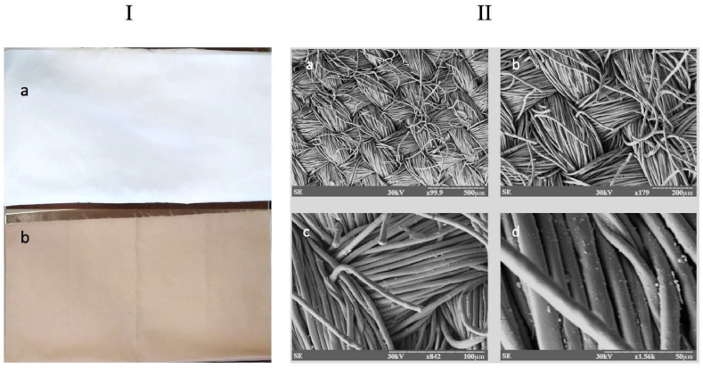
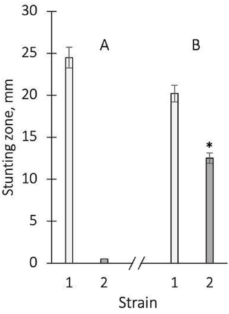
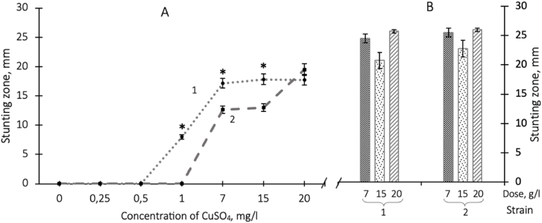
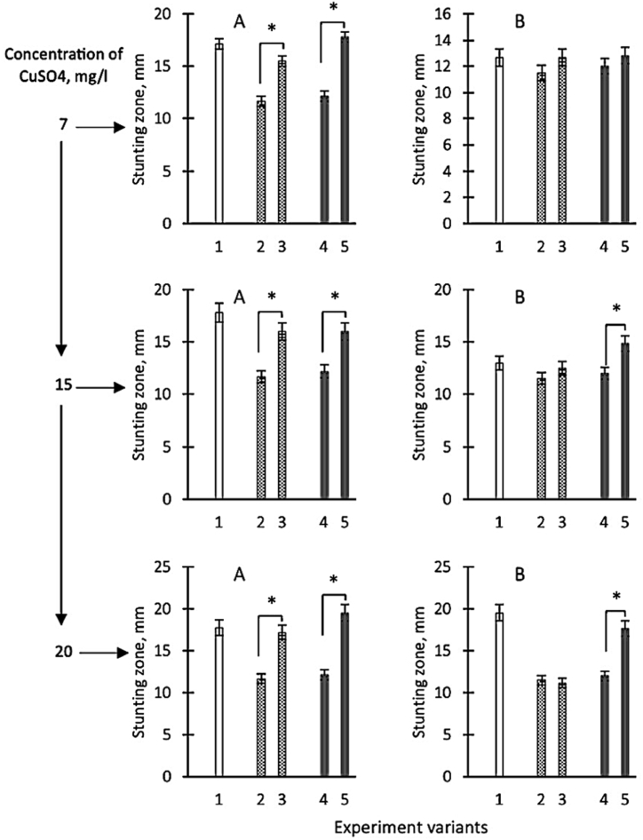
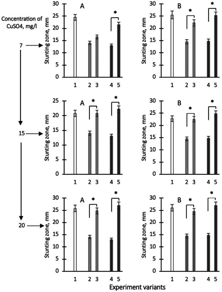
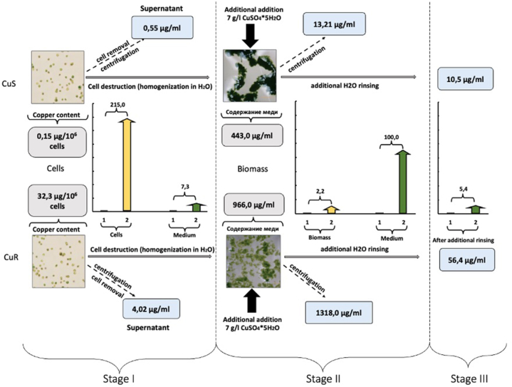
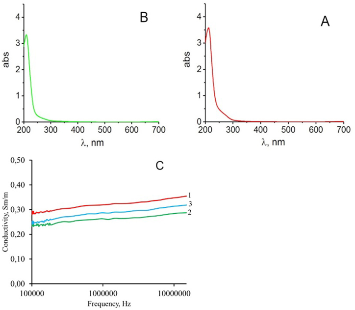
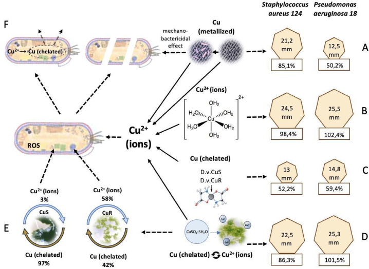
The hypothesis that the antibacterial effect of copper depends on its state was tested. It was studied the antibacterial effect of copper applied to the fabric, copper in chelated and free (ionic) forms on the growth intensity of Staphylococcus aureus 124 and Pseudomonas aeruginosa 18 in the in vitro system after a single or "primary" contact. Classical microbiology methods were used. Copper was applied to the fabric by magnetron and arc planar discharge systems, and the culture of microalgae Dunaliella viridis, resistant to the action of high concentrations of copper, was used to obtain copper in chelated form. It was shown that a thin layer of copper (3 μm) applied to the fabric showed pronounced antibacterial activity against Staphylococcus (85 % compared to the antibiotic meropenem) and less pronounced activity against Pseudomonas, which is resistant to meropenem. Copper in ionic form inhibited the growth of Staphylococcus aureus 124 as well as the antibiotic, and also effectively inhibited the growth of Pseudomonas aeruginosa 18 i.e., copper ions did not have species specificity like the antibiotic. Components of Dunaliella viridis microalgae cells had weakly expressed antibacterial effect to these types of bacteria, and supplementary addition of copper sulfate to the biomass of microalgae led to an increase of their antibacterial activity and this is more pronounced for microalgae culture in which the ratio « chelated/ionic » forms of copper is shifted to the ionic form. It was shown that the antibacterial activity of microalgae biomass after the first introduction into the tested bacterial cultures depends on the amount of free or "weakly bound" with cell components copper ions. It is suggested that the antibacterial effect of fabric with a thin layer of copper may be determined by two mechanisms: the action of copper ions and mechano-bactericidal effects, while chelated forms of copper may have a prolonged effect on bacterial cultures.


Conflict of interest statement

The work was carried out within the framework of the project of the Ministry of Education and Science of Ukraine No. 0123U101860. The authors declare that they have no known competing financial interests or personal relationships that could have appeared to influence the work reported in this paper.

Figures

Fig. 1
Fig. 1
The appearance of the fabric base (a) on which a layer of copper 3 μm thick is applied (b); II - the image of the surface relief of metalized fabric with copper at various magnifications (а – х99.9, b – х179, c – х842, d – х1.56k).
Fig. 2
Fig. 2
Zones of stunted growth of S. aureus 124 (1) and P. aeruginosa 18 (2) using the disk diffusion test with the antibiotic meropenem (A), as well as fabric with a 3 μm thick copper layer applied (B). The average values from 3 to 5 experiments and their standard errors are presented. ∗ - differences between antibiotic and fabric for variants are significant with P < 0.05.
Fig. 3
Fig. 3
Zones of stunted growth of S. aureus 124 (1) and P. aeruginosa 18 (2) with applying of copper sulfate to paper disks in different doses after cultivation for 24 h at a temperature of 37 °C (A) and with applying of copper sulfate to agar wells in doses: 7, 15 and 20 g/l– for S. aureus 124 (1) and P. aeruginosa 18 (2) (B). The means and their standard errors from three independent experiments are presented. ∗ - the difference between variants S. aureus 124 and P. aeruginosa 18 is significant at P < 0.05.
Fig. 4
Fig. 4
Growth inhibition zones of Staphylococcus aureus 124 (A) and Pseudomonas aeruginosa 18 (B) when tested on paper discs after applying copper sulfate solution (ionic form) at different concentrations to the disc: 7; 15 and 20 g/l (shown on an additional scale) respectively they are shown on the ordinate axis as variants 1, variants 2 - zones of growth retardation in case of application to discs of homogenates of D.v-Cu.S cells characterised by low copper content in chelate form, variants 3 - zones of growth retardation after application of homogenates of D.v-Cu. cells. S with the additional, preliminary introduction of sulfuric acid copper in cell homogenates respectively 7; 15 and 20 g/l of sulfuric acid copper, variant 4 - zones of growth retardation after application on discs of homogenates of cells D.v-Cu. R cells (which initially contained a relatively large amount of copper in the chelate form) and variant 5 - growth retardation zones after application of D.v-Cu.R cells homogenates on discs with additional preliminary introduction of 7; 15, and 20 g/l of copper sulfate into homogenates, respectively. The average values from three experiments and their standard errors are presented. ∗ - differences between options 2–3 and 4–5 are significant with P < 0.05.
Fig. 5
Fig. 5
Growth inhibition zones of Staphylococcus aureus 124 (A) and Pseudomonas aeruginosa 18 (B) when tested by diffusion (well method) after application of copper sulfate solution (ionic form) at different concentrations: 7; 15 and 20 g/l (shown on an additional scale) respectively they are shown on the ordinate axis as variants 1, variants 2 - zones of growth retardation in case of application of homogenates of D.v-Cu.S cells on discs, which are characterised by the low copper content in chelate form, variants 3 - zones of growth retardation after application of homogenates of D.v-Cu. S with the additional, preliminary introduction of copper sulphoxide into cell homogenates respectively 7; 15, and 20 g/l of copper sulphoxide, variant 4 - zones of growth retardation after application on discs of cell homogenates D.v-Cu. R cells (which initially contained a relatively large amount of copper in the chelate form) and variant 5 - zones of growth retardation after applying homogenates of D.v-Cu.R cells to discs with additional preliminary introduction of 7; 15 and 20 g/l of copper sulfate into homogenates, respectively. The average values from three experiments and their standard errors are presented. ∗ - differences between options 2–3 and 4–5 are significant with P < 0.05.
Fig. 6
Fig. 6
The stages of determining the content of copper ions in the studied samples. At Stage I, the number of copper ions in native cells D.v-Cu.S and D.v-Cu.R was determined (the number of cells of the two strains in the suspension was the same - 3.9 × 109 and the volume of cell suspensions was equalized) and the number of copper ions that were present in their culture medium. At Stage II, the cells were destroyed, and 7 g/l of copper sulfate was added to the obtained biomass (its amount was the same for the two strains), followed by determination of the amount of copper bound to the D.v-Cu.S and D.v-Cu.R biomass and the amount of remaining – unbound copper in the medium. At Stage III, the amount of easily removed (“weakly bound”) copper with biomass D.v-Cu.S and D.v-Cu.R was determined by water extraction. The squares represent the average values of the amount of copper from three determinations, and the histograms represent the comparative differences in the amount of copper ions in the studied samples between D.v-Cu.S and D.v-Cu.R.
Fig. 7
Fig. 7
Absorption spectra in the UV region of the water medium after rinsing the D.v-Cu.S (A) and D.v-Cu.R (B) cells, the electrical conductivity of the water medium after rinsing the D.v-Cu.S (1) and D.v-Cu cells. R (2) (C) and copper content in the water medium after rinsing these strains. To rinse the cells from the salts contained in the culture medium, 10 ml of sterile distilled water were added to cell sediments of equal weight, stirred for 5 min, precipitated again by centrifugation at 3000g, and the studied parameters were determined in the water phase.
Fig. 8
Fig. 8
Appearance of formed aggregates from biomass components D.v-Cu.S (A) and D.v-Cu.R (B) after their homogenization under the same conditions, absorption spectra of components of the water medium after sedimentation of the cell biomass components D.v-Cu.S (C) and D.v- Cu.R(D). To rinse the cells from the salts contained in the culture medium, 10 ml of sterile distilled water was added to cell sediments of equal weight, stirred for 5 min, precipitated again by centrifugation at 3000g, and the spectral characteristics were determined in the water phase.
Fig. 9
Fig. 9
Absorption spectra of extractive substances from the biomass of cells D.v-Cu.S (A) and D.v-Cu.R (B), which were obtained after supplementary addition of copper sulfate to them at a final concentration of 7 g/l and subsequent sedimentation of the cell biomass by centrifugation at 15,000 g and electrical conductivity of the aquatic medium after sedimentation of the cell biomass components D.v-Cu.S (1) and D.v-Cu.R (2), as well as the electrical conductivity of a solution of copper sulfate (3) at a concentration of 7 g/l (C). Typical results for this series of experiments are presented.
Fig. 10
Fig. 10
Scheme that demonstrates the effectiveness of inhibiting the growth of microorganisms S. aureus 124 and P. aeruginosa 18 when copper in metalized form - A, ionic form - B, was introduced into the bacterial cultures, the effect of components of microalgae D.v-Cu.S and D.v-Cu.R, which contained a small amount of copper (32.2 μg/106 cells) in chelate form - C, the effect of components of the biomass of these microalgae with supplementary added copper sulfate at a concentration of 7 g/l – D, the ratios between the chelate and ionic forms of copper at D.v-Cu.S and D.v-Cu.R - E, as well as a hypothetical mechanism of antibacterial action of copper ions, which demonstrates both the death of most bacteria due to the action on their cell walls and pro-oxidant action of copper ions and possible resistance in a small part of bacteria, which are able to provide effective chelation and its excretion from cells - F.

References

    1. Zhu Y., Huang W.E., Yang Q. Clinical perspective of antimicrobial resistance in bacteria. Infect. Drug Resist. 2022:735–746. doi: 10.2147/IDR.S345574. - DOI - PMC - PubMed
    1. Årdal C., Balasegaram M., et al. Antibiotic development—economic, regulatory and societal challenges. Nat. Rev. Microbiol. 2020;18(5):267–274. doi: 10.1038/s41579-019-0293-3. - DOI - PubMed
    1. Durand G.A., Raoult D., Dubourg G. Antibiotic discovery: history, methods and perspectives. Int. J. Antimicrob. Agents. 2019;53(4):371–382. doi: 10.1016/j.ijantimicag.2018.11.010. - DOI - PubMed
    1. Wohlleben W., Mast Y., et al. Antibiotic drug discovery. Microb. Biotechnol. 2016;9(5):541–548. doi: 10.1111/1751-7915.12388. - DOI - PMC - PubMed
    1. Godoy-Gallardo M., Eckhard U., Delgado L.M., et al. Antibacterial approaches in tissue engineering using metal ions and nanoparticles: from mechanisms to applications. Bioact. Mater. 2021;6(12):4470–4490. doi: 10.1016/j.bioactmat.2021.04.033. - DOI - PMC - PubMed

LinkOut - more resources