. 2024 Dec;13(12):e70018.
doi: 10.1002/jev2.70018.

Dopamine-conjugated extracellular vesicles induce autophagy in Parkinson's disease

Affiliations

Dopamine-conjugated extracellular vesicles induce autophagy in Parkinson's disease

Jae Hoon Sul et al. J Extracell Vesicles. 2024 Dec.

Erratum in

Abstract

The application of extracellular vesicles (EVs) as vehicles for anti-Parkinson's agents represents a significant advance, yet their clinical translation is hampered by challenges in efficient brain delivery and complex blood-brain barrier (BBB) targeting strategies. In this study, we engineered dopamine onto the surface of adipose-derived stem cell EVs (Dopa-EVs) utilizing a facile, two-step cross-linking approach. This engineering enhanced neuronal uptake of the EVs in primary neurons and neuroblastoma cells, a process shown to be competitively inhibited by dopamine pretreatment and dopamine receptor antibodies. Notably, Dopa-EVs demonstrated increased brain accumulation in mouse Parkinson's disease (PD) models. Therapeutically, Dopa-EVs administration led to the rescue of dopaminergic neuronal loss and amelioration of behavioural deficits in both 6-hydroxydopamine (6-OHDA) and α-Syn PFF-induced PD models. Furthermore, we observed that Dopa-EVs stimulated autophagy evidenced by the upregulation of Beclin-1 and LC3-II. These findings collectively indicate that surface modification of EVs with dopamine presents a potent strategy for targeting dopaminergic neurons in the brain. The remarkable therapeutic potential of Dopa-EVs, demonstrated in PD models, positions them as a highly promising candidate for PD treatment, offering a significant advance over current therapeutic modalities.

Keywords: Parkinson's disease; autophagy; dopamine; exosome; extracellular vesicles.


Conflict of interest statement

The authors declare the following competing financial interest(s): The authors declare the following competing interests: D.‐G. Jo and J. H. Park are stockholders of ExoStemTech Inc. The other authors declare no competing financial interests. The graphical abstract was created with BioRender.com.

Figures

FIGURE 1
FIGURE 1
Schematic illustration of dopamine engineered ADSC‐EVs (Dopa‐EVs) for brain‐targeted delivery and their anti‐Parkinson's mechanisms. (a) Dopamine was cross‐linked onto the surface of the ADSC‐EVs (Dopa‐EVs) using EDC/NHS‐ester. (b) Intravenous administration of Dopa‐EVs selectively targeted dopaminergic neurons via dopamine receptor‐mediated endocytosis leading to enhanced brain accumulation. Dopa‐EVs stimulated autophagy induction by upregulation of Beclin‐1, Parkin and LC3‐II expression. Benefiting from this effect, Dopa‐EVs treatment eliminated pathological α‐synuclein, upregulated TH expression, and improved dopaminergic neuronal loss and motor dysfunction in 6‐hydroxydopamine and α‐synuclein‐induced PD mice. ADSC, adipose‐derived mesenchymal stem cells; EVs, extracellular vesicles; PD, Parkinson's disease; TH, tyrosine‐hydroxylase.
FIGURE 2
FIGURE 2
Characterization of EVs. (a) Schematic diagram of conjugating dopamine to carboxyl groups of ADSC‐EVs by a two‐step coupling reaction. (b) Representative western blot analysis of cell lysates, ADSC‐EVs, and Dopa‐EVs. (c) TEM images of ADSC‐EVs and Dopa‐EVs. (d) Size distribution and (e) average diameter of ADSC‐EVs and Dopa‐EVs measured by NTA. Data represent mean ± SD. ADSC, adipose‐derived mesenchymal stem cells; EVs, extracellular vesicles; NTA, nanoparticle tracking analysis.
FIGURE 3
FIGURE 3
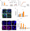
Neuronal delivery of Dopa‐EVs via dopamine receptors in vitro (a) Quantification of dopamine concentration in EVs using ELISA. The dotted line indicates the LOQ. (b) Schematic illustration of the binding affinity assay for Dopa‐EVs using biolayer interferometry with dopamine antibody. (c) Binding kinetics of ADSC‐EVs, Dopa‐EVs, and Dopa‐EVs with dopamine‐conjugated BSA (Dopa‐BSA) on dopamine antibody derivatized biosensors. Cellular uptake of Cy5.5‐labelled ADSC‐EVs or Dopa‐EVs in primary mouse neurons at 1 and 3 h (d), with corresponding fluorescence quantification (e). Scale bar, 20 µm. Cellular uptake of Cy5.5‐labelled ADSC‐EVs or Dopa‐EVs in SH‐SY5Y neuroblastoma cells pre‐incubated with a nonspecific isotype control (IgG Ab) or monoclonal antibodies against D1R and D2R (DR Ab) (f), with corresponding fluorescence quantification (g). Scale bar, 50 µm. Unpaired Student's t‐test. *p < 0.05, **p < 0.01, and ***p < 0.001. Data represent mean ± SD. ADSC, adipose‐derived mesenchymal stem cells; EVs, extracellular vesicles; LOQ, limit of quantitation.
FIGURE 4
FIGURE 4
Brain accumulation of Dopa‐EVs in PD mice (a) Representative fluorescence images of biodistribution of Free dye, Cy5.5‐labelled ADSC‐EVs, and Dopa‐EVs in 6‐OHDA‐induced PD mice as a function of time. (b) Quantification of in vivo fluorescence intensity of brain as a function of time (n = 4). (c) Representative ex vivo fluorescence images of major organs in α‐Syn PFF‐induced PD mice (n = 3). (d) Quantification of ex vivo fluorescence intensity of major organs. (e) Representative ex vivo fluorescence images of brains (n = 3). (f) Quantification of fluorescence intensity of brains (g) Quantification of fluorescence of brain homogenates. Data in (b) and (d) are analyzed by two‐way ANOVA and data in (f) and (g) analyzed by unpaired Student's t‐test. *p < 0.05, **p < 0.01, and ***p < 0.001. Data represent mean ± SEM. α‐Syn PFF, α‐synuclein preformedfibril; ADSC, adipose‐derived mesenchymal stem cells; EVs, extracellular vesicles; PD, Parkinson's disease.
FIGURE 5
FIGURE 5
Improvement of dopaminergic nigrostriatal degeneration by Dopa‐EVs in vivo PD mice (a) Schematic illustration of experimental design. (b) Pole test for discriminating PD induced mice 3 days after unilateral injection of 6‐OHDA. (c–f) Behavioural assessment of Sham (n = 5), Vehicle (6‐OHDA, n = 11) and Dopa‐EVs (6‐OHDA + Dopa‐EVs, n = 9) injected mice after 7 consecutive days of intravenous injection. Representative double immunostaining for GFAP (green) and tyrosine hydroxylase (TH, red) in the (g) striatum and (i) substantia nigra. Scale bar, 500 µm. (h) Quantification of the relative TH‐immunoreactive area in the ipsilateral region of the striatum compared to the contralateral region (n = 4). (j) Western blot analysis of TH and LC3 in the ipsilateral region of mice striatum. Quantification of TH (k) and total LC3‐II (li) protein expression levels in the ipsilateral region of mice striatum (n = 3). One‐way ANOVA followed by Tukey's post hoc test. *p < 0.05, **p < 0.01, and ***p < 0.001. Data represent mean ± SEM. ADSC, adipose‐derived mesenchymal stem cells; EVs, extracellular vesicles; TH, tyrosine‐hydroxylase.
FIGURE 6
FIGURE 6
Neuroprotective effect of Dopa‐EVs on 6‐OHDA in vitro via autophagy induction (a) Cell viability analysis using WST‐8 in primary mouse neuron. (b–c) Representative western blot analysis and quantification results of the expression levels of TH, Beclin‐1 and LC3‐II in primary neurons treated as indicated. (d) Representative double immunostaining for TH (green) and LC3 (red) in the primary neurons. Scale bar, 500 µm. (e) Quantification of the relative LC3 expression in TH+ area in primary neurons treated as indicated. One‐way ANOVA followed by Tukey's post hoc test. n = 4, *p < 0.05, **p < 0.01, and ***p < 0.001. Data represent mean ± SD. TH, tyrosine‐hydroxylase.
FIGURE 7
FIGURE 7
Therapeutic effect of Dopa‐EVs on α‐Syn PFF in vitro via autophagy induction. (a) Schematic diagram illustrating the treatment of Dopa‐EVs in primary mouse neurons induced with α‐Syn PFF. (b) Amelioration of α‐Syn PFF‐induced neuronal death in primary mouse neurons treated with Dopa‐EVs, as determined by the WST‐8 cell viability assay. (c) Representative Western blot analysis showing the expression levels of Parkin and LC3 in primary neurons treated as indicated. (d) Inhibition of α‐Syn PFF‐induced propagation and phosphorylation of α‐Syn (green) by ADSC‐EVs and Dopa‐EVs in primary neurons. Scale bar: 100 µm. (e) Quantitative analysis of the intensity of p‐α‐Syn over time (n = 3). (f) Schematic diagram showing the treatment of Dopa‐EVs or sonicated Dopa‐EVs in α‐Syn PFF‐induced primary mouse neurons. (g) Representative phospho‐α‐Syn (Ser129) immunostaining (green) in the Dopa‐EVs or sonicated Dopa‐EVs treated α‐Syn PFF‐induced primary mouse neurons. Scale bar, 100 µm. One‐way ANOVA followed by Tukey's post hoc test. n = 4, *p < 0.05, and **p < 0.01. Data represent mean ± SD. α‐Syn PFF, α‐synuclein preformedfibril; ADSC, adipose‐derived mesenchymal stem cells; EVs, extracellular vesicles.

References

    1. Anraku, Y. , Kuwahara, H. , Fukusato, Y. , Mizoguchi, A. , Ishii, T. , Nitta, K. , Matsumoto, Y. , Toh, K. , Miyata, K. , Uchida, S. , Nishina, K. , Osada, K. , Itaka, K. , Nishiyama, N. , Mizusawa, H. , Yamasoba, T. , Yokota, T. , & Kataoka, K. (2017). Glycaemic control boosts glucosylated nanocarrier crossing the BBB into the brain. Nature Communications, 8(1), 1001. 10.1038/s41467-017-00952-3 - DOI - PMC - PubMed
    1. Bahn, G. , Park, J.‐S. , Yun, U. J. , Lee, Y. J. , Choi, Y. , Park, J. S. , Baek, S. H. , Choi, B. Y. , Cho, Y. S. , Kim, H. K. , Han, J. , Sul, J. H. , Baik, S.‐H. , Lim, J. , Wakabayashi, N. , Bae, S. H. , Han, J.‐W. , Arumugam, T. V. , Mattson, M. P. , & Jo, D.‐G. (2019). NRF2/ARE pathway negatively regulates BACE1 expression and ameliorates cognitive deficits in mouse Alzheimer's models. Proceedings of the National Academy of Sciences, 116(25), 12516–12523. 10.1073/pnas.1819541116 - DOI - PMC - PubMed
    1. Burbulla, L. F. , Song, P. , Mazzulli, J. R. , Zampese, E. , Wong, Y. C. , Jeon, S. , Santos, D. P. , Blanz, J. , Obermaier, C. D. , Strojny, C. , Savas, J. N. , Kiskinis, E. , Zhuang, X. , Krüger, R. , Surmeier, D. J. , & Krainc, D. (2017). Dopamine oxidation mediates mitochondrial and lysosomal dysfunction in Parkinson's disease. Science, 357(6357), 1255–1261. 10.1126/science.aam9080 - DOI - PMC - PubMed
    1. Burns, J. A. , Kroll, D. S. , Feldman, D. E. , Kure Liu, C. , Manza, P. , Wiers, C. E. , Volkow, N. D. , & Wang, G.‐J. (2019). Molecular imaging of opioid and dopamine systems: Insights into the pharmacogenetics of opioid use disorders. Frontiers in Psychiatry, 10, 626. 10.3389/fpsyt.2019.00626 - DOI - PMC - PubMed
    1. Carlsson, A. , Lindqvist, M. , & Magnusson, T. (1957). 3,4‐Dihydroxyphenylalanine and 5‐hydroxytryptophan as reserpine antagonists. Nature, 180(4596), 1200. 10.1038/1801200a0 - DOI - PubMed

LinkOut - more resources