. 2025 Jan 7;13(1):e0216324.
doi: 10.1128/spectrum.02163-24. Epub 2024 Dec 6.

A unique Helicobacter pylori strain to study gastric cancer development

Affiliations

A unique Helicobacter pylori strain to study gastric cancer development

Jeannette M Whitmire et al. Microbiol Spectr. .

Abstract

Helicobacter pylori colonizes a majority of the human population worldwide and can trigger development of a variety of gastric diseases. Since the bacterium is classified as a carcinogen, elucidation of the characteristics of H. pylori that influence gastric carcinogenesis is a high priority. To this end, the Mongolian gerbil infection model has proven to be an important tool to study gastric cancer progression. However, only a small number of H. pylori strains have been evaluated in the gerbil model. Thus, to identify additional strains able to colonize and induce disease in this model, several H. pylori strains were used to infect Mongolian gerbils, and stomachs were harvested at multiple timepoints to assess colonization and gastric pathology. The USU101 strain reproducibly colonized Mongolian gerbils and induced gastric inflammation in the majority of the animals 1 month after infection. Adenocarcinoma or dysplasia was observed in the majority of gerbils by 2 months post-infection. To define the contribution of key virulence factors to this process, isogenic strains lacking cagA or vacA, along with restorant strains containing a wild-type (WT) copy of the genes, were studied. The ΔcagA USU101 strain colonized gerbils at levels similar to WT, but did not induce comparable levels of inflammation or disease. In contrast, the ΔvacA USU101 strain did not colonize gerbils, and the stomach pathology resembled that of the mock-infected animals. The restorant USU101 strains expressed the CagA and VacA proteins in vitro, and in vivo experiments with Mongolian gerbils showed a restoration of colonization levels and inflammation scores comparable to those observed in WT USU101. Our studies indicate that the USU101 strain is a valuable tool to study H. pylori-induced disease.IMPORTANCEGastric cancer is the fifth leading cause of cancer-related death globally; the majority of gastric cancers are associated with Helicobacter pylori infection. Infection of Mongolian gerbils with H. pylori has been shown to result in induction of gastric cancer, but few H. pylori strains have been studied in this model; this limits our ability to fully understand gastric cancer pathogenesis in humans because H. pylori strains are notoriously heterogenous. Our studies reveal that USU101 represents a unique H. pylori strain that can be added to our repertoire of strains to study gastric cancer development in the Mongolian gerbil model.

Keywords: CagA; H. pylori; Mongolian gerbils; VacA; gastric adenocarcinoma.


Conflict of interest statement

The authors declare no conflict of interest.

Figures
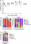
Fig 1
Fig 1
Gerbil infections with USU101 and J166. (A) Colonization levels (CFU/g stomach tissue) of J166 (black circles) and USU101 (open triangles) at five timepoints post-infection. (B) Percent of gerbils (n = 5) displaying the indicated gastric histologic diagnoses for each strain at five timepoints post-infection. (C) Percent of gerbils (n = 10) displaying the indicated gastric histologic diagnoses for USU101 at three timepoints post-infection in a study conducted at a different institution (University of South Carolina School of Medicine).
Fig 2
Fig 2
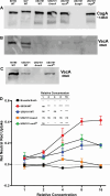
Western blot analysis. (A) CagA Western blot of the indicated strains. WT 60190 and WT G27 strains are shown for comparison. (B) VacA Western blot of the indicated strains. WT 60190 and WT G27 strains are shown for comparison. (C) VacA Western blot of broth culture supernatants from the indicated strains. The WT 60190 strain was included for comparison. (cagAR = cagA restorant strain; vacAR = vacA restorant strain). (D) Cell vacuolation induced by broth culture supernatants from the indicated strains as indicated by neutral red uptake. The inset shows statistical significance as determined by an ordinary one-way analysis of variance with a Dunnett’s multiple comparison test. The Brucella broth control at each of the relative concentrations was used as the comparison group to each of the other indicated strains and the symbols denote the following: -- the comparator group, ns no significance, * P < 0.05, ** P < 0.005, *** P < 0.0005, and **** P < 0.0001.
Fig 3
Fig 3
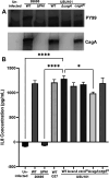
CagA translocation and IL-8 induction. (A) Western blots of AGS cells infected with the indicated strains and probed with PY99 to detect phosphorylated CagA and anti-CagA to detect total CagA. The 26695 WT strain and 26695 ∆cag PAI mutant are included for comparison. (B) Induction of IL-8 production by AGS cells following infection with the indicated strains. The y-axis shows IL-8 concentrations in supernatants from AGS cells. WT 26695, 26695 ∆cag PAI mutant, and WT G27 were included for comparison. Statistical significance was analyzed by comparing each group to WT USU101 using a one-way analysis of variance with a Dunnett’s multiple test correction; significant differences are noted on the graph. (cagAR = cagA restorant strain; vacAR = vacA restorant strain) (*P < 0.05; ****P < 0.0001).
Fig 4
Fig 4
Colonization levels of USU101 and isogenic strains. Colonization levels (CFU/g stomach tissue) for the indicated strains are plotted at the indicated timepoints post-infection. Each point indicates the colonization level of an individual gerbil, and the bars indicate the mean for the animals in the group at each timepoint. The dashed line demarcates the limit of detection (LOD) of 10–15 CFU, and non-colonized gerbils are plotted at the LOD. Statistical significance at each timepoint relative to the USU101 WT colonization level at the same timepoint was assessed using the Kruskal–Wallis test with Dunn’s correction for multiple comparisons. Significant differences are indicated as follows: * P < 0.05, ** P < 0.01, *** P ≤ 0.001.
Fig 5
Fig 5
Inflammation scores. Inflammation scores were generated by combining the scores obtained from the antrum and corpus and are plotted for the indicated strains at each of the noted timepoints post-infection. Each point indicates the combined inflammation score for an individual gerbil, and the bars indicate the mean for the animals in the group at each timepoint. A two-way analysis of variance with multiple comparisons was performed to identify statistically significant differences between groups. (**P < 0.01; ****P < 0.0001).
Fig 6
Fig 6
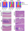
Gastric pathology determination. (A) Percent of gerbils (n = 10) displaying the indicated gastric histologic diagnoses for each strain at three timepoints post-infection. (B) Representative images of stomach tissue stained with hematoxylin and eosin for each strain at 3 months post-infection. All images were taken at 100× magnification.

References

    1. Ernst PB, Gold BD. 2000. The disease spectrum of Helicobacter pylori: the immunopathogenesis of gastroduodenal ulcer and gastric cancer. Annu Rev Microbiol 54:615–640. doi:10.1146/annurev.micro.54.1.615 - DOI - PubMed
    1. Asaka M, Kudo M, Kato M, Sugiyama T, Takeda H. 1998. Review article: Long-term Helicobacter pylori infection--from gastritis to gastric cancer. Aliment Pharmacol Ther 12 Suppl 1:9–15. doi:10.1111/j.1365-2036.1998.00007.x - DOI - PubMed
    1. International Agency for Research on Cancer . 1994. Infection with Helicobacter pylori, p 177–240. In Monographs on the evaluation of carcinogenic risks to humans. Vol. 61. - PMC - PubMed
    1. Ansari S, Yamaoka Y. 2019. Helicobacter pylori virulence factors exploiting gastric colonization and its pathogenicity. Toxins (Basel) 11:677. doi:10.3390/toxins11110677 - DOI - PMC - PubMed
    1. Baj J, Forma A, Sitarz M, Portincasa P, Garruti G, Krasowska D, Maciejewski R. 2020. Helicobacter pylori virulence factors-mechanisms of bacterial pathogenicity in the gastric microenvironment. Cells 10:27. doi:10.3390/cells10010027 - DOI - PMC - PubMed

MeSH terms

LinkOut - more resources