. 2024 Dec 2;65(14):15.
doi: 10.1167/iovs.65.14.15.

Elevation of Granulocyte Colony Stimulating Factor in Human AMD Donor RPE-Choroid

Affiliations

Elevation of Granulocyte Colony Stimulating Factor in Human AMD Donor RPE-Choroid

Kelly Mulfaul et al. Invest Ophthalmol Vis Sci. .

Abstract

Purpose: Choroidal inflammation, complement deposition, and accumulation of C-reactive protein (CRP) are involved in age-related macular degeneration (AMD) pathology. The pro-inflammatory signals that regulate immune cell recruitment in the choroid of patients with AMD remain to be determined. We performed cytokine profiling of human AMD and age-matched control donor tissue to identify inflammatory molecules upregulated in AMD tissue.

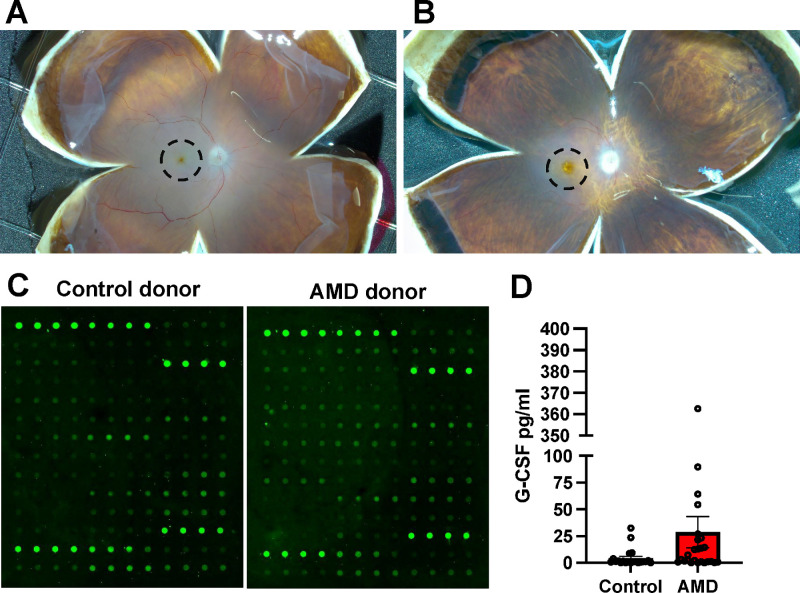
Methods: Protein was isolated from 25 AMD and 21 control donor RPE/choroid macular punches. Total protein was quantified, and 50 µg assayed for expression of 40 cytokines using an inflammation array. We validated the elevated expression of granulocyte colony stimulating factor (G-CSF) protein by ELISA in a second cohort of 22 control and 26 AMD donors. To identify an AMD associated stressor responsible for upregulating G-CSF we assayed for changes in G-CSF protein secretion in RPE/choroid organ cultures treated with the monomeric (m)CRP, an inflammatory protein elevated in AMD.

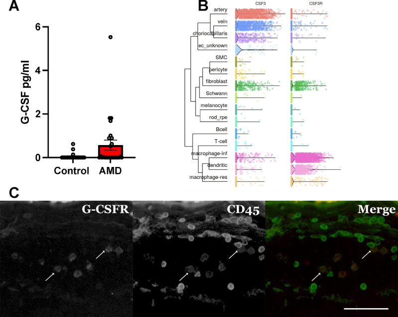
Results: Using a multiplex array, we identified elevated G-CSF protein in the choroid of AMD donors compared to age-matched non-AMD controls. Differential expression of G-CSF was confirmed via ELISA in an independent cohort of samples (P = 0.01). The mCRP, which is deposited in AMD choroids, increased G-CSF protein secretion in RPE/choroid organ cultures. Single nuclei RNA sequencing identified choroidal endothelial cells and fibroblasts as the primary cell types responsible for increased G-CSF secretion in response to mCRP. The G-CSF receptor is expressed primarily by choroidal macrophages and dendritic cells and anti-G-CSFR colocalizes with anti-CD45 and anti-CD68 in human donor choroid tissue.

Conclusions: Elevated G-CSF expression in AMD donor tissue as a result of increased levels of mCRP may be involved in immune cell recruitment in AMD contributing to inflammatory stress in the choroid.


Conflict of interest statement

Disclosure: K. Mulfaul, None; A.H. Khan, None; S.G. Schwarte, None; A.P. Voigt, None; R.F. Moore, None; L.A. Potempa, None; K. Wang, None; T.E. Scheetz, None; E.M. Stone, None; B.A. Tucker, None; R.F. Mullins, None

Figures

Figure 1.
Figure 1.
Protein was isolated from macular RPE/choroid punches collected from 21 age-matched control (A) and 25 AMD donor (B) eyes. Cytokine array analysis of bulk protein detected altered cytokine levels in control and AMD donor RPE/choroid tissue (C). The chemokine G-CSF showed a trend toward elevation (6.5-fold) in AMD donor tissue P value = 0.06.
Figure 2.
Figure 2.
Elevation of G-CSF cytokine was validated by ELISA in an additional cohort of 22 control and 26 AMD donor protein samples P value = 0.01 (A). The gene encoding for G-CSF, CSF3, is expressed by artery, vein, choriocapillaris, and fibroblasts and the G-CSF receptor, CSF3R is expressed on fibroblasts, macrophage-inflammatory and dendritic cells in human single cell RNA sequencing datasets (B). G-CSFR was visualized on a subset of CD45 positive cells in human donor choroid (C). Scale bar = 50 µm.
Figure 3.
Figure 3.
Organ cultures of human RPE-choroid were collected as pairwise punches during donor eye dissection illustrated in schematic (A) the optic nerve head is indicated by an open circle and the macula by a circle labeled MAC. Tissue was cultured in the presence of either PBS as vehicle or with 20 µg/mL of monomeric CRP. After 22 hours of incubation, media samples were collected, and levels of G-CSF were measured in paired punches in 6 human donors using ELISA punch 1 = P value = 0.03, punch 2 = P value = 0.004, and punch 3 = P value = 0.04 (B, C, D). An average value was taken per donor per treatment condition and showed a 2-fold increase in G-CSF in response to monomeric CRP P value = 0.009 (E). Single nuclei RNA seq on a human RPE-Choroid punch identified fibroblasts and choriocapillaris endothelial cells as the primary cell types responsible for increased G-CSF expression (F).

References

    1. Mullins RF, Johnson MN, Faidley EA, Skeie JM, Huang J.. Choriocapillaris vascular dropout related to density of drusen in human eyes with early age-related macular degeneration. Invest Ophthalmol Vis Sci. 2011; 52(3): 1606–1612. - PMC - PubMed
    1. Lutty GA, McLeod DS, Bhutto IA, Edwards MM, Seddon JM.. Choriocapillaris dropout in early age-related macular degeneration. Exp Eye Res. 2020; 192: 107939. - PMC - PubMed
    1. Kar D, Corradetti G, Swain TA, et al. .. Choriocapillaris impairment is associated with delayed rod-mediated dark adaptation in age-related macular degeneration. Invest Ophthalmol Vis Sci. 2023; 64(12): 41. - PMC - PubMed
    1. McLeod DS, Bhutto I, Edwards MM, Silver RE, Seddon JM, Lutty GA.. Distribution and quantification of choroidal macrophages in human eyes with age-related macular degeneration. Invest Ophthalmol Vis Sci. 2016; 57(14): 5843–5855. - PMC - PubMed
    1. Bhutto IA, McLeod DS, Jing T, Sunness JS, Seddon JM, Lutty GA.. Increased choroidal mast cells and their degranulation in age-related macular degeneration. Br J Ophthalmol. 2016; 100(5): 720–726. - PMC - PubMed

Substances