

ILAE genetic literacy series: Focal cortical dysplasia
- PMID: 39641771
- PMCID: PMC11829622
- DOI: 10.1002/epd2.20308
ILAE genetic literacy series: Focal cortical dysplasia
Abstract
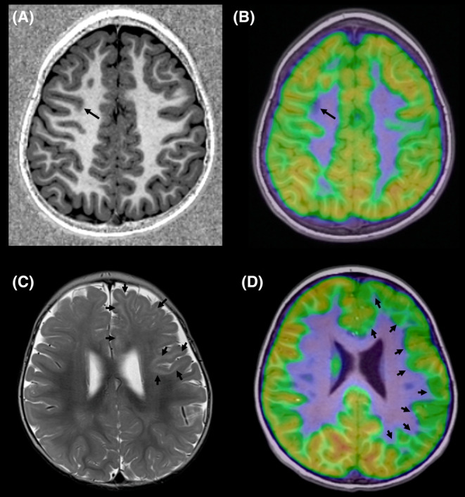
Focal cortical dysplasia (FCD) is a common cause of drug-resistant focal epilepsy in children and young adults and is often surgically remediable. The genetics of FCD are increasingly understood due to the ability to perform genomic testing including deep sequencing of resected FCD tissue specimens. There is clear evidence that FCD type II occurs secondary to both germline and somatic mTOR pathway variants, while emerging literature supports the role of SLC35A2, a glycosylation gene, in mild malformation of cortical development with oligodendroglial hyperplasia and epilepsy (MOGHE). Herein, we provide a review of FCDs focusing on their clinical phenotypes, genetic basis, and management considerations when performing genetic testing in this patient group.
Keywords: GATOR1; bottom‐of‐sulcus dysplasia; familial focal epilepsy; focal epilepsy genetics; mTOR.
© 2024 The Author(s). Epileptic Disorders published by Wiley Periodicals LLC on behalf of International League Against Epilepsy.
Conflict of interest statement
Authors have no conflicts of interest to disclose.
Figures
References
-
- Bernasconi A, Cendes F, Theodore WH, Gill RS, Koepp MJ, Hogan RE, et al. Recommendations for the use of structural magnetic resonance imaging in the care of patients with epilepsy: a consensus report from the international league against epilepsy neuroimaging task force. Epilepsia. 2019;60(6):1054–1068. - PubMed
-
- Blumcke I, Spreafico R, Haaker G, Coras R, Kobow K, Bien CG, et al. Histopathological findings in brain tissue obtained during epilepsy surgery. N Engl J Med. 2017;377(17):1648–1656. - PubMed
-
- Cepeda C, André VM, Vinters HV, Levine MS, Mathern GW. Are cytomegalic neurons and balloon cells generators of epileptic activity in pediatric cortical dysplasia? Epilepsia. 2005;46(s5):82–88. - PubMed
-
- Stephenson SEM, Maixner WJ, Barton SM, D'Arcy C, Mandelstam SA, MacGregor D, et al. Resection of tuber centers only for seizure control in tuberous sclerosis complex. Epilepsy Res. 2021;171:106572. - PubMed
Publication types
MeSH terms
Grants and funding
LinkOut - more resources
Full Text Sources
Miscellaneous
