. 2024 Dec 10;121(50):e2413011121.
doi: 10.1073/pnas.2413011121. Epub 2024 Dec 6.

Understanding paralogous epilepsy-associated GABAA receptor variants: Clinical implications, mechanisms, and potential pitfalls

Affiliations

Understanding paralogous epilepsy-associated GABAA receptor variants: Clinical implications, mechanisms, and potential pitfalls

Anthony S H Kan et al. Proc Natl Acad Sci U S A. .

Abstract

Recent discoveries have revealed that genetic variants in γ-aminobutyric acid type A (GABAA) receptor subunits can lead to both gain-of-function (GOF) and loss-of-function (LOF) receptors. GABAA receptors, however, have a pseudosymmetrical pentameric assembly, and curiously diverse functional outcomes have been reported for certain homologous variants in paralogous genes (paralogous variants). To investigate this, we assembled a cohort of 11 individuals harboring paralogous M1 proline missense variants in GABRA1, GABRB2, GABRB3, and GABRG2. Seven mutations (α1P260L, α1P260S, β2P252L, β3P253L, β3P253S, γ2P282A, and γ2P282S) in α1β2/3γ2 receptors were analyzed using electrophysiological examinations and molecular dynamics simulations. All individuals in the cohort were diagnosed with developmental and epileptic encephalopathy, with a median seizure onset age of 3.5 mo, and all exhibited global developmental delay. The clinical data for this cohort aligned with established GABAA receptor GOF but not LOF cohorts. Electrophysiological assessments revealed that all variants caused GOF by increasing GABA sensitivity by 3- to 23-fold. In some cases, this was accompanied by LOF traits such as reduced maximal current amplitude and enhanced receptor desensitization. The specific subunit mutated and whether the mutation occurred in one or two subunits within the pentamer influenced the overall effects. Molecular dynamics simulations confirmed similar structural changes from all mutations, but with position-dependent asymmetry. These findings establish that paralogous variants affecting the 100% conserved proline residue in the M1 transmembrane helix of GABAAR subunits all lead to overall GOF traits. The unexpected asymmetric and mixed effects on receptor function have broader implications for interpreting functional analyses for multimeric ion-channel proteins.

Keywords: GABRA1; GABRB3; GABRG2; epilepsy; neurodevelopmental disorders.


Conflict of interest statement

Competing interests statement:P.C. is Executive Vice President, Research at the company Saniona in Denmark. The remaining authors declare no competing interests.

Figures

Fig. 1.
Fig. 1.
The M1 proline residue is fully conserved in GABAAR subunits. (A) Top-Left: Cryo-EM structure of a pentameric GABAAR viewed from the extracellular side (PDB: 6HUP). Right: Cryo-EM structure of an α1 subunit viewed along the plasma membrane illustrating the M1 (purple) and M2 (teal) transmembrane domain (TMD) helices, and the position of the conserved M1 proline (red) and presumed pathogenic variants. ECD: extracellular domain; ICD: intracellular domain. (B) Amino acid sequence alignment of the M1 and M2 TMD helixes of α(1-5), β(1-3), γ(1-2), and δ subunits with the conserved proline bracketed in red. Based on a literature review, variants with published functional data are highlighted bold in yellow, with most of them located in fully (dark gray) or highly (light gray) conserved residues (–16).
Fig. 2.
Fig. 2.
Clinical features of individuals with M1 proline variants in GABRA1, GABRB2, GABRB3, and GABRG2. (A) Distribution of age of seizure onset, epileptic syndromes, and eight key clinical features in the M1 proline cohort are shown. Solid line and dotted lines in the violin plot depict the median and interquartile range, respectively. EIDEE: Early infantile developmental and epileptic encephalopathy; IESS: Infantile epileptic spasms syndrome; DEE: Developmental and epileptic encephalopathy. Movement disorders* include dystonia, dyskinesia, choreoathetosis, and athetosis. For detailed information, see Dataset S1. (B) Age of seizure onset of the M1 proline cohort (n = 11) was compared against the published LOF (n = 74) and GOF (n = 48) cohorts from GABRA1/B2/B3 (–5). The age of onset in the M1 proline cohort was lower than the LOF cohort (Mann–Whitney test, P < 0.0001) but statistically insignificant to the GOF cohort (P = 0.87). Survival analysis also showed that the M1 proline cohort had a significantly higher seizure risk earlier in life than the LOF cohort (Mantel-Cox test, P < 0.0001), but no different to the GOF cohort (P = 0.17). Frequency comparisons of five clinical features showed that both M1 and GOF cohorts share similar phenotypic frequencies which differ significantly from those observed in the LOF cohort. OR comparisons were subsequently performed. ORs were displayed along with 95% CI, and statistical significance was determined using two-tailed Fisher’s exact test. OR could not be determined (ND) when either the lower or upper limit of the OR was 0 or infinite, respectively. Lilac circles indicate that the clinical feature is more prevalent in the M1 proline cohort, and red indicates that it is more prevalent in the LOF cohort. Gray indicates no difference in prevalence between the cohorts.
Fig. 3.
Fig. 3.
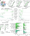
Functional properties of GABAARs containing M1 proline mutations in the α1 subunit. Variant receptors are compared to the wildtype (wt) receptor. Statistical analyses results are presented as **P < 0.01; ***P < 0.001; ****P < 0.0001. (A) α1 subunit mutations are introduced in the 3rd and 5th construct positions of the concatenated receptor. Middle and Right: GABA concentration–response relationships are presented as mean ± SD for α1β3γ2 GABAARs containing either one or two α1 P260S mutation(s) with mean differences in LogEC50 (ΔLogEC50) between variant (n = 11-20) and wt (n = 28) receptors. (B) Changes in GABA sensitivity [n = 11-20 (variants) and n = 156 (wt)] and maximal GABA-evoked current amplitudes [n = 24-31 (variants) and n = 332 (wt)] are depicted for receptors containing one or two α1 P260S or P260L mutations. For detailed information, see Datasets S2–S4. (C) Desensitization properties are presented as representative traces, current decay rates, and steady-state currents for variant and wt receptors (n = 21-26).
Fig. 4.
Fig. 4.
Functional properties of GABAARs containing M1 proline mutations in the β2 or β3 subunits. See Fig. 3 legend for a description of figure layout and statistical methods. (A) β3 subunit mutations are introduced in the 2nd and 4th construct positions of the concatenated receptor. GABA concentration–response relationships are presented for α1β3γ2 GABAARs containing either one or two β3 P253L mutation(s) with indicated mean difference in LogEC50 (ΔLogEC50) between the variant (n = 9-15) and wt (n = 29) receptors. (B) Changes in GABA sensitivity [n = 9-28 (variants) and n = 156 (wt)] and maximum GABA-evoked current amplitude [n = 22-48 (variants) n = 332 (wt)] are depicted for receptors containing either one or two β2 P252L, β3 P253L, or P253S mutations. For detailed information, see Datasets S2–S4. (C) Desensitization properties are presented as representative traces, current decay rates, and steady-state currents for variant and wt receptors (n = 22-28).
Fig. 5.
Fig. 5.
Functional properties of GABAARs containing M1 proline mutations in the γ2 subunit. See Fig. 3 legend for a detailed description of the figure layout and statistical methods. (A) γ2 subunit mutations are introduced in the 1st construct position of the concatenated receptor. GABA concentration–response relationships are displayed for α1β3γ2 GABAARs containing γ2 P282A (n = 14) or P282S (n = 21) mutations and wildtype receptors (n = 47). (B) Changes in GABA sensitivity [n = 14-21 (variants) and n = 156 (wt)] and maximum GABA-evoked current amplitude [n = 36-48 (variants) and n = 332 (wt)] are depicted for receptors containing the γ2 P282A or P282S mutation. For detailed information, see Datasets S2–S4. (C) Desensitization properties are presented as representative traces, current decay rates, and steady-state currents for variant and wt receptors (n = 20-27).
Fig. 6.
Fig. 6.
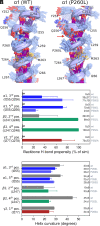
Analysis of the M1 helix curvature from 500 ns MD simulations of the α1β2γ2 GABAAR. (A) An ensemble of simulation frames for the α1 subunit M1 helix in the 3rd position with wildtype sequence (Left) and with the P260L mutant (Right), 10 frames are captured uniformly over the last 300 ns of three simulation replicas (30 frames in total). Backbone H-bonds are shown as purple lines, protein sidechain atoms except for P260 (Left) and P260L (Right) are faded for clarity. The red arrow highlights the ability of Q256 to form backbone H-bond in the M1 helix containing P260L but not wildtype M1 helix. (B) H-bond propensity was measured as percentage of simulation frames where H-bonds occurred from the backbone carbonyl oxygen of α1 I255/Q256, β2 L247/Q248, and γ2 I277/Q278 to NH atoms below them. H-bonds were measured as an interaction to either but not both residues, # denotes near-zero value (no H-bonds observed). (C) Helix curvature around α1 I255, β2 L247, and γ2 I277. For both plots, means ± SD are obtained from the last 300 ns of triplicate MD simulations.

References

    1. Chua H. C., Chebib M., GABA(A) receptors and the diversity in their structure and pharmacology. Adv. Pharmacol. 79, 1–34 (2017). - PubMed
    1. Maljevic S., et al. , Spectrum of GABAA receptor variants in epilepsy. Curr. Opin. Neurol. 32, 183–190 (2019). - PubMed
    1. Musto E., et al. , GABRA1-related disorders: From genetic to functional pathways. Ann. Neurol. 95, 27–41 (2024). - PubMed
    1. Mohammadi N. A., et al. , Distinct neurodevelopmental and epileptic phenotypes associated with gain- and loss-of-function GABRB2 variants. EBioMedicine 106, 105236 (2024). - PMC - PubMed
    1. Absalom N. L., et al. , Gain-of-function and loss-of-function GABRB3 variants lead to distinct clinical phenotypes in patients with developmental and epileptic encephalopathies. Nat. Commun. 13, 1822 (2022). - PMC - PubMed