. 2024 Dec 6;23(1):270.
doi: 10.1186/s12943-024-02184-8.

Chimeric antigen receptor macrophages targeting c-MET(CAR-M-c-MET) inhibit pancreatic cancer progression and improve cytotoxic chemotherapeutic efficacy

Affiliations

Chimeric antigen receptor macrophages targeting c-MET(CAR-M-c-MET) inhibit pancreatic cancer progression and improve cytotoxic chemotherapeutic efficacy

Huaijin Zheng et al. Mol Cancer. .

Abstract

Background: Pancreatic ductal adenocarcinoma (PDAC) is one of the most malignant tumors. Macrophages are abundant in the tumor microenvironment, making them an attractive target for therapeutic intervention. While current immunotherapies, including immune checkpoint inhibition (ICI) and chimeric antigen receptor T (CAR-T) cells, have shown limited efficacy in pancreatic cancer, a novel approach involving chimeric antigen receptor macrophages (CAR-M) has, although promising, not been explored in pancreatic cancer. In this study, we first investigated the role of CAR-M cells targeting c-MET in pancreatic cancer.

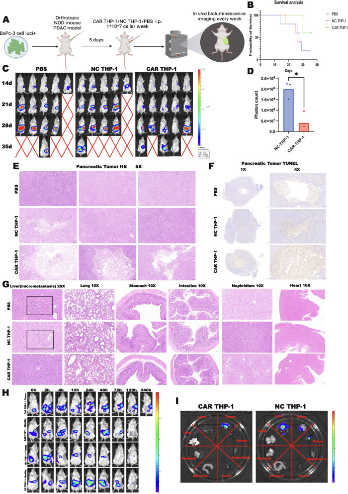
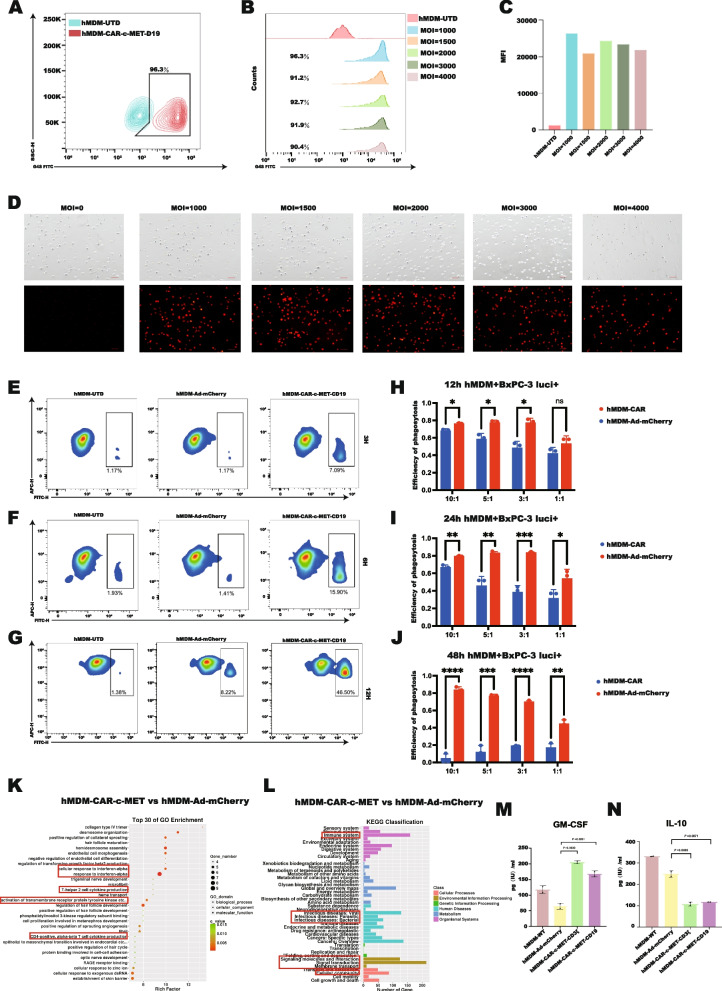
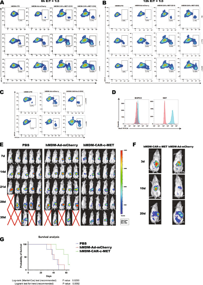
Methods: The effectiveness and rationality of c-MET as a target for CAR-M in pancreatic cancer were validated through bioinformatic analyses and immunohistochemical staining of samples from pancreatic cancer patients. We utilized flow cytometry and bioluminescence detection methods to demonstrate the specific binding and phagocytic killing effect of CAR-M on pancreatic cancer cells. Additionally, we observed the process of CAR-M engulfing pancreatic cancer cells using confocal microscopy and a long-term fluorescence live cell imaging system. In an in situ tumor model transplanted into NOD/SCID mice, we administered intraperitoneal injections of CAR-M to confirm its inhibitory function on pancreatic cancer. Furthermore, we validated these findings in human monocyte-derived macrophages (hMDM).

Results: Bioinformatics and tumor tissue microarray analyses revealed significantly higher expression levels of c-MET in tumor tissues, compared to the paired peritumoral tissues, and higher c-MET expression correlated with worse patient survival. CAR-M cells were engineered using human monocytic THP-1 cell line and hMDM targeting c-MET (CAR-M-c-MET). The CAR-M-c-MET cells demonstrated highly specific binding to pancreatic cancer cells and exhibited more phagocytosis and killing abilities than the pro-inflammatory polarized control macrophages. In addition, CAR-M-c-MET cells synergized with various cytotoxic chemotherapeutic drugs. In a NOD/SCID murine model, intraperitoneally injected CAR-M-c-MET cells rapidly migrated to tumor tissue and substantially inhibited tumor growth, which did not lead to obvious side effects. Cytokine arrays and mRNA sequencing showed that CAR-M-c-MET produced higher levels of immune activators than control macrophages.

Conclusions: This study provides compelling evidence for the safety and efficacy of CAR-M therapy in treating pancreatic cancer. The results demonstrate that CAR-M-c-MET significantly suppresses pancreatic cancer progression and enhances the effectiveness of cytotoxic chemotherapy. Remarkably, no discernible side effects occur. Further clinical trials are warranted in human pancreatic cancer patients.

Keywords: Chemotherapy; Chimeric antigen receptor macrophage; Pancreatic cancer; c-MET.


Conflict of interest statement

Declarations. Ethics approval and consent to participate: The Institutional Review Board of Peking Union Medical College Hospital approved this animal study (February 15th, 2023, XHDW-2022–126). Human blood for this study was ethically sourced and analyzed with approval from the Roc Rock Biotechnology Ethics Committee (approval number 2022,002). Research adhered to medical standards and regulations. Competing interests: The authors declare no competing interests.

Figures

Fig. 1
Fig. 1
A Bioinformatic analysis showed the different expression levels of c-MET in pancreatic cancer tissues compared to normal pancreatic tissues. B Bioinformatic analysis showed correlations between c-MET expression and overall survival in pancreatic cancer patients. C Bioinformatic analysis showed correlation between c-MET expression and progression-free survival in pancreatic cancer patients. D Representative IHC staining of c-MET expression in tumor tissue and peritumoral tissues. E Representative IHC staining of different levels of c-MET expression in tumor tissue. F Higher IHC score of c-MET predicted poorer overall survival of patients after surgery. G Spearman analysis showed a positive correlation between CA19-9 and c-MET. H the details of the IHC score of c-MET in all 76 cases of tumor tissue and its paired peritumoral tissue. I A paired t-test was used to test the c-MET IHC scores of tumor and peritumoral tissue. J FCM results showed that the BxPC-3 cell line had the highest expression of c-MET (***, p < 0.001;****, p < 0.0001)
Fig. 2
Fig. 2
A Schematic diagram of the CAR structure and CAR THP-1 construction, induction, phagocytosis, and killing effect. B Purification of CAR THP-1 by FCM sorting. C FCM showed that CAR THP-1 could effectively bind His-tag hu-c-MET recombinant protein. D CAR THP-1 or NC THP-1 was co-cultured with BxPC-3 mcherry + at a ratio of 5:1, and the co-culture system was detected by FCM at 30 min, 2 h, 4 h, and 6 h. E CAR THP-1 or NC THP-1 was co-cultured with BxPC-3 mcherry + at a ratio of 10:1 for 24 h, and residual BxPC-3 mcherry + cells were detected by FCM. F CAR THP-1 or NC THP-1 was co-cultured with BxPC-3 mcherry + at a ratio of 5:1 for 24 h; residual BxPC-3 mcherry + cells were detected by FCM
Fig. 3
Fig. 3
A CAR THP-1 or NC THP-1 and BxPC-3 luci + cells were co-cultured for 24 h at different E: T ratios and bioluminescence imaging was performed in the co-culture system. B CAR THP-1 or NC THP-1 and BxPC-3 luci + cells were co-cultured at 5:1 for different times, and bioluminescence imaging was performed in the co-culture system. C CAR THP-1 or NC THP-1 and BxPC-3 were co-cultured for 24 h at different E: T ratios, and LDH content in the supernatant of the co-culture system was detected. D The co-culture system was photographed by fluorescence confocal microscope, and the process of macrophages extending pseudopodia, endocytosis, and lysis of BxPC-3 cells.(**, p < 0.01;***, p < 0.001)
Fig. 4
Fig. 4
A Schematic diagram of the flow of CAR THP-1 combined with cytotoxic chemotherapy drugs to intervene in BxPC-3 luci + cells. B BxPC-3 luci + cells were treated with CAR THP-1 or NC THP-1 combined with different cytotoxic chemotherapy drugs, and the residual BxPC-3 luci + cells were detected by bioluminescence imaging. C LDH content test in the supernatant of the treatment systems (*, p < 0.05;**, p < 0.01;***, p < 0.001;****, p < 0.0001)
Fig. 5
Fig. 5
A Co-culture of BxPC-3 cells with CAR THP-1 or NC THP-1, macrophages were sorted out by FCM and subjected to RNA-seq, and the co-culture supernatant was detected by cytokine microarray. B Quantitative detection and T-test of GM-CSF in co-culture supernatant. C Quantitative detection and T-test of IL-10 in co-culture supernatant. D Volcano map of differentially expressed genes by RNA-seq of CAR THP-1/NC THP-1. E Heat map of CAR THP-1/NC THP-1 RNA-seq. F-I GO and KEGG enrichment of differentially expressed genes. (J-K) CAR THP-1 and NC THP-1 coculture with or without BxPC-3 were sorted for qPCR (**, p < 0.01;***, p < 0.001)
Fig. 6
Fig. 6
A Flow chart of establishment of orthotopic pancreatic transplantation tumor model in NOD/SCID mice and verification of anti-tumor effect of CAR THP-1 i.p. B Survival curves of orthotopic pancreatic transplantation tumor model. C Bioluminescence image of orthotopic pancreatic transplantation tumor model. D Quantitative analysis and t-test of tumor burden on the last bioluminescence images of orthotopic pancreatic transplantation tumor model. E HE staining of tumor tissue. F TUNEL detection of tumor tissues. G HE staining of liver (micrometastasis), lung, stomach, intestine, kidney and heart. (H) CAR THP-1 luci + or NC THP-1 luci + cells were intraperitoneally injected into orthotopically tumor-bearing mice and healthy mice, and the biodistribution of THP-1 cells was detected by bioluminescence. I CAR THP-1 luci + /NC THP-1 luci + cells were intraperitoneally injected into orthotopically tumor-bearing mice and healthy mice for 5 days, and the distribution of THP-1 cells in various vital organs of mice was detected by bioluminescence (*, p < 0.05)
Fig. 7
Fig. 7
A-C FCM analysis of the G4S tag of CAR-M-hMDM represents transfection efficiency. D The positive rate of hMDM mcherry + under different MOI transfection conditions was photographed by fluorescence microscope. E-G FCMdetected the proportion of double positive macrophages in the co-culture system after 3,6,12 h of CAR-M-hMDM and control macrophages co-cultured with BxPC-3 cells. H-J Residual tumor cells in the co-culture system was detected by bioluminescence after CAR-M-hMDM and control macrophages co-cultured with BxPC-3 luci + cells for 12,24,48 h, and the phagocytosis efficiency was calculated. K, L After co-cultured with BxPC-3 cells, RNA-seq was performed on macrophages. GO and KEGG enrichment of differentially expressed genes. M, N Quantitative detection and T test of GM-CSF and IL-10 in CAR-M-hMDM, hMDM-UTD and hMDM-Ad-mCherry co-culture supernatant (*, p < 0.05; **, p < 0.01;***, p < 0.001)
Fig. 8
Fig. 8
A, B hMDM-UTD,hMDM-Adv,hMDM-CAR coculture with BxPC-3, Mia Paca 2,PANC-1 cell for 6 h or 12 h at a ratio of 1:3. C hMDM-UTD,hMDM-Adv and hMDM-CAR-HER2 coculture with BxPC-3 and N87 for 12 h at a ratio of 1:3. D BxPC-3, N87 HER2 expression detection. E the dynamic tumor burden after different treatments. F the biodistribution of hMDM-CAR after i.p. G CAR-c-MET-hMDM could prolong the survival of the orthotopic tumor bearing mice

References

    1. Bray, F., et al., Global cancer statistics 2022: GLOBOCAN estimates of incidence and mortality worldwide for 36 cancers in 185 countries. CA Cancer J Clin, 2024. - PubMed
    1. Zhao Y, et al. Chinese expert consensus on minimally invasive radical surgery for pancreatic ductal adenocarcinoma (version 2022). Journal of Pancreatology. 2022;5(3):111–7. - PubMed
    1. Wu W, et al. Real-world study of surgical treatment of pancreatic cancer in China: annual report of China Pancreas Data Center (2016–2020). Journal of Pancreatology. 2022;5(1):1–9.
    1. Conroy T, et al. FOLFIRINOX versus gemcitabine for metastatic pancreatic cancer. N Engl J Med. 2011;364(19):1817–25. - PubMed
    1. Xu ZH, et al. Insight of pancreatic cancer: recommendations for improving its therapeutic efficacy in the next decade. Journal of Pancreatology. 2022;5(2):58–68.

MeSH terms

Substances

LinkOut - more resources